

Early MRI and intraoperative findings in rapidly destructive osteoarthritis of the hip: A case report
- PMID: 25603485
- PMCID: PMC4353947
- DOI: 10.1016/j.ijscr.2015.01.009
Early MRI and intraoperative findings in rapidly destructive osteoarthritis of the hip: A case report
Abstract
Introduction: The pathophysiology of rapidly destructive hip osteoarthritis (OA) of the hip is still unclear. Also, there have been only few reports on the initial stage of the disease. We report a case of an initial-stage rapidly destructive hip OA, documented by magnetic resonance imaging and intraoperative findings.
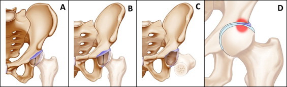
Presentation of case: A 77-year-old woman reported left hip pain without any antecedent trauma. Initial radiographs showed no obvious abnormality. After 4 months of conservative therapy, radiographs showed progressive joint-space narrowing and T1-weighted magnetic resonance images revealed a bone-marrow edema pattern not only on the femoral head but also on the lateral side of the acetabulum. Then during total hip arthroplasty, we found extensive inversion of the anterosuperior portion of the acetabular labrum, and the location was mostly consistent with the bone-marrow edema lesions in the femoral head and acetabulum.
Discussion: Several theories for the etiology of rapidly destructive hip OA have been proposed, including idiopathic chondrolysis, abnormal immunoreaction, intra-articular deposition of hydroxyapatite crystals, and subchondral insufficiency fracture. One of the reasons rapidly destructive hip OA is still considered idiopathic is the lack of reports regarding the initial stage of the disease. Our report is the first to demonstrate magnetic resonance imaging for initial-stage disease with intraoperative findings before collapse of the femoral head.
Conclusion: Inversion of the acetabular labrum may be a mechanism of rapidly destructive hip OA.
Keywords: Bone marrow edema; Early findings; Inverted acetabular labrum; Rapidly destructive hip osteoarthritis.
Copyright © 2015 The Authors. Published by Elsevier Ltd.. All rights reserved.
Figures


Similar articles
-
Inversion of the acetabular labrum triggers rapidly destructive osteoarthritis of the hip: representative case report and proposed etiology.J Arthroplasty. 2014 Dec;29(12):2468-72. doi: 10.1016/j.arth.2014.06.017. Epub 2014 Jun 28. J Arthroplasty. 2014. PMID: 25081509
-
Rapidly progressive hip disease-A rare entity in Korean population.Int J Surg Case Rep. 2018;53:486-489. doi: 10.1016/j.ijscr.2018.11.055. Epub 2018 Nov 24. Int J Surg Case Rep. 2018. PMID: 30567076 Free PMC article.
-
Early MRI findings of rapidly destructive coxarthrosis.Skeletal Radiol. 2002 Jan;31(1):35-8. doi: 10.1007/s00256-001-0445-0. Epub 2001 Nov 28. Skeletal Radiol. 2002. PMID: 11807591
-
Current Research on Subchondral Insufficiency Fracture of the Femoral Head.Clin Orthop Surg. 2022 Dec;14(4):477-485. doi: 10.4055/cios22175. Epub 2022 Nov 14. Clin Orthop Surg. 2022. PMID: 36518923 Free PMC article. Review.
-
Rapid destructive arthritis of the hip revisited.Eur J Orthop Surg Traumatol. 2015 Oct;25(7):1115-20. doi: 10.1007/s00590-015-1676-4. Epub 2015 Aug 5. Eur J Orthop Surg Traumatol. 2015. PMID: 26242861 Review.
Cited by
-
The Presentation, Clinical Diagnosis, Risk Factors, and Management of Rapidly Progressive Hip Osteoarthritis: A Narrative Literature Review.J Clin Med. 2024 Oct 17;13(20):6194. doi: 10.3390/jcm13206194. J Clin Med. 2024. PMID: 39458144 Free PMC article. Review.
-
Rapidly Progressive Osteoarthritis of the Hip: A Prospective Study.J Clin Med. 2024 Apr 23;13(9):2467. doi: 10.3390/jcm13092467. J Clin Med. 2024. PMID: 38730996 Free PMC article.
-
Clinico-radiological diagnosis and grading of rapidly progressive osteoarthritis of the hip.Medicine (Baltimore). 2017 Mar;96(12):e6395. doi: 10.1097/MD.0000000000006395. Medicine (Baltimore). 2017. PMID: 28328832 Free PMC article.
-
Bilateral spontaneous simultaneous femoral neck occult fracture in a middle-aged man due to osteoporosis and vitamin D deficiency osteomalacia: A case report and literature review.Int J Surg Case Rep. 2019;60:358-362. doi: 10.1016/j.ijscr.2019.06.058. Epub 2019 Jun 28. Int J Surg Case Rep. 2019. PMID: 31295706 Free PMC article.
-
A case of rapidly destructive osteoarthritis of the hip with onset of less than six weeks.J Clin Orthop Trauma. 2017 Aug;8(Suppl 1):S72-S75. doi: 10.1016/j.jcot.2017.02.005. Epub 2017 Feb 24. J Clin Orthop Trauma. 2017. PMID: 28878546 Free PMC article.
References
-
- Bock G.W., Garcia A., Weisman M.H., Major P.A., Lyttle D., Haghighi P. Rapidly destructive hip disease: clinical and imaging abnormalities. Radiology. 1993;186:461–466. - PubMed
-
- Lequesne M. Rapid destructive coxarthritis. Rhumatologie. 1970;2:51–63. [In French] - PubMed
-
- Postel M., Kerboull M. Total prosthetic replacement in rapidly destructive arthrosis of the hip joint. Clin. Orthopaedics Relat. Res. 1970;72:138–144. - PubMed
-
- Rosenberg Z.S., Shankman S., Steiner G.C., Kastenbaum D.K., Norman A., Lazansky M.G. Rapidly destructive osteoarthritis: clinical, radiographic, and pathologic features. Radiology. 1992;182:213–216. - PubMed
-
- Irwin L.R., Roberts J.A. Rapidly progressive osteoarthrosis of the hip. J. Arthroplasty. 1998;13:642–646. - PubMed
LinkOut - more resources
Full Text Sources
Other Literature Sources
Research Materials
