Review
. 2015 Jun 15;593(12):2617-25.
doi: 10.1113/jphysiol.2014.287672. Epub 2015 Feb 16.

KCNQ1 channel modulation by KCNE proteins via the voltage-sensing domain

Affiliations
Review

KCNQ1 channel modulation by KCNE proteins via the voltage-sensing domain

Koichi Nakajo et al. J Physiol. .

Abstract

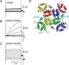
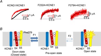
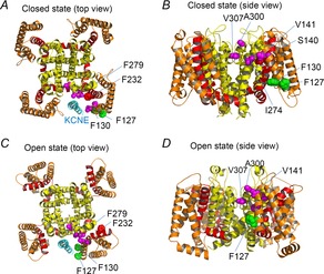
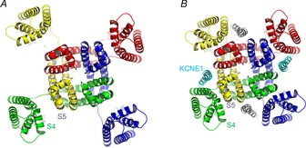
The gating of the KCNQ1 potassium channel is drastically regulated by auxiliary subunit KCNE proteins. KCNE1, for example, slows the activation kinetics of KCNQ1 by two orders of magnitude. Like other voltage-gated ion channels, the opening of KCNQ1 is regulated by the voltage-sensing domain (VSD; S1-S4 segments). Although it has been known that KCNE proteins interact with KCNQ1 via the pore domain, some recent reports suggest that the VSD movement may be altered by KCNE. The altered VSD movement of KCNQ1 by KCNE proteins has been examined by site-directed mutagenesis, the scanning cysteine accessibility method (SCAM), voltage clamp fluorometry (VCF) and gating charge measurements. These accumulated data support the idea that KCNE proteins interact with the VSDs of KCNQ1 and modulate the gating of the KCNQ1 channel. In this review, we will summarize recent findings and current views of the KCNQ1 modulation by KCNE via the VSD. In this context, we discuss our recent findings that KCNE1 may alter physical interactions between the S4 segment (VSD) and the S5 segment (pore domain) of KCNQ1. Based on these findings from ourselves and others, we propose a hypothetical mechanism for how KCNE1 binding alters the VSD movement and the gating of the channel.


Figures

Figure 1
Figure 1
KCNE proteins modulate the gating behaviour of KCNQ1 by being located in the open space between VSDs A–C, representative ionic current traces from KCNQ1, KCNQ1 + KCNE1 and KCNQ1 + KCNE3 expressed in Xenopus oocytes 3 days after RNA injection. The membrane potential was stepped from −100 mV to +60 mV in 20 mV steps for 2 s from the holding potential of −90 mV, and then stepped to −30 mV for tail current measurement. Dotted lines indicate zero current level. D, structural model of the open state KCNQ1 tetramer with KCNE1 (Smith et al. ; Kang et al. 2008). Each KCNQ1 subunit is in a different colour. The location and number of KCNE1 subunits (cyan and grey) are hypothetical. Each segment in the green subunit is labelled (S1–S6).
Figure 2
Figure 2
KCNE1 alters the VSD movement and the opening of KCNQ1 channels A, G–V relationships (black curves) and F–V relationships (red curves) for KCNQ1 (dashed curves) and KCNQ1 + KCNE1 (continuous curves). The G–V relationship of KCNQ1 is shifted in the positive direction by KCNE1. The F–V relationship is split into two components (F1 and F2) by KCNE1 (Osteen et al. ; Barro-Soria et al. 2014). The larger F1 component is shifted well in the negative direction while the smaller F2 component is shifted in the positive direction, as is the G–V relationship. B, S4 segments (red bars with ‘+’) of the KCNQ1/KCNE1 channel sit in the down state (closed state) at negative membrane potential. Depolarization moves the S4 segments to the pre-open state (F1), in which all S4 segments are in the up state with the pore domain still closed. Higher or longer depolarization induces the channel to enter the open state (F2).
Figure 3
Figure 3
Two bulky phenylalanine residues hamper the opening of the KCNQ1/KCNE1 channel A, simultaneous recording of VCF (VSD movement; red traces) and ionic current (black traces) from the wild-type KCNQ1/KCNE1 channel, F232A with KCNE1 and F279A with KCNE1. The wild-type KCNQ1/KCNE1 channel shows a delay of ionic current after the VSD movement; on the other hand, VSD movement and ionic currents from the two F–A mutants are almost synchronized (data from Nakajo & Kubo, Nature Communications, 2014). Red bars indicate 0.5% increase from the basal fluorescence of Alexa488 maleimide attached to G219C on the S3–S4 linker. B, schematic diagrams showing how Phe232 and Phe279 interact with each other during activation. Phe232 on the S4 segment moves by depolarization, up to the pre-open state (F1). Further depolarization rearranges the S4 a little more; however, physical interaction between Phe232 and Phy279 prevents the channel from entering the open state (F2). This would hamper the opening after the VSD movement in the wild-type KCNQ1/KCNE1 channels (A) (Nakajo & Kubo, 2014).
Figure 4
Figure 4
The putative interaction sites between KCNE protein and the S1/S5 segments A–D, structural models of closed (A and B) and open (C and D) states (Smith et al. 2007). A and C are top views from the extracellular side. B and D are side views. The transmembrane region of KCNE1 (cyan) (Kang et al. 2008) is also depicted in A and C. S1–S3, S4 & S4–S5 linker and S5–S6 are coloured in orange, red and yellow, respectively. The side chain of amino acid residues whose mutation causes short QT syndrome or gain-of-function (S140, V141, I274, A300 and V307) are coloured in magenta (Smith et al. 2007). The amino acid residues responsible for the constitutive activity induced by KCNE3 (F127 and F130) are coloured in green (Nakajo et al. 2011). The side chain of F232 (red) and F279 (yellow) are also depicted. Only side chains around KCNE protein (cyan) are depicted.
Figure 5
Figure 5
Hypothetical role of KCNE1 in packing the VSDs and the pore domain A, VSDs move laterally around the pore domain. In this situation the distance between the S4 and S5 segments would fluctuate depending on the location of the VSD. B, KCNE1 proteins (cyan and grey) pack the VSDs and the pore domain. In tightly packed channels with KCNE1, the interaction between the S4 and the S5 segments may be facilitated and that may affect the gating of KCNQ1 channels.

Similar articles

Cited by

References

    1. Abbott GW, Sesti F, Splawski I, Buck ME, Lehmann MH, Timothy KW, Keating MT. Goldstein SA. MiRP1 forms IKr potassium channels with HERG and is associated with cardiac arrhythmia. Cell. 1999;97:175–187. - PubMed
    1. Akabas MH, Stauffer DA, Xu M. Karlin A. Acetylcholine receptor channel structure probed in cysteine-substitution mutants. Science. 1992;258:307–310. - PubMed
    1. Angelo K, Jespersen T, Grunnet M, Nielsen MS, Klaerke DA. Olesen SP. KCNE5 induces time- and voltage-dependent modulation of the KCNQ1 current. Biophys J. 2002;83:1997–2006. - PMC - PubMed
    1. Barhanin J, Lesage F, Guillemare E, Fink M, Lazdunski M. Romey G. KVLQT1 and lsK (minK) proteins associate to form the IKs cardiac potassium current. Nature. 1996;384:78–80. - PubMed
    1. Barro-Soria R, Rebolledo S, Liin SI, Perez ME, Sampson KJ, Kass RS. Larsson HP. KCNE1 divides the voltage sensor movement in KCNQ1/KCNE1 channels into two steps. Nat Commun. 2014;5:3750. - PMC - PubMed

Publication types

MeSH terms

Substances

LinkOut - more resources