Case Reports
. 2015 Oct;23(10):1301-7.
doi: 10.1038/ejhg.2014.293. Epub 2015 Jan 21.

RMND1 deficiency associated with neonatal lactic acidosis, infantile onset renal failure, deafness, and multiorgan involvement

Affiliations
Case Reports

RMND1 deficiency associated with neonatal lactic acidosis, infantile onset renal failure, deafness, and multiorgan involvement

Alexandre Janer et al. Eur J Hum Genet. 2015 Oct.

Abstract

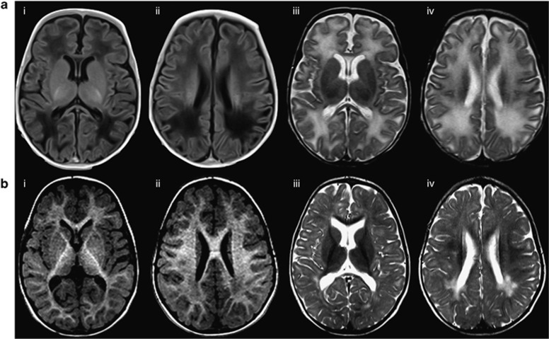
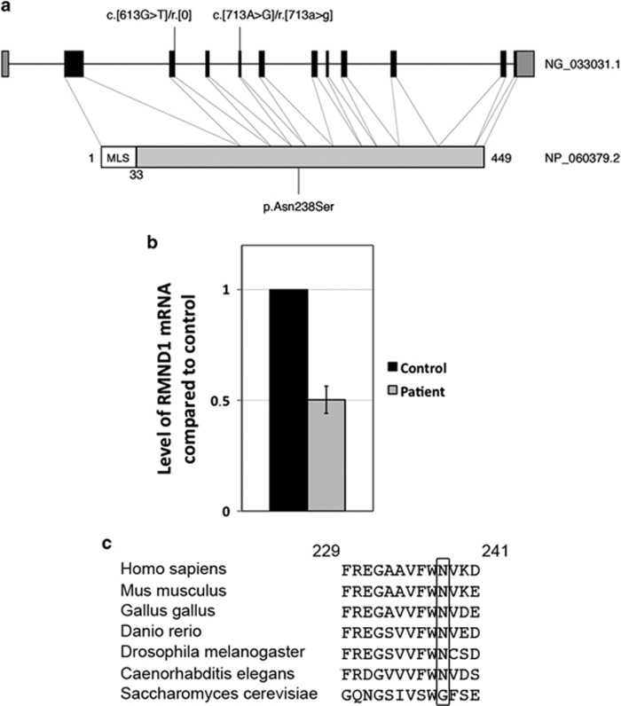
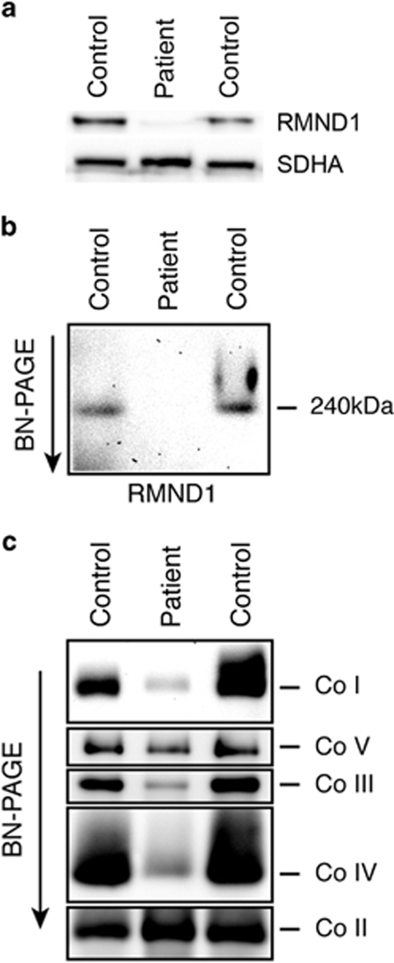
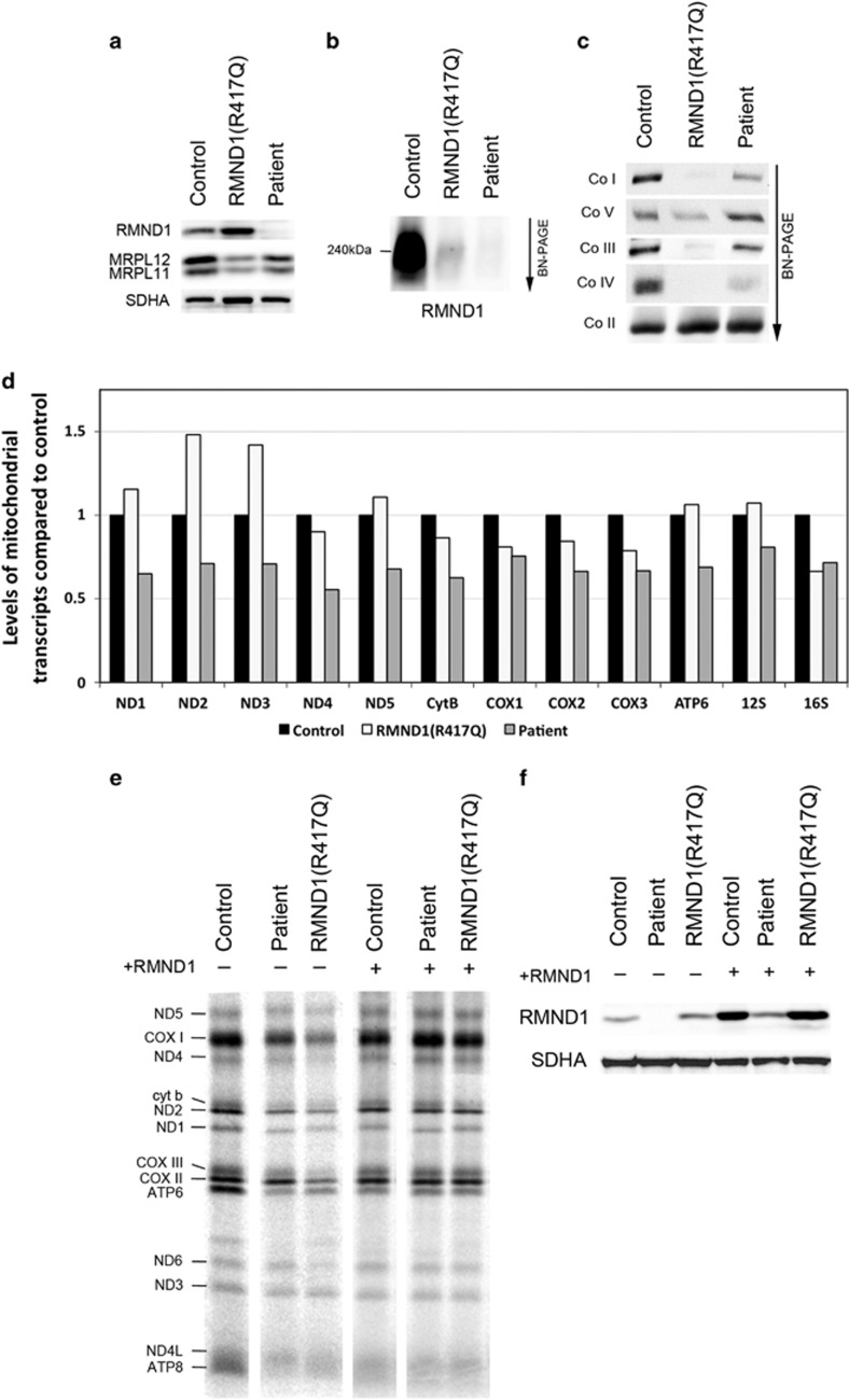
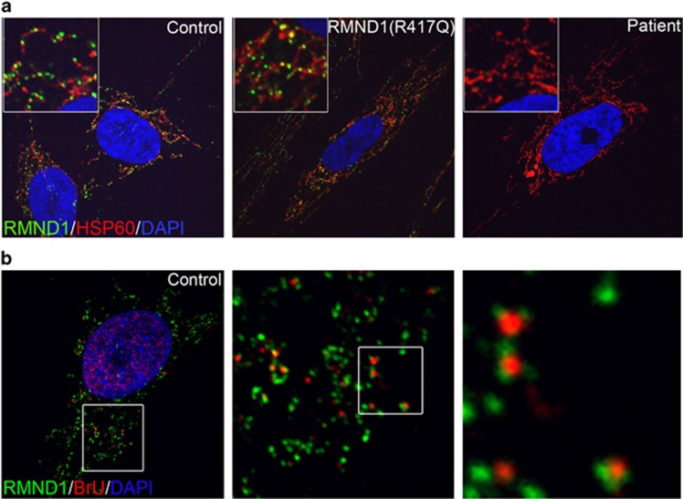
RMND1 is an integral inner membrane mitochondrial protein that assembles into a large 240 kDa complex to support translation of the 13 polypeptides encoded on mtDNA, all of which are essential subunits of the oxidative phosphorylation (OXPHOS) complexes. Variants in RMND1 produce global defects in mitochondrial translation and were first reported in patients with severe neurological phenotypes leading to mortality in the first months of life. Using whole-exome sequencing, we identified compound heterozygous RMND1 variants in a 4-year-old patient with congenital lactic acidosis, severe myopathy, hearing loss, renal failure, and dysautonomia. The levels of mitochondrial ribosome proteins were reduced in patient fibroblasts, causing a translation defect, which was rescued by expression of the wild-type cDNA. RMND1 was almost undetectable by immunoblot analysis in patient muscle and fibroblasts. BN-PAGE analysis showed a severe combined OXPHOS assembly defect that was more prominent in patient muscle than in fibroblasts. Immunofluorescence experiments showed that RMND1 localizes to discrete foci in the mitochondrial network, juxtaposed to RNA granules where the primary mitochondrial transcripts are processed. RMND1 foci were not detected in patient fibroblasts. We hypothesize that RMND1 acts to anchor or stabilize the mitochondrial ribosome near the sites where the mRNAs are matured, spatially coupling post-transcriptional handling mRNAs with their translation, and that loss of function variants in RMND1 are associated with a unique constellation of clinical phenotypes that vary with the severity of the mitochondrial translation defect.


Figures

Figure 1
Figure 1
Brain imaging (MRI) of the patient. This analysis shows significantly delayed myelination and abnormal posterior periventricular white matter signal. White matter signal is abnormally increased on T2 and reduced on T1. Myelination of the internal capsules is relatively preserved. There is thinning of the corpus callosum. (a) Axial MRI, age 2 months. ai, aii: Axial T1-weighted MRI without gadolinium (TR/TE 6100/ 12 ms, 4 mm); aiii, aiv: Axial T2-weighted MRI (TR/TE 4000/ 101 ms, 4 mm). (b) Axial MRI, age 2 years. bi, bii: Axial T1 MRI without gadolinium (TR/TE 1990/3.35 ms, 1 mm); biii, biv: Axial T2-weighted MRI (TR/TE 4000/ 101 ms, 5 mm).
Figure 2
Figure 2
Analysis of RMND1 variants. (a) Schematic representation of RMND1 gene (NG_0333031.1) and protein (NP_060379.2) showing the position of the variants in the patient (MLS is the mitochondrial localization signal). (b) Quantitative RT-PCR measurements of RMND1 mRNA levels in the patient compared with control (n=4). Expressed as mean±SEM. (c) Alignment of the amino-acid sequences of RMND1 homologs in different species. The asparagine at position 238 is shown in the black rectangle.
Figure 3
Figure 3
Characterization of the molecular defects in patient muscle. (a) Immunoblot analysis of control and patient muscle tissues showing very low levels of RMND1 in the patient. The 70 kDa subunit of the complex II (SDHA) was used as a loading control. (b) BN-PAGE analysis of control and patient muscle tissues showing that the 240 kDa RMND1 complex does not assemble in the patient. (c) BN-PAGE analysis of the OXPHOS complexes in control and patient muscle tissues. Each of the five OXPHOS complexes (I–V) was visualized with a subunit specific antibody that recognizes the native complexes as follows: Co I (NDUFA9), Co II (SDHA), Co III (UQCRC1), Co IV (COX4), and Co V (ATP5A1). Co II is the loading control.
Figure 4
Figure 4
Characterization of molecular defects in patient fibroblasts and rescue of the phenotype by expression of wild-type RMND1. (a) Immunoblot analysis of fibroblasts from control, the previously described RMND1(R417Q) patient, and the patient, with antibodies against RMND1 and the mitochondrial ribosomal proteins MRPL11 and MRPL12. The 70 kDa subunit of the complex II (SDHA) was used as a loading control. (b) BN-PAGE analysis of the RMND1 complex in control, RMND1(R417Q), and the patient. The 240 kDa RMND1 complex does not assemble either in RMND1(R417Q) or in the patient. (c) BN-PAGE analysis of the OXPHOS complexes in fibroblasts from control, RMND1(R417Q), and the patient. (d) Quantitative RT-PCR measurements of mitochondrial transcript levels in the patient compared with control. mRNAs coding for five subunits of the complex I (ND), one subunit of the complex III (cyt b), three subunits of the complex IV (COX), one subunit of the complex V (ATP), and mitochondrial ribosomal RNAs are indicated. (e) Analysis of mitochondrial translation by pulse labeling of mtDNA-encoded polypeptides in control, RMND1(R417Q), and the patient showing a generalized protein synthesis defect that is rescued by expression of the wild-type RMND1 cDNA. The seven subunits of the complex I (ND), one subunit of the complex III (cyt b), three subunits of the complex IV (COX), and two subunits of the complex V (ATP) are indicated. (f) Immunoblot analysis of RMND1 protein levels in fibroblasts from control, RMND1(R417Q), and the patient before and after overexpression of the wild-type RMND1 cDNA. The 70 kDa subunit of the complex II (SDHA) was used as a loading control.
Figure 5
Figure 5
Sub-mitochondrial localization of RMND1. (a) Immunofluorescence analysis of fibroblasts from control, RMND1(R417Q), and the patient. Endogenous RMND1 staining is shown in green, mitochondria are stained in red (HSP60), and the nucleus is counterstained in blue with DAPI. (b) Spatial organization of RMND1 in relation to newly synthesized mitochondrial RNA. Endogenous RMND1 staining is shown in green, BrU staining in red, and the nucleus is counterstained in blue with DAPI.

References

    1. 1Coenen MJ, Antonicka H, Ugalde C et al: Mutant mitochondrial elongation factor G1 and combined oxidative phosphorylation deficiency. N Engl J Med 2004; 351: 2080–2086. - PubMed
    1. 2Hallberg BM, Larsson NG: Making proteins in the powerhouse. Cell Metab 2014; 20: 226–240. - PubMed
    1. 3Weraarpachai W, Antonicka H, Sasarman F et al: Mutation in TACO1, encoding a translational activator of COX I, results in cytochrome c oxidase deficiency and late-onset Leigh syndrome. Nat Genet 2009; 41: 833–837. - PubMed
    1. 4Janer A, Antonicka H, Lalonde E et al: An RMND1 Mutation causes encephalopathy associated with multiple oxidative phosphorylation complex deficiencies and a mitochondrial translation defect. Am J Hum Genet 2012; 91: 737–743. - PMC - PubMed
    1. 5Antonicka H, Ostergaard E, Sasarman F et al: Mutations in C12orf65 in patients with encephalomyopathy and a mitochondrial translation defect. Am J Hum Genet 2010; 87: 115–122. - PMC - PubMed

Publication types