. 2015 Jan 21:5:7916.
doi: 10.1038/srep07916.

Angiogenin secretion from hepatoma cells activates hepatic stellate cells to amplify a self-sustained cycle promoting liver cancer

Affiliations

Angiogenin secretion from hepatoma cells activates hepatic stellate cells to amplify a self-sustained cycle promoting liver cancer

Cristina Bárcena et al. Sci Rep. .

Abstract

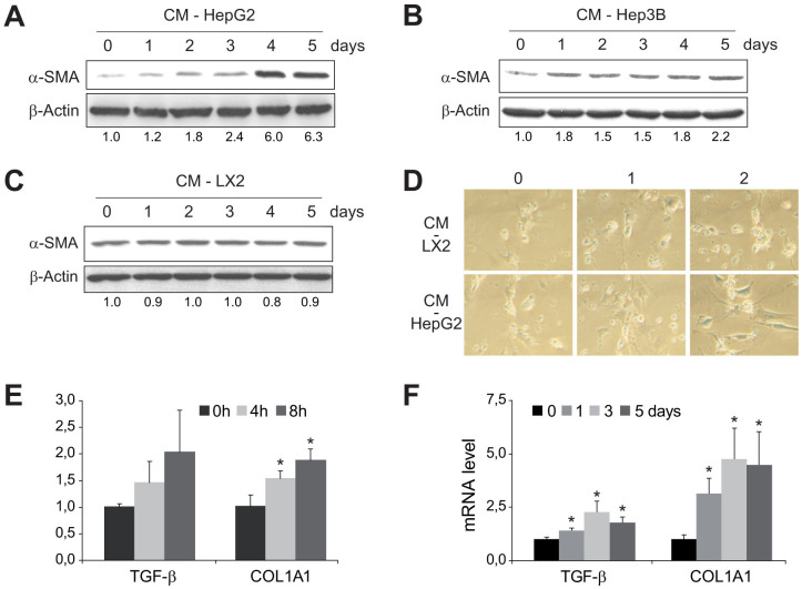
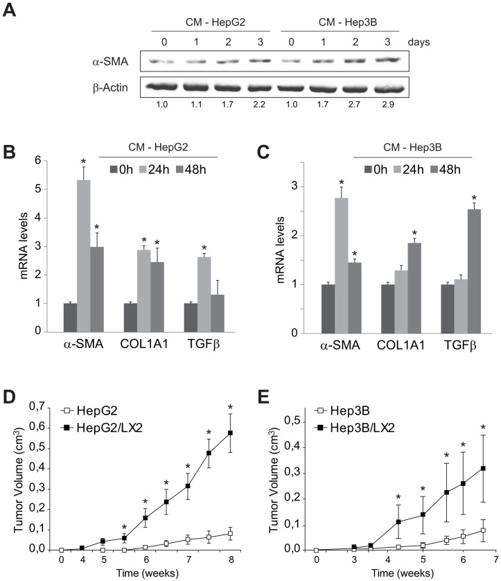
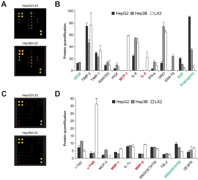
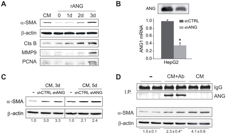
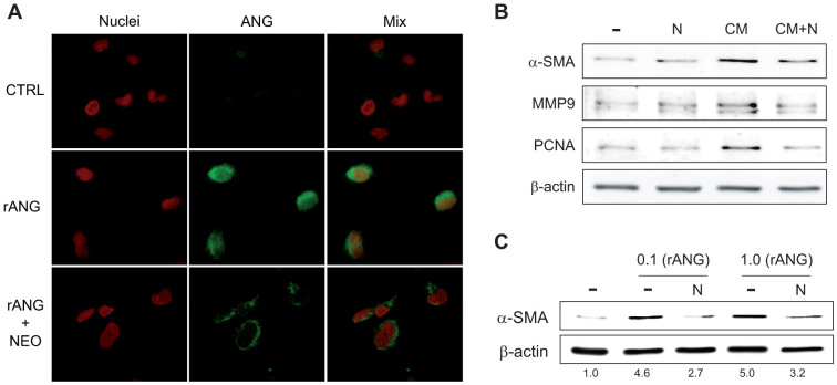
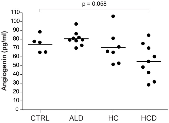
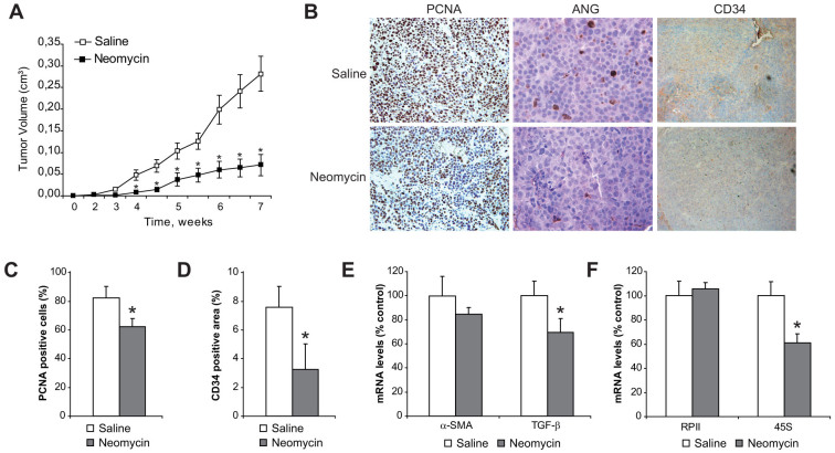
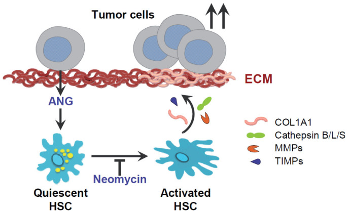
Hepatocellular carcinoma (HCC) frequently develops in a pro-inflammatory and pro-fibrogenic environment with hepatic stellate cells (HSCs) remodeling the extracellular matrix composition. Molecules secreted by liver tumors contributing to HSC activation and peritumoral stromal transformation remain to be fully identified. Here we show that conditioned medium from HCC cell lines, Hep3B and HepG2, induced primary mouse HSCs transdifferentiation, characterized by profibrotic properties and collagen modification, with similar results seen in the human HSC cell line LX2. Moreover, tumor growth was enhanced by coinjection of HepG2/LX2 cells in a xenograft murine model, supporting a HCC-HSC crosstalk in liver tumor progression. Protein microarray secretome analyses revealed angiogenin as the most robust and selective protein released by HCC compared to LX2 secreted molecules. In fact, recombinant angiogenin induced in vitro HSC activation requiring its nuclear translocation and rRNA transcriptional stimulation. Moreover, angiogenin antagonism by blocking antibodies or angiogenin inhibitor neomycin decreased in vitro HSC activation by conditioned media or recombinant angiogenin. Finally, neomycin administration reduced tumor growth of HepG2-LX2 cells coinjected in mice. In conclusion, angiogenin secretion by HCCs favors tumor development by inducing HSC activation and ECM remodeling. These findings indicate that targeting angiogenin signaling may be of potential relevance in HCC management.


Figures

Figure 1
Figure 1. HSCs are activated by conditioned medium (CM) from HepG2 and Hep3B cells.
Representative western blot showing α-SMA activation primary murine HSCs (day 7), previously exposed to conditioned medium from HepG2 (A), Hep3B (B) or LX2 cells (C) for 0–5 days, using β-actin levels as a control. D, Microscopic images of morphological changes in HSCs preteated with CM from HepG2 or LX2 during 0, 1 or 2 days. E and F, mRNA quantification of TGF-β and COL1A1 in 7-days-old HSCs after previous CM-HepG2 addition for the indicated periods of time. (n = 3). *, p ≤ 0.05, specific time vs. 0 time point.
Figure 2
Figure 2. LX2 cells are activated by conditioned medium (CM) from HepG2 and Hep3B cells and promote tumor growth in mice.
A, Representative western blot showing α-SMA activation, using β-actin levels as a control, of human LX2 cells previously exposed to conditioned medium from HepG2 and Hep3B. B and C, mRNA levels of genes associated to HSC activation measured in LX2 cells exposed to conditioned medium from HepG2 and Hep3B. D and E, Evolution of tumor growth in a murine subcutaneous model of Hep3B and HepG2 cells injected alone or combined with LX2 cells. (n = 3). *, p ≤ 0.05, Hep3B/LX2 and HepG2/LX2 vs. Hep3B and HepG2, respectively.
Figure 3
Figure 3. Differential profile of angiogenesis-related proteins in the secretome of hepatoma (HepG2 and Hep3B) and human HSC activated (LX2) cells.
A and B, Representative merged images of antibody microarrays for protein detection in conditioned medium (CM) from HepG2 or Hep3B (green) and compared to CM-LX2 (red) protein pattern. B and D, protein quantification was measured after calibration with internal standards and background controls. (n = 2). Indicated in red the proteins more expressed in LX2 respect to hepatoma cells, and in green the proteins more expressed in Hep3B/HepG2 compared to LX2.
Figure 4
Figure 4. Recombinant angiogenin activates HSCs and angiogenin depletion from conditioned medium reduces HSC activation.
A, Primary murine HSCs were exposure to recombinant angiogenin (rANG, 1 μg/ml) for several days (0 to 3) and phenotypic transformation was detected by changes in α-SMA, PCNA, CtsB or MMP9 expression at day 7. B, Angiogenin protein (up) and mRNA (down) levels of HepG2 cells stably transfected with shRNA control (shCTRL) or against angiogenin (shANG). C, α-SMA protein expression in HSCs after 4 or 5 days of exposure to control medium (-) or conditioned medium from HepG2 cells with shCTRL or shANG transfection. D, α−SMA protein expression after HSC treatment (day 7) with control medium (−), CM-HepG2 (CM) and CM-HepG2 where angiogenin content was previously depleted by immunoprecipitation (CM+Ab), as denoted by angiogenin detection in agarose beads.
Figure 5
Figure 5. Angiogenin nuclear deposition and HSC activation is reduced by neomycin administration.
A, Angiogenin subcellular location (green) was visualized by confocal immunofluorescence in HSCs treated with rANG (1 μg/ml) and/or neomycin preincubation (100 μM) by nuclear (red) co-staining with Hoechst 33258. Representatives images of α-SMA protein expression: B, analyzed after HSC treatment with different doses of angiogenin (0.1 and 1 μg/ml) and/or neomycin (100 μM) for 3 days; and C, after HSC treatment with control medium (−), or CM-HepG2 (CM) and/or neomycin (N) for 5 days.
Figure 6
Figure 6. Angiogenin serum levels are not increased in ALD.
Angiogenin serum levels were measured in control individuals (n = 5) and patients with liver pathologies: ALD, alcoholic liver disease (n = 9, age: 48.1 ± 3.8), HC, hepatic cirrhosis (n = 7, age: 58.1 ± 2.4, MELD: 10.3 ± 1.6), and HCD, hepatic cirrhosis decompensated (n = 9 age: 51.3 ± 2.2, MELD: 14.3 ± 1.8). Additional data of patients is provided in Suppl. Fig. 2.
Figure 7
Figure 7. Neomycin administration reduces HCC/HSC tumor growth by blocking angiogenin nuclear translocation and HCC development.
A, measurement of tumors from nude mice subcutaneously injected with HepG2/LX2 cells and treated intraperitonealy with saline or neomycin for eight weeks. B, Representative images of tumor cell proliferation by PCNA detection (40×), angiogenin levels (80×) and CD34 (40×) were visualized in tumor samples from mice treated with neomycin or saline. C, Quantification of PCNA positive cells in tumor slides. D, Quantification of CD34 positive areas in tumor slides. E, mRNA quantification of α-SMA and TGF-β in tumors, using β-actin as control. (n ≥ 6). *, p ≤ 0.05, neomycin vs. saline-injected animals. F, RNA quantification of RNA polymerase II (RPII) and 45S ribosomal RNA in tumors, as above.
Figure 8
Figure 8. Schematic representation of angiogenin role in HCC/HSC crosstalk.
Angiogenin secreted from hepatoma cells is an inducer of HSC transformation by changing numerous proteins involved in ECM remodeling. Among them, specific fibrillar components such as collagen, type I, alpha 1, encoded by the COL1A1 gene, are induced, as well as abnormal expression of enzymes that degrade type IV and V collagens and other extracellular matrix proteins, such us MMP9, tissue inhibitors of metalloproteinases (TIMP) or cysteine proteinases such as cathepsin B. Consequently, physiologic ECM formation is altered due to HSC induction, providing a profibrogenic environment that facilitates tumor growth. In summary, liver tumor promotes its own development via angiogenin-dependent HSC activation, and antagonism of angiogenin signaling, as neomycin does, may be an interesting approach to halter liver cancer progression.

References

    1. Hanahan D. & Coussens L. M. Accessories to the crime: functions of cells recruited to the tumor microenvironment. Cancer Cell 21, 309–322 (2012). - PubMed
    1. Hernandez-Gea V., Toffanin S., Friedman S. L. & Llovet J. M. Role of the microenvironment in the pathogenesis and treatment of hepatocellular carcinoma. Gastroenterology 144, 512–527 (2013). - PMC - PubMed
    1. Schrader J. & Iredale J. P. The inflammatory microenvironment of HCC - the plot becomes complex. J. Hepatol. 54, 853–855 (2011). - PubMed
    1. Friedman S. L. Hepatic stellate cells: protean, multifunctional, and enigmatic cells of the liver. Physiol. Rev. 88, 125–172 (2008). - PMC - PubMed
    1. Friedman S. L., Sheppard D., Duffield J. S. & Violette S. Therapy for fibrotic diseases: nearing the starting line. Sci. Transl. Med. 5, 167sr161, 10.1126/scitranslmed.3004700 (2013). - DOI - PubMed

Publication types

MeSH terms

LinkOut - more resources