. 2015 Feb 20;6(5):2827-42.
doi: 10.18632/oncotarget.3090.

Folate-mediated mitochondrial targeting with doxorubicin-polyrotaxane nanoparticles overcomes multidrug resistance

Affiliations

Folate-mediated mitochondrial targeting with doxorubicin-polyrotaxane nanoparticles overcomes multidrug resistance

He Wang et al. Oncotarget. .

Abstract

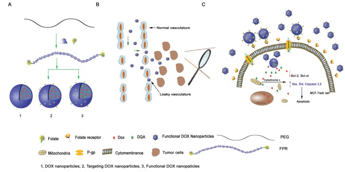
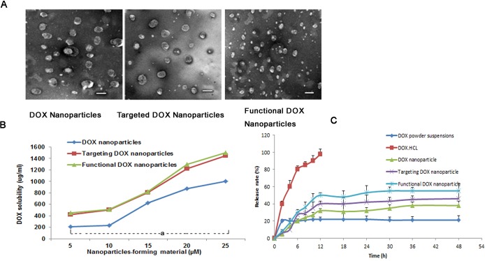
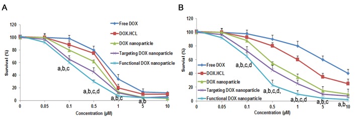
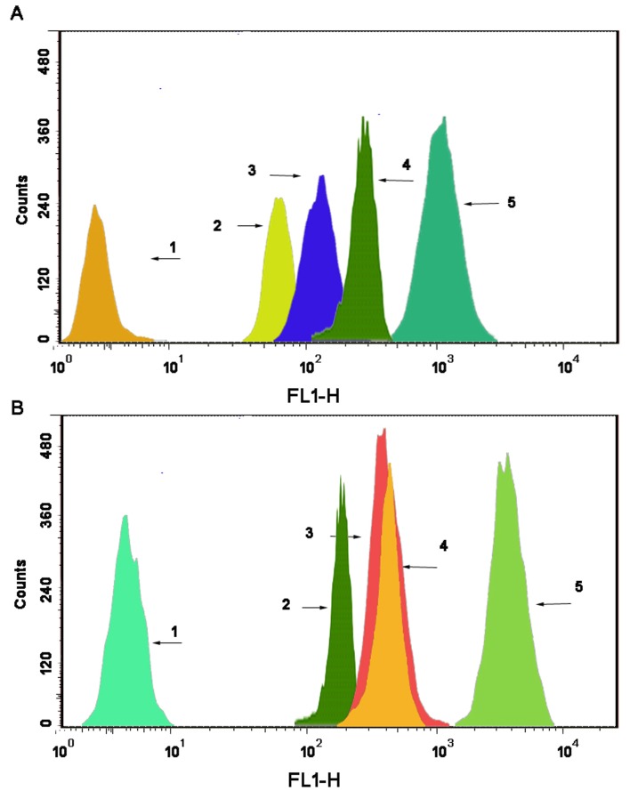
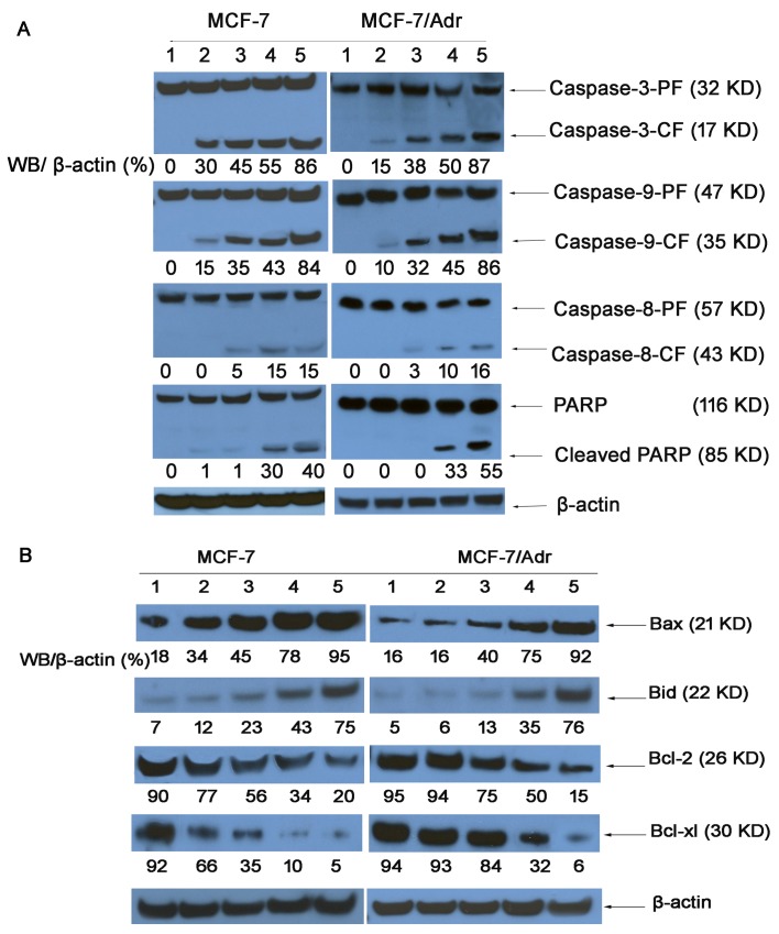
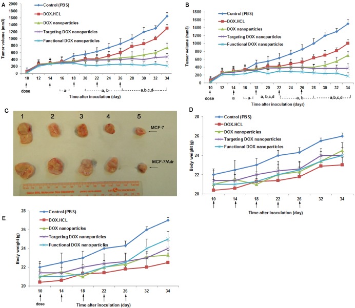
Resistance to treatment with anticancer drugs is a significant obstacle and a fundamental cause of therapeutic failure in cancer therapy. Functional doxorubicin (DOX) nanoparticles for targeted delivery of the classical cytotoxic anticancer drug DOX to tumor cells, using folate-terminated polyrotaxanes along with dequalinium, have been developed and proven to overcome this resistance due to specific molecular features, including a size of approximately 101 nm, a zeta potential of 3.25 mV and drug-loading content of 18%. Compared with free DOX, DOX hydrochloride, DOX nanoparticles, and targeted DOX nanoparticles, the functional DOX nanoparticles exhibited the strongest anticancer efficacy in vitro and in the drug-resistant MCF-7/Adr (DOX) xenograft tumor model. More specifically, the nanoparticles significantly increased the intracellular uptake of DOX, selectively accumulating in mitochondria and the endoplasmic reticulum after treatment, with release of cytochrome C as a result. Furthermore, the caspase-9 and caspase-3 cascade was activated by the functional DOX nanoparticles through upregulation of the pro-apoptotic proteins Bax and Bid and suppression of the antiapoptotic protein Bcl-2, thereby enhancing apoptosis by acting on the mitochondrial signaling pathways. In conclusion, functional DOX nanoparticles may provide a strategy for increasing the solubility of DOX and overcoming multidrug-resistant cancers.


Conflict of interest statement

Conflict of interest

The authors declare no conflict of interest.

Figures

Figure 1
Figure 1. Schematic representations of DOX nanoparticles, targeted DOX nanoparticles and functional DOX nanoparticles
(A) and the mechanism of functional DOX nanoparticles' effect on cells with MDR (B, C). The functional DOX nanoparticles, which consisted of FPRs, DQA, and DOX, overcame DOX resistance in vitro by targeting the mitochondria of MCF-7/Adr cells.
Figure 2
Figure 2. DOX nanoparticles and characterization
(A) TEM image of different types of nanoparticles, scale bar = 150 nm. (B) Effect of the nanoparticle-forming material's total concentration on the solubilized DOX concentration in the nanoparticles. aP<0.05, compared with DOX nanoparticles. (C) DOX release rates (%) of five different formulations of DOX in PBS containing 10% FBS at 37°C (pH 7.4). The data are presented as the mean ± SD (n=3).
Figure 3
Figure 3. Inhibitory effect of five DOX-containing formulations on the proliferation of MCF-7 cells
(A) and MCF-7/Adr cells (B) in vitro. The data are presented as the mean ± SD (n=3). Notes: aP<0.05, compared with free DOX; bP<0.05, compared with DOX·HCL; cP<0.05, compared with DOX nanoparticles; dP<0.05, compared with targeted DOX nanoparticles.
Figure 4
Figure 4. Uptake after incubation with varying formulations
Uptake of drugs by MCF-7 cells and MCF-7/Adr cells (A). The data are presented as the mean ± SD (n=3). Notes: aP<0.05, compared with blank control; bP<0.05, compared with free 6-coumarin; cP<0.05, compared with 6-coumarin nanoparticles;dP<0.05, compared with targeted 6-coumarin nanoparticles. Uptake of different rhodamine 123-containing formulations by MCF-7 cells (B, left) and MCF-7/Adr cells (B, right). Notes: The blue color denotes the nuclei of MCF-7 or MCF-7/Adr cells stained with Hoechst 33342. The green color denotes rhodamine 123. The overlapping images show both Hoechst 33342 and rhodamine 123 in MCF-7 or MCF-7/Adr cells. (C) Intracellular accumulation in several cytoplasmic organelles in MCF-7 (C, left) and MCF-7/Adr (C, right) cells after treatment with free 6-coumarin, 6-coumarin nanoparticles, targeted 6-coumarin nanoparticles, or functional 6-coumarin nanoparticles. Notes: The blue and green colors denote Hoechst 33342 and 6-coumarin, respectively. The red color denotes lysosomes, the ER, the GA, and mitochondria stained with LysoTracker Red DND-99, ER-Tracker Red, BODIPY TR ceramide complexed to BSA, or MitoTracker Deep Red 633, respectively. The yellow color (merged image) indicates 6-coumarin in different organelles.
Figure 5
Figure 5. Drug content of the mitochondrial fraction in MCF-7 cells
(A) and MCF-7/Adr cells (B) after applying different formulations, as determined by flow cytometry. The abscissa indicates the fluorescence intensity of a 6-coumarin formulation internalized by the cancer cells, and the ordinate represents the cell counts. Notes: A1 and B1, blank control; A2 and B2, free 6-coumarin; A3 and B3, 6-coumarin nanoparticles; A4 and B4, targeted 6-coumarin nanoparticles; A5 and B5, functional 6-coumarin nanoparticles.
Figure 6
Figure 6. Immunohistochemical staining of cytochrome C translocated from mitochondria to the cytosol in MCF-7/Adr cells after incubation with different formulations, including a blank control
(A), free DOX (B), DOX·HCL (C), DOX nanoparticles (D), targeted DOX nanoparticles (E), and functional DOX nanoparticles (F).
Figure 7
Figure 7. The cell apoptosis rate was detected by flow cytometry
MCF-7 cells (A) and MCF-7/Adr cells (B) were treated with different formulations each containing a total DOX concentration of 2 μM for 24 h. Notes: A1 and B1, control (PBS); A2 and B2, DOX solution; A3 and B3, DOX·HCL; A4 and B4, DOX nanoparticles; A5 and B5, targeted DOX nanoparticles; A6 and B6, functional DOX nanoparticles.
Figure 8
Figure 8. Expression of proteins involved in the apoptosis signaling pathways in MCF-7 and MCF-7/Adr cells, as determined by western blotting
(1) Control (PBS); (2) free DOX; (3) DOX·HCL; (4) DOX nanoparticles; (5) targeted DOX nanoparticles; and (6) functional DOX nanoparticles. Activity ratios of caspase-3 and caspase-9 (A) and expression ratios of the pro-apoptotic proteins Bax and Bid and the anti-apoptotic proteins Bcl-2 and Bcl-xl (B) in MCF-7 and MCF-7/Adr cells after incubation with varying formulations. β-actin was also assessed by western blotting. All protein levels were quantified densitometrically and normalized to β-actin.
Figure 9
Figure 9. Anticancer efficacy of functional DOX nanoparticles in MCF-7- or MCF-7/Adr-bearing Balb/c mice
Mice were injected intravenously with DOX·HCL, DOX nanoparticles, targeted DOX nanoparticles, or functional DOX nanoparticles at doses equivalent to 5 mg DOX per kg on days 10, 14, 18, 22, and 26 (PBS was used as a control). The changes in (A, B) tumor volume and (D, E) body weight in the MCF-7- or MCF-7/Adr-bearing Balb/c mice after administration are shown. (C) After 34 days, the tumors were excised from the MCF-7- or MCF-7/Adr-bearing mice in each group. The data are presented as the mean ± SD (n=5). aP<0.05, compared with control; bP<0.05, compared with DOX·HCL; cP<0.05, compared with DOX nanoparticles; dP<0.05, compared with targeted DOX nanoparticles.

References

    1. Tiwari G, Tiwari R, Sriwastawa B, Bhati L, Pandey S, Pandey P, Bannerjee SK. Drug delivery systems: An updated review. International journal of pharmaceutical investigation. 2012;2(1):2–11. - PMC - PubMed
    1. Ozben T. Mechanisms and strategies to overcome multiple drug resistance in cancer. FEBS letters. 2006;580(12):2903–2909. - PubMed
    1. Wong ST, Goodin S. Overcoming drug resistance in patients with metastatic breast cancer. Pharmacotherapy. 2009;29(8):954–965. - PubMed
    1. Green DR, Reed JC. Mitochondria and apoptosis. Science. 1998;281(5381):1309–1312. - PubMed
    1. Brenner C, Kroemer G. Apoptosis. Mitochondria--the death signal integrators. Science. 2000;289(5482):1150–1151. - PubMed

Publication types

MeSH terms