. 2015 Apr 15;593(8):1809-27.
doi: 10.1113/jphysiol.2014.283028. Epub 2015 Feb 19.

The use of murine-derived fundic organoids in studies of gastric physiology

Affiliations

The use of murine-derived fundic organoids in studies of gastric physiology

Michael A Schumacher et al. J Physiol. .

Abstract

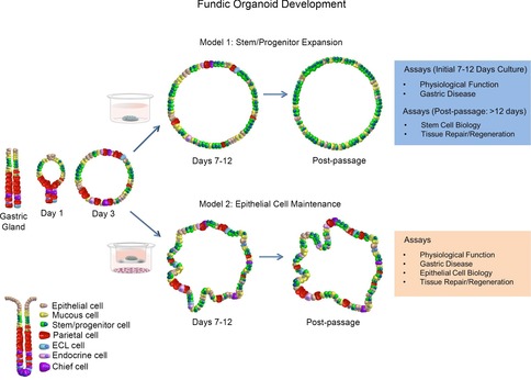
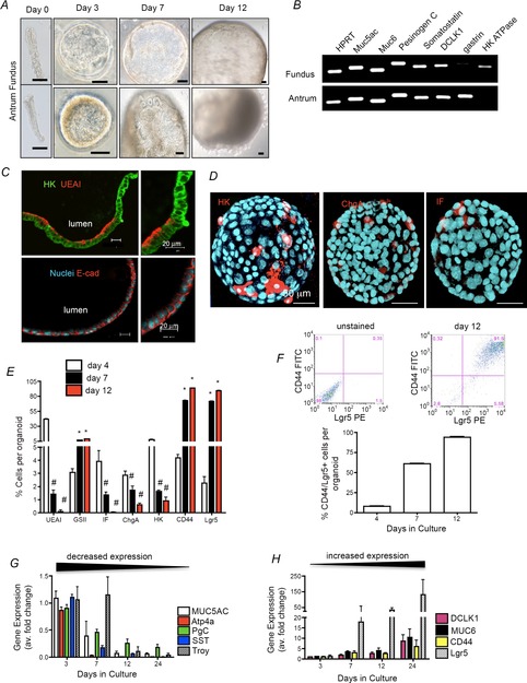
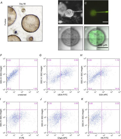
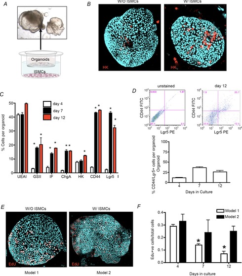
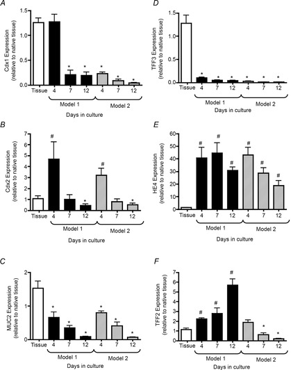
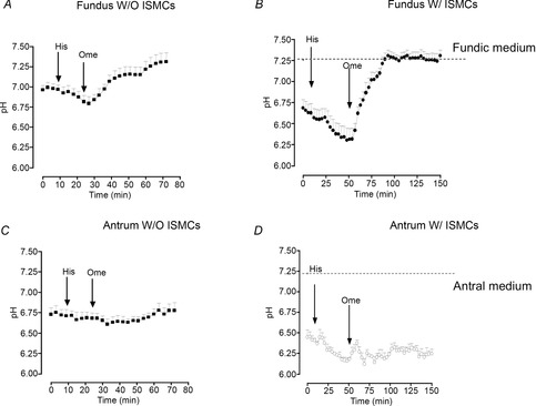
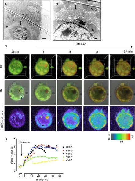
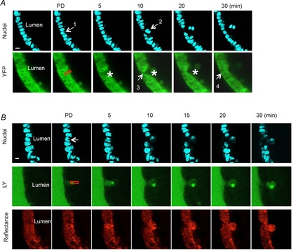
Key points: An in vitro approach to study gastric development is primary mouse-derived epithelium cultured as three-dimensional spheroids known as organoids. We have devised two unique gastric fundic-derived organoid cultures: model 1 for the expansion of gastric fundic stem cells, and model 2 for the maintenance of mature cell lineages. Organoids maintained in co-culture with immortalized stomach mesenchymal cells express robust numbers of surface pit, mucous neck, chief, endocrine and parietal cells. Histamine induced a significant decrease in intraluminal pH that was reversed by omeprazole in fundic organoids and indicated functional activity and regulation of parietal cells. Localized photodamage resulted in rapid cell exfoliation coincident with migration of neighbouring cells to the damaged area, sustaining epithelial continuity. We report the use of these models for studies of epithelial cell biology and cell damage and repair.

Abstract: Studies of gastric function and disease have been limited by the lack of extended primary cultures of the epithelium. An in vitro approach to study gastric development is primary mouse-derived antral epithelium cultured as three-dimensional spheroids known as organoids. There have been no reports on the use of organoids for gastric function. We have devised two unique gastric fundic-derived organoid cultures: model 1 for the expansion of gastric fundic stem cells, and model 2 for the maintenance of mature cell lineages. Both models were generated from single glands dissociated from whole fundic tissue and grown in basement membrane matrix (Matrigel) and organoid growth medium. Model 1 enriches for a stem cell-like niche via simple passage of the organoids. Maintained in Matrigel and growth medium, proliferating organoids expressed high levels of stem cell markers CD44 and Lgr5. Model 2 is a system of gastric organoids co-cultured with immortalized stomach mesenchymal cells (ISMCs). Organoids maintained in co-culture with ISMCs express robust numbers of surface pit, mucous neck, chief, endocrine and parietal cells. Histamine induced a significant decrease in intraluminal pH that was reversed by omeprazole in fundic organoids and indicated functional activity and regulation of parietal cells. Localized photodamage resulted in rapid cell exfoliation coincident with migration of neighbouring cells to the damaged area, sustaining epithelial continuity. Thus, we report the use of these models for studies of epithelial cell biology and cell damage and repair.


Figures

Figure 1
Figure 1. Schematic diagram showing the development of two organoid culture systems that may be used for studies of gastric physiological function and disease
Model 1 represents a system whereby stem/progenitor cells are expanded for studies in stem cell biology and tissue repair/regeneration. Model 2 represents a culture system of maintained epithelial cells for studies of physiological function, gastric disease and epithelial cell biology.
Figure 2
Figure 2. Fundic and antral dissection
Stomachs were opened along the greater curvature, and washed in ice-cold DPBS. Stomachs were pinned and muscle was stripped using dissecting scissors and a microscope. Images before and after muscle stripping are shown for fundus and antrum. Tissue collected for gland isolation is shown by dotted lines, demonstrating collection of distinct regions for fundus and antrum for organoid preparation.
Figure 3
Figure 3. Determination of optimal growth conditions for fundic gastric organoids
A, the requirement for growth factors is demonstrated in representative images at day 7 of culture. B, fundic organoid growth efficiency (percentage organoids per glands seeded). C, size (at day 7) was assayed in primary cultures by removing each individual growth factor from growth medium. *P < 0.05 compared to complete.
Figure 4
Figure 4. Model 1: fundic gastric organoids used in the expansion of stem/progenitor cells
A, cyst-like 3D structures grown from fundic or antral gastric glands isolated from mouse. Images from 0, 3, 7 and 12 day cultures. Scale bars = 50 μm. B, RT-PCR analysis of gastric lineage markers at culture day 12. Both fundic and antral organoids expressed mRNA for mucin 5AC (surface mucous pit cells), mucin 6 (mucous neck cells), pepsinogen C (zymogen/chief cells) and somatostatin (D cells). In contrast, the expression of gastrin (G cells) was specific to antral organoids whereas H+,K+-ATPase (parietal cells) was specific to fundic organoids. C, organoid sections immunostained for H+,K+-ATPase (HK, green) and UEAI (red), and E-cadherin (red) and Hoechst (nuclear, blue) Scale bars = 20 μm. D, organoids immunostained for HK (red), chromogranin A (chgA, red), intrinsic factor (IF, red) and Hoechst (nuclear, blue). Scale bars = 50 μm. E, flow cytometric analysis using fundic organoids at 4, 7 and 12 days in culture. F, 2D flow cytometric histogram of gated cells co-expressing CD44 and Lgr5. G and H, qRT-PCR of cell lineage and gastric stem cell markers using RNA isolated from organoids cultured for 3, 7 and 12 days. #P < 0.05 compared with day 4, n = 4 individual organoid preparations.
Figure 5
Figure 5. Transepithelial permeability and flow cytometric analysis of fundic organoids
A, fundic gastric organoid maintained in culture for 90 days. B–E, injection and retention of Lucifer yellow (LY) within the organoid lumen after injection. Luminal retention of microinjected Lucifer yellow over 24 h confirmed low transepithelial permeability of fundic organoids. Stereoscopic images of organoids during LY injection in bright-field (B) or fluorescence (C), and confocal images of organoids 1 day after LY injection in bright-field (D) or fluorescence plus bright-field (E). F–K, representative flow cytometric dot plots showing the gating scheme and cell distribution of UEAI (surface pit), GSII (mucous neck), IF (intrinsic factor, chief), ChgA (endocrine) and HK (H+,K+-ATPase, parietal) cells in gastric organoids.
Figure 6
Figure 6. Model 2: Maintenance of epithelial cells within fundic gastric organoids co-cultured with ISMCs
A, organoid/ISMC co-culture Transwell system showing morphological changes in organoids. B, organoids in whole mount immunostained for H+,K+-ATPase (HK, red), and Hoechst (nuclear, blue) co-cultured without (W/O) or with (W/) ISMCs. C, flow cytometric analysis using fundic organoids co-cultured with ISMCs for 4, 7 and 12 days in culture. D, 2D flow cytometric histogram of gated cells co-expressing CD44 and Lgr5. E and F, EdU immunostaining (EdU: red, nuclear: blue) (E) followed by quantification of Edu+ nuclei/total cell number (F).
Figure 7
Figure 7. Expression of metaplastic markers in cultures of gastric organoids of model 1 and model 2
qRT-PCR of metaplastic markers Cdx1 (A), Cdx2 (B), MUC2 (C), TFF3 (D), HE4 (E) and TFF2 (F) using RNA isolated from organoids cultured for 4, 7 and 12 days. *P < 0.05, significantly decreased compared with native tissue; # P < 0.05, significantly increased compared with native tissue; n = 3 individual organoid preparations.
Figure 8
Figure 8. Organoid-derived parietal cell functional assay
Intraluminal pH response to histamine (His) and omeprazole (Ome) using fundic organoids cultured without (W/O) ISMCs (A) or with (W/) ISMCs (B). Intraluminal pH response to His and Ome using antral organoids cultured W/O ISMCs (C) or W/ ISMCs (D). n = 6 individual organoids.
Figure 9
Figure 9. Presence of parietal cells in the fundic gastric organoids
A and B, observation of parietal cells within the fundic organoid at low (A) or high (B) magnification by transmission electron microscopy. N, nuclear; M, mitochondria. C, confocal images of a fundic organoid labelled with Acridine Orange before and after histamine (100 μm). Images shows 3D, 2D or pseudocolour of F458 (red)/F488 (green), respectively. D, changes in the ratio F458/F488 in response to histamine from five individual cells indicated in C.
Figure 10
Figure 10. Fundic gastric organoids used to study epithelial restitution
A, two-photon damage (at site indicated by arrow 1 and red box) results in cell exfoliation into the lumen and restoration of the damaged epithelium within 30 min. Blue = nuclear stain; green = endogenous YFP cytoplasmic fluorescence of fundic organoids from YC transgenic mouse cells. B, maintenance of epithelial barrier following cell damage. Two-photon damage (at site indicated by arrow 1 and red box) results in cell exfoliation into the lumen and restoration of the damaged epithelium within 30 min. The entrance of Lucifer yellow-containing extraluminal medium into the lumen of the organoid is limited. Blue = nuclear stain; green = Lucifer yellow; Red = reflectance.

References

    1. Aihara E, Hentz CL, Korman AM, Perry NP, Prasad V, Shull GE. Montrose MH. In vivo epithelial wound repair requires mobilization of endogenous intracellular and extracellular calcium. J Biol Chem. 2013;288:33585–33597. - PMC - PubMed
    1. Aruffo A, Stamenkovic I, Melnick M, Underhill CB. Seed B. CD44 is the principal cell surface receptor for hyaluronate. Cell. 1990;61:1303–1313. - PubMed
    1. Barker N, Huch M, Kujala P, van de Wetering M, Snippert H, van Es JH, Sato T, Stange DE, Begthel H, van den Born M, Danenberg E, van den Brink S, Korving J, Abo A, Peters PJ, Wright N, Poulsom R. Clevers H. Lgr5(+ve) stem cells drive self-renewal in the stomach and build long-lived gastric units in vitro. Cell Stem Cell. 2010;6:25–36. - PubMed
    1. Bartfeld S, Bayram T, van de Wetering M, Huch M, Begthel H, Kujala P, Vries R, Peters PJ. Clevers H. In vitro expansion of human gastric epithelial stem cells and their responses to bacterial infection. Gastroenterology. 2015;148:126–136. - PMC - PubMed
    1. Bell SM, Schreiner CM, Wert SE, Mucenski ML, Scott WJ. Whitsett JA. R spondin 2 is required for normal laryngeal–tracheal, lung and limb morphogenesis. Development. 2008;135:1049–1058. - PubMed

Publication types