. 2015 Jan 21:15:12.
doi: 10.1186/s12885-015-1007-5.

Microfluidic model of ductal carcinoma in situ with 3D, organotypic structure

Affiliations

Microfluidic model of ductal carcinoma in situ with 3D, organotypic structure

Lauren L Bischel et al. BMC Cancer. .

Abstract

Background: Ductal carcinoma in situ (DCIS) is a non-invasive form of breast cancer that is thought to be a precursor to most invasive and metastatic breast cancers. Understanding the mechanisms regulating the invasive transition of DCIS is critical in order to better understand how some types of DCIS become invasive. While significant insights have been gained using traditional in vivo and in vitro models, existing models do not adequately recapitulate key structure and functions of human DCIS well. In addition, existing models are time-consuming and costly, limiting their use in routine screens. Here, we present a microscale DCIS model that recapitulates key structures and functions of human DCIS, while enhancing the throughput capability of the system to simultaneously screen numerous molecules and drugs.

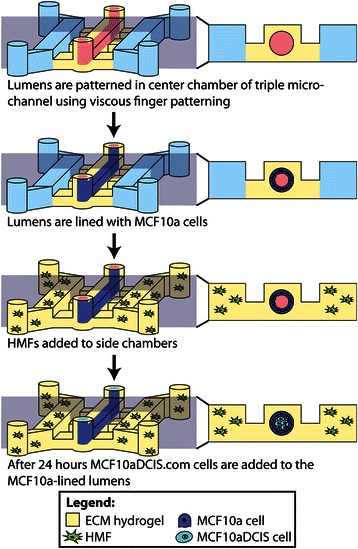
Methods: Our microscale DCIS model is prepared in two steps. First, viscous finger patterning is used to generate mammary epithelial cell-lined lumens through extracellular matrix hydrogels. Next, DCIS cells are added to fill the mammary ducts to create a DCIS-like structure. For coculture experiments, human mammary fibroblasts (HMF) are added to the two side channels connected to the center channel containing DCIS. To validate the invasive transition of the DCIS model, the invasion of cancer cells and the loss of cell-cell junctions are then examined. A student t-test is conducted for statistical analysis.

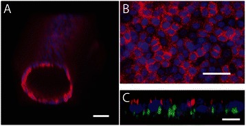
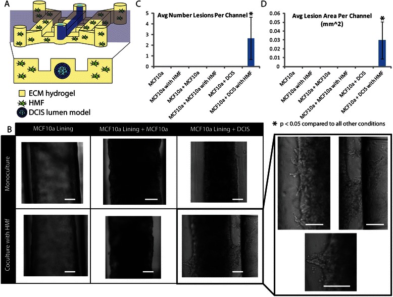
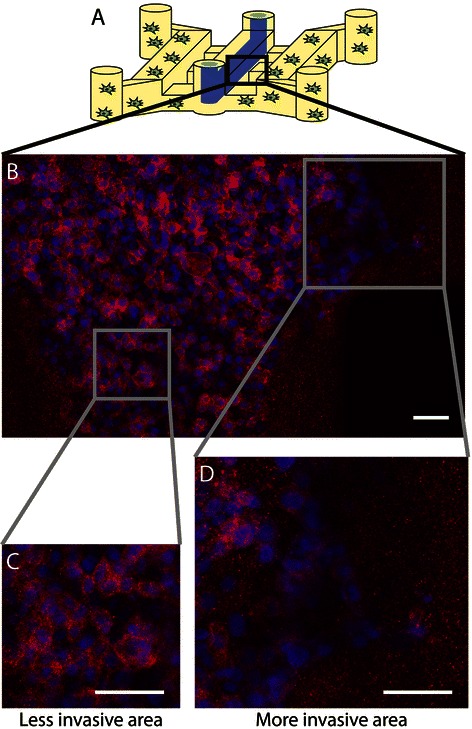
Results: We demonstrate that our DCIS model faithfully recapitulates key structures and functions of human mammary DCIS and can be employed to study the mechanisms involved in the invasive progression of DCIS. First, the formation of cell-cell junctions and cell polarity in the normal mammary duct, and the structure of the DCIS model are characterized. Second, coculture with HMF is shown to induce the invasion of DCIS. Third, multiple endpoint analyses are demonstrated to validate the invasion.

Conclusions: We have developed and characterized a novel in vitro model of normal and DCIS-inflicted mammary ducts with 3D lumen structures. These models will enable researchers to investigate the role of microenvironmental factors on the invasion of DCIS in more in vivo-like conditions.


Figures

Figure 1
Figure 1
Schematic overview of model patterning methods. As previously described, a lumen can be patterned through a collagen I hydrogel in the center chamber of a triple microchannel using viscous finger patterning [22]. The collagen I lumen is then coated with a Matrigel lining. Following polymerization, MCF10a cells are used to line the lumen and mimic a mammary duct structure. HMF cells in a collagen I hydrogel can be added to the side chambers. To model DCIS, MCF10aDCIS.com cells are flowed into the lumen after 24 hours.
Figure 2
Figure 2
Mammary duct model with 3D lumen structure. A) Volume-rendered confocal image of MCF10a cells lining a lumen patterned through a collagen I hydrogel and coated with Matrigel. Cells were cultured for 72 hours and then stained for e-cadherin (red) and nuclei (blue). Scale bar represents 50 μm. B) Confocal slice of bottom of the lumen to show e-cadherin localization around cell junctions. Scale bar represents 5 μm. C) Volume-rendered confocal image of MCF10a cells lining a lumen and stained for laminin V (green), nuclei (blue) and GM130 (red) to demonstrate cell polarization. Scale bar represents 1 μm.
Figure 3
Figure 3
To model ductal carcinoma in situ MCF10aDCIS.com cells were added to the center of MCF10alined lumens after 24 hours. Volume-rendered (A) and confocal slice (B) images are shown. Cells were stained with cell tracker green (MCF10a) and cell tracker blue (MCF10DCIS.com) and cultured for 72 hours after the addition of the DCIS cells. Scale bars represent 100 μm.
Figure 4
Figure 4
Coculture with HMF induces invasive transition in the DCIS model. A) Schematic image of coculture experiments with the DCIS model in the center chamber of a triple microchannel and HMFs in the side chambers. B) Bright-field images of mammary duct, mammary duct filled with MCF10a cells, and mammary duct filled with DCIS cells either alone or in coculture with HMFs. Scale bars represent 100 μm (main chart) and 50 μm (close up image at the right). The average number (C) and area (D) of invasive lesions in the different culture conditions were quantified across three independent experiments with at least three channels per experiment. A student Student’s t-test was used to determine significant differences between culture conditions (p < 0.05 is considered to be significant).
Figure 5
Figure 5
Invasive lesions show decreased e-cadherin. Schematic (A) and confocal slice (B-D) of bottom of DCIS model cocultured with HMFs for 5 days and stained for e-cadherin (red) and nuclei (blue). A magnified view of a less invasive region (C) compared to a more invasive region (D) indicate that e-cadherin is down regulated in cells that have undergone the invasive transition. Scale bars represent 50 μm.
Figure 6
Figure 6
Second Harmonic Generation (SHG) imaging of collagen modifications by invasive clusters. Multiphoton image of a DCIS model cocultured with HMFs. Cells were stained with phalloidin to mark f-actin (shown in red). Using SHG imaging we are able to identify increased collagen I (show in white) modification near the invasive region compared to non-invasive regions. Scale bar represents 100 μm.

References

    1. Burstein HJ, Polyak K, Wong JS, Lester SC, Kaelin CM. Ductal carcinoma in situ of the breast. N Engl J Med. 2004;350(14):1430–41. doi: 10.1056/NEJMra031301. - DOI - PubMed
    1. Espina V, Liotta LA. What is the malignant nature of human ductal carcinoma in situ? Nat Rev Cancer. 2011;11(1):68–75. doi: 10.1038/nrc2950. - DOI - PMC - PubMed
    1. Bane A. Ductal carcinoma in situ: what the pathologist needs to know and why. Int J Breast Cancer. 2013;2013:914053. doi: 10.1155/2013/914053. - DOI - PMC - PubMed
    1. Espina V, Mariani BD, Gallagher RI, Tran K, Banks S, Wiedemann J, et al. Malignant precursor cells Pre-exist in human breast DCIS and require autophagy for survival. PLoS One. 2010;5(4):e10240. doi: 10.1371/journal.pone.0010240. - DOI - PMC - PubMed
    1. Kumar AS, Bhatia V, Henderson IC. Overdiagnosis and overtreatment of breast cancer: rates of ductal carcinoma in situ: a US perspective. Breast Cancer Res BCR. 2005;7(6):271–5. doi: 10.1186/bcr1346. - DOI - PMC - PubMed

Publication types