. 2015 Mar;61(3):514-22.
doi: 10.1373/clinchem.2014.235457. Epub 2015 Jan 20.

Brain tumor mutations detected in cerebral spinal fluid

Affiliations

Brain tumor mutations detected in cerebral spinal fluid

Wenying Pan et al. Clin Chem. 2015 Mar.

Abstract

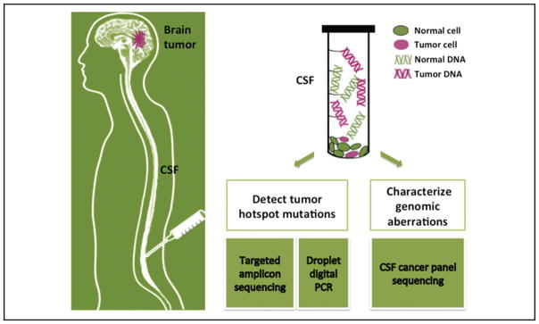
Background: Detecting tumor-derived cell-free DNA (cfDNA) in the blood of brain tumor patients is challenging, presumably owing to the blood-brain barrier. Cerebral spinal fluid (CSF) may serve as an alternative "liquid biopsy" of brain tumors by enabling measurement of circulating DNA within CSF to characterize tumor-specific mutations. Many aspects about the characteristics and detectability of tumor mutations in CSF remain undetermined.

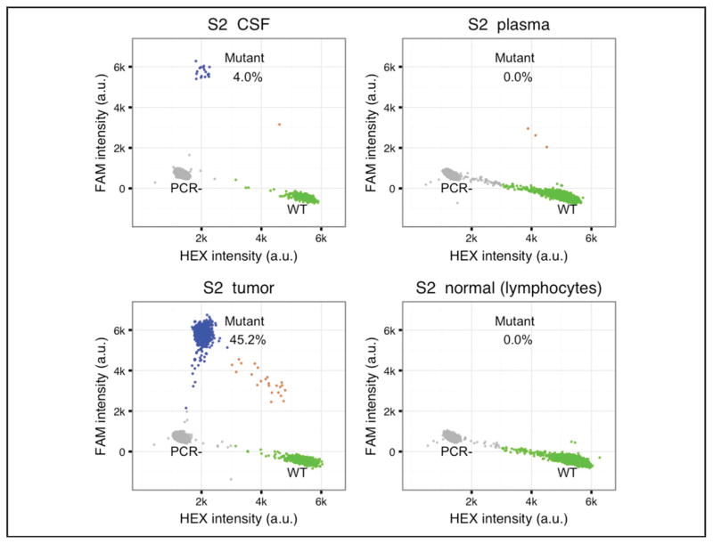
Methods: We used digital PCR and targeted amplicon sequencing to quantify tumor mutations in the cfDNA of CSF and plasma collected from 7 patients with solid brain tumors. Also, we applied cancer panel sequencing to globally characterize the somatic mutation profile from the CSF of 1 patient with suspected leptomeningeal disease.

Results: We detected tumor mutations in CSF samples from 6 of 7 patients with solid brain tumors. The concentration of the tumor mutant alleles varied widely between patients, from <5 to nearly 3000 copies/mL CSF. We identified 7 somatic mutations from the CSF of a patient with leptomeningeal disease by use of cancer panel sequencing, and the result was concordant with genetic testing on the primary tumor biopsy.

Conclusions: Tumor mutations were detectable in cfDNA from the CSF of patients with different primary and metastatic brain tumors. We designed 2 strategies to characterize tumor mutations in CSF for potential clinical diagnosis: the targeted detection of known driver mutations to monitor brain metastasis and the global characterization of genomic aberrations to direct personalized cancer care.


Conflict of interest statement

Authors’ Disclosures or Potential Conflicts of Interest: No authors declared any potential conflicts of interest.

Figures

Fig. 1
Fig. 1. Brain tumor genome analysis and diagnosis from CSF DNA
Two strategies are used for CSF DNA analysis: (1) The cancer hotspot mutations are detected with target amplicon sequencing or droplet digital PCR. (2) The cancer genomic aberrations are globally characterized by CSF cancer panel sequencing.
Fig. 2
Fig. 2. The characteristics of CSF DNA
(A), An example of the size distribution of CSF total DNA (from L3, a patient with leptomeningeal disease from breast cancer), as measured by Fragment Analyzer. The CSF total DNA was composed of cfDNA and cellular genomic DNA (gDNA). RFU, relative fluorescence unit. (B), cfDNA concentrations of CSF and plasma samples from patients S1–S7. The cfDNA concentration values are included in Table 1. The black dots in the plot indicate the values of individual samples. (C), The correlation of mutant allele fraction (AF) measured by ddPCR and targeted amplicon sequencing (AmpSeq). The dots in the plot represent the AF of S1_CSF [NF2 (neurofibromin 2 [merlin]) p.R57*], S1_plasma (NF2 p.R57*), S5_CSF (KRAS p.G12D and TP53 p.R282W), S5_plasma (KRAS p.G12D and TP53 p.R282W), S6_CSF (EGFR p.L585R), S7_CSF (EGFR p.E746_A750del), and S7_plasma (EGFR p.E746_A750del). The AF values are included in Table 1.
Fig. 3
Fig. 3. An example of ddPCR results
The TaqMan PCR probes were used to detect wild-type (WT) and mutant DNA in CSF, plasma, tumor, and blood cell samples of patient S2. AKT WT probe was labeled with HEX fluorophore and AKT p.E17K probe was labeled with FAM fluorophore. Each dot in the plot represents a droplet. The x axis is HEX fluorescence intensity in arbitrary units (a.u.); the y axis is FAM fluorescence intensity in a.u. Blue, droplets with mutant DNA; green, droplets with wild-type DNA; gray, droplets without target DNA templates; brown, droplets have both mutant and wild-type DNA.
Fig. 4
Fig. 4. Quantification of tumor cfDNA in CSF and plasma
The y axis represents mutant allele concentrations (copies/mL) of both CSF and plasma samples in log10 scale. The demonstrated results were from digital PCR measurement.

References

    1. Murtaza M, Dawson S-J, Tsui DWY, Gale D, Forshew T, Piskorz AM, et al. Non-invasive analysis of acquired resistance to cancer therapy by sequencing of plasma DNA. Nature. 2013;497:108–12. - PubMed
    1. Bettegowda C, Sausen M, Leary RJ, Kinde I, Wang Y, Agrawal N, et al. Detection of circulating tumor DNA in early- and late-stage human malignancies. Sci Transl Med. 2014;6:224ra24. - PMC - PubMed
    1. Weston CL, Glantz MJ, Connor JR. Detection of cancer cells in the cerebrospinal fluid: current methods and future directions. Fluids Barriers CNS. 2011;8:14. - PMC - PubMed
    1. Shi W, Lv C, Qi J, Zhao W, Wu X, Jing R, et al. Prognostic value of free DNA quantification in serum and cerebro-spinal fluid in glioma patients. J Mol Neurosci. 2012;46:470–5. - PubMed
    1. Chen WW, Balaj L, Liau LM, Samuels ML, Kotsopoulos SK, Maguire CA, et al. BEAMing and droplet digital PCR analysis of mutant IDH1 mRNA in glioma patient serum and cerebrospinal fluid extracellular vesicles. Mol Ther Nucleic Acids. 2013;2:e109. - PMC - PubMed