. 2015 Feb 18;43(3):1869-82.
doi: 10.1093/nar/gkv013. Epub 2015 Jan 20.

Alternative splicing regulates the expression of G9A and SUV39H2 methyltransferases, and dramatically changes SUV39H2 functions

Affiliations

Alternative splicing regulates the expression of G9A and SUV39H2 methyltransferases, and dramatically changes SUV39H2 functions

Oriane Mauger et al. Nucleic Acids Res. .

Abstract

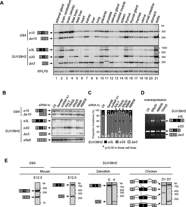
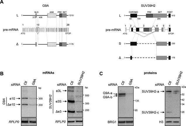
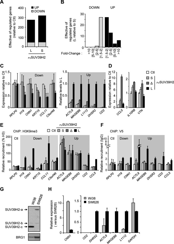
Alternative splicing is the main source of proteome diversity. Here, we have investigated how alternative splicing affects the function of two human histone methyltransferases (HMTase): G9A and SUV39H2. We show that exon 10 in G9A and exon 3 in SUV39H2 are alternatively included in a variety of tissues and cell lines, as well as in a different species. The production of these variants is likely tightly regulated because both constitutive and alternative splicing factors control their splicing profiles. Based on this evidence, we have assessed the link between the inclusion of these exons and the activity of both enzymes. We document that these HMTase genes yield several protein isoforms, which are likely issued from alternative splicing regulation. We demonstrate that inclusion of SUV39H2 exon 3 is a determinant of the stability, the sub-nuclear localization, and the HMTase activity. Genome-wide expression analysis further revealed that alternative inclusion of SUV39H2 exon 3 differentially modulates the expression of target genes. Our data also suggest that a variant of G9A may display a function that is independent of H3K9 methylation. Our work emphasizes that expression and function of genes are not collinear; therefore alternative splicing must be taken into account in any functional study.


Figures

Figure 1.
Figure 1.
Alternative splicing regulates the expression of G9A and SUV39H2. (A) Analysis in 21 human tissues of inclusion of G9A exon 10 (e10, exon 10 included; Δe10, exon 10 skipped) and SUV39H2 exon 3 (e3L, exon 3 fully included; e3S, exon 3 partially included; and Δe3, exon 3 skipped). Semi-quantitative RT-PCR was performed on total RNA using radiolabeled primers. PCR products are indicated on the left, and level of RPLP0 transcripts were used as control. (B) Regulation of G9A exon 10 and SUV39H2 exon 3 by splicing factors. The alternative inclusion of G9A exon 10 and SUV39H2 exon 3 was assessed in HeLa cells by siRNA-mediated depletion of indicated factors. Various spliced isoforms were amplified as in the panel A, while SUV39H2 exons 5 and 6 were amplified as control (bottom). (C) Ratio of SUV39H2 spliced isoforms in HeLa cells were quantified by capillary electrophoresis. Data display an average of two experiments, and stars indicate significant changes of isoforms in three cell lines (see also Supplementary Figure S1I) estimated by Wilcoxon-ranked test (P-value < 0.05). (D) Overexpression in HeLa cells of indicated splicing factors affects SUV39H2 exon 3 splicing. SUV39H2 isoforms were amplified by RT-PCR and analyzed in agarose gel stained with ethidium bromide. Splicing patterns displayed a representative example of three experiments. (E) Analysis in different organisms of alternative splicing decisions for orthologous G9A exon 10 and SUV39H2 exon 3. Semi-quantitative RT-PCR was performed with radiolabeled primers and total RNA extracted from: E12.5 mouse embryos, Zebrafish embryos (E) and adult (A), chicken embryos at day 1 (D1) and day 7 (D7). For chicken, the various alternative splicing events of SUV39H2 exon 3 are indicated with sharp lines. Inclusion of orthologous G9A exon 10 was only tested in mouse as the structure of its gene is not conserved in other species.
Figure 2.
Figure 2.
G9A and SUV39H2 produce several protein isoforms. (A) Schematic view displaying the protein isoforms encoded by the commonly defined transcript (-L) or versions that artificially reproduced the skipping of G9A exon 10 (-Δ) and SUV39H2 exon 3 (-S and -Δ for partial and total skipping respectively). For pre-mRNA, introns are drawn with bold lines (), exons with boxes, alternative exons of interest with gray boxes, alternative splicing events of pre-mRNA are drawn with sharp lines and the correspondence on protein isoforms are indicated with dotted lines. Known protein domains are reported as follows: nuclear localization signal (NLS), ankyrin (ANK), pre-SET (PRE), SET (SET), post-SET (POST), chromodomain (CHROMO). Symbols and dotted lines were reported on the pre-mRNA to indicate correspondences. (B) and (C) Analysis in HeLa cells of mRNAs and proteins resulting from G9A and SUV39H2 expression. The specificity of signals detected by radiolabeled RT-PCR (B) or western blot (C) was checked by the analysis of HeLa cells transfected with siRNA: SUV39H2, G9A or control (Ctl). SiRNA were designed to target constitutive exons of G9A or SUV39H2 transcripts. Transcripts levels were assessed 3 days after transfection and proteins levels were detected after 3 days for G9A and 5 days for SUV39H2. RPLP0 transcripts and BRG1 or H3 proteins were used as controls.
Figure 3.
Figure 3.
Protein stability and sub-nuclear localization of SUV39H2 isoforms are regulated by alternative inclusion of exon 3. (A) Expression in HEK 293T cells of ectopic tagged G9A and SUV39H2 protein isoforms. Cells were transduced with the pLVX vector containing Flag-V5 tags and SUV39H2 or G9A cDNA to express exogenous protein isoforms (FVSUV39H2 or FVG9A). Cells transduced with pLVX empty vector were used as controls (Ctl). The ectopic proteins were revealed by western blot using the V5 antibody. (B) and (C) Stability analysis of each G9A and SUV39H2 protein isoform. FVG9A and FVSUV39H2 isoforms were assessed by western blot in HeLa cells treated with cycloheximide (CHX) (B) or MG132 (C) for the indicated times in hours (h). The signal detected with V5 antibody was normalized to histone H3 levels. (D) Localization of FVG9A and FVSUV39H2 isoforms in HeLa cells. Exogenous proteins were revealed with V5 antibody (red), while DNA was counterstained with DAPI (blue). (E) and (F) Analysis of FVG9A and FVSUV39H2 and the endogenous SUV39H2 isoforms after fractionation of HeLa cells. (E) Scheme displaying the cell fractionation procedure. (F) Protein isoforms were detected in whole cell extracts (WCE) and fractions by western blot using the V5 or SUV39H2 antibodies. Endogenous tubulin, HP1α and H3 proteins were analyzed as markers of the cell fractions.
Figure 4.
Figure 4.
Inclusion of SUV39H2 exon 3 is required to encode an active histone methyltransferase. (A) In vitro analysis of the methyltransferase activity associated to each FVG9A and FVSUV39H2 isoforms. Scheme of the in vitro histone methyltransferase (HMT) assay procedure (left panel). Various quantities of purified FVG9A and FVSUV39H2 (right panel) were incubated with the recombinant human histone H3.1 and S-adénosylméthionine (SAM) producing methylated H3.1 and S-adenosylhomocysteine (SAH). Histone H3 and H3K9me3 were analyzed by western blot using specific antibodies (right panel). Control reactions were supplemented with FVTomato, water (-) or sample issued from blank purification procedure (Ctl). Levels of recombinant proteins were estimated using V5 antibody. (B) and (C) H3K9me3 was assessed in HeLa cells after expression of FVG9A and FVSUV39H2 isoforms. (B) DNA was counterstained with DAPI (blue), H3K9me3 was immunostained with a specific antibody (red) and cells expressing FVG9A and FVSUV39H2 were revealed with ZsGreen1 Fluorescence Protein (ZsGFP panel; labeled with white arrows in H3K9me3 panel). (C) Analysis of H3K9me3, H3K9me2 and H3 levels by western blot in total protein extracts of HeLa cells expressing FVG9A and FVSUV39H2 isoforms.
Figure 5.
Figure 5.
Alternative inclusion of SUV39H2 exon 3 modulates the expression of target genes. (A) Graph showing the number of genes that are up- or down-regulated in HeLa cells expressing FVSUV39H2-L or FVSUV39H2-S. Gene expression levels were assessed by high throughput RNA sequencing and hits were retained with a threshold of 1.5 fold-change (P-value < 0.05) when compared to control (Ctl, virus generated with empty construct). (B) Graph displaying the differential effect on gene expression of FVSUV39H2-L versus FVSUV39H2-S. Number of genes is displayed in function of expression fold change. (C and D) Validation of 13 genes differentially affected in their transcription by SUV39H2 isoforms. Transcription levels were analyzed by RT-qPCR and quantification was displayed as means ± s.e.m. of three experimental replicate. (C) Five genes down-regulated by the over-expression of FVSUV39H2-L (Down) were shown relative to control (Ctl). RPLP0 gene is shown as unaffected gene. Five genes up-regulated by the over-expression of FVSUV39H2-L (Up) were presented relative to the expression in presence of FVSUV39H2-L (set to 1). (D) Expression analysis of three genes up-regulated specifically by FVSUV39H2-S and –Δ isoforms. Transcriptional levels were expressed relative to control (Ctl). (E and F) Analysis of H3K9me3 and SUV39H2 isoforms at promoters of target genes. Chromatin from cells expressing FVSUV39H2 isoforms or not (Ctl) was immunoprecipated with antibodies to H3, H3K9me3, V5 tag or nonimmune (IgG). Relative enrichments were measured by qPCR using primer sets targeting promoters of indicated genes. Values are means ± s.e.m. of three independent experiments. Amounts of H3K9me3 are expressed in percent of H3 (E), and amounts of FVSUV39H2 isoforms are expressed relatively to IgG control (F). (G) Endogenous SUV39H2 isoforms expressed in WI38 and SW626 cell lines were detected by western blot. (H) Relative gene expression in WI38 and SW626 cells of seven hits validated in panel 5C were quantified by RT-qPCR. CD2 gene expression is not detectable in both cell lines.

References

    1. Pan Q., Shai O., Lee L.J., Frey B.J., Blencowe B.J. Deep surveying of alternative splicing complexity in the human transcriptome by high-throughput sequencing. Nat. Genet. 2008;40:1413–1415. - PubMed
    1. Wang E.T., Sandberg R., Luo S., Khrebtukova I., Zhang L., Mayr C., Kingsmore S.F., Schroth G.P., Burge C.B. Alternative isoform regulation in human tissue transcriptomes. Nature. 2008;456:470–476. - PMC - PubMed
    1. Jurica M.S., Moore M.J. Pre-mRNA splicing: awash in a sea of proteins. Mol. Cell. 2003;12:5–14. - PubMed
    1. Wahl M.C., Will C.L., Lührmann R. The spliceosome: design principles of a dynamic RNP machine. Cell. 2009;136:701–718. - PubMed
    1. Krainer A.R., Maniatis T., Ruskin B., Green M.R. Normal and mutant human beta-globin pre-mRNAs are faithfully and efficiently spliced in vitro. Cell. 1984;36:993–1005. - PubMed

Publication types

Substances