. 2015 Feb 3;112(5):1571-6.
doi: 10.1073/pnas.1416418112. Epub 2015 Jan 20.

Microtubule-targeting agents augment the toxicity of DNA-damaging agents by disrupting intracellular trafficking of DNA repair proteins

Affiliations

Microtubule-targeting agents augment the toxicity of DNA-damaging agents by disrupting intracellular trafficking of DNA repair proteins

Marianne S Poruchynsky et al. Proc Natl Acad Sci U S A. .

Abstract

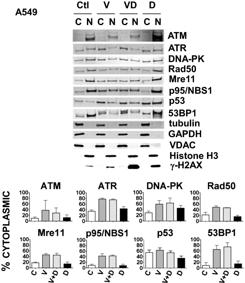
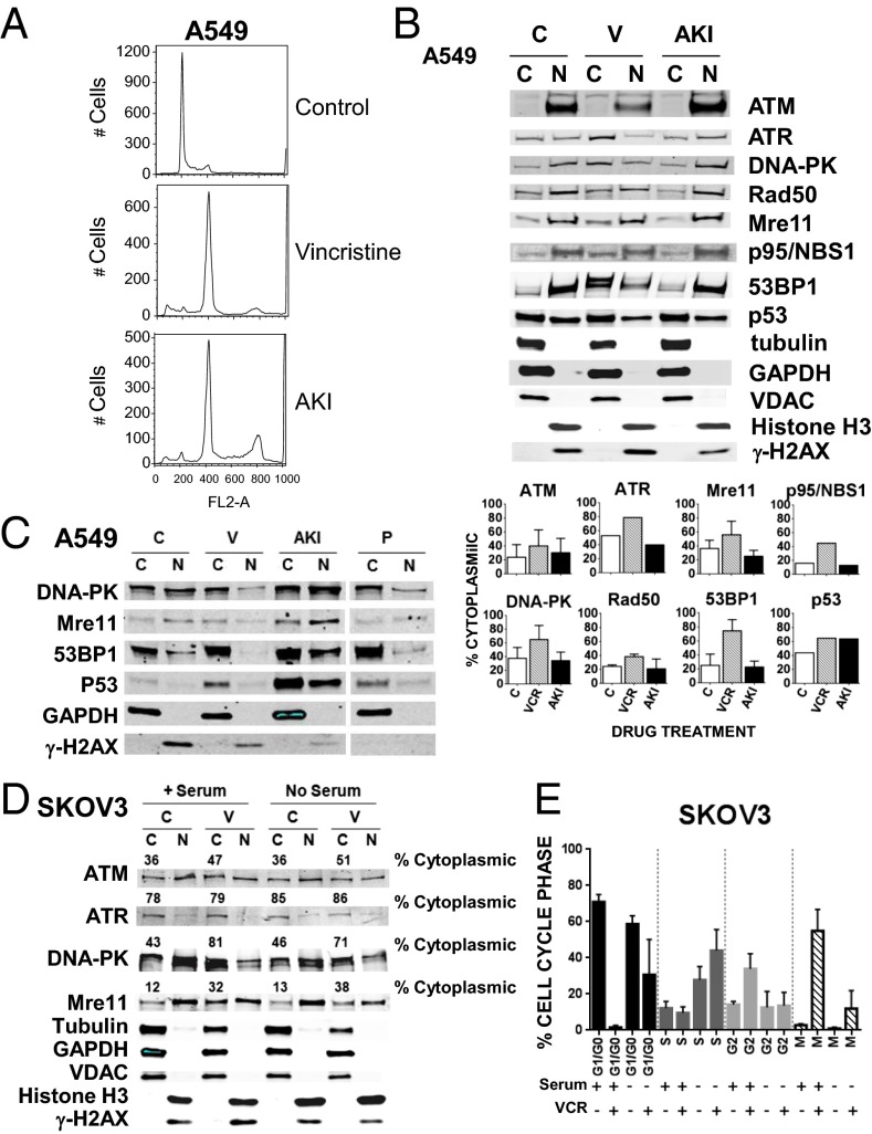
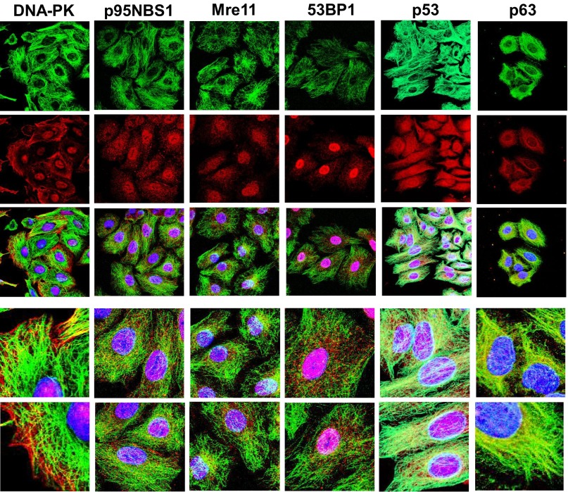
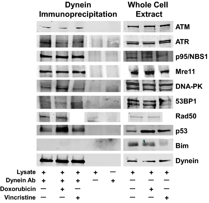
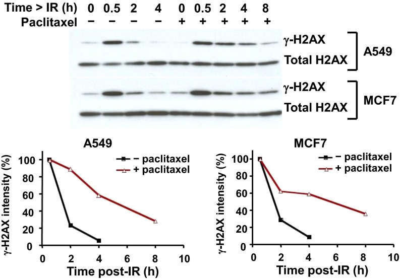
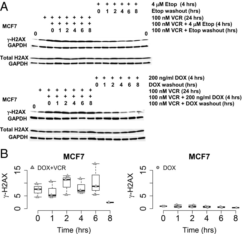
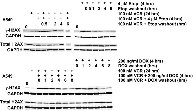
The paradigm that microtubule-targeting agents (MTAs) cause cell death via mitotic arrest applies to rapidly dividing cells but cannot explain MTA activity in slowly growing human cancers. Many preferred cancer regimens combine a MTA with a DNA-damaging agent (DDA). We hypothesized that MTAs synergize with DDAs by interfering with trafficking of DNA repair proteins on interphase microtubules. We investigated nine proteins involved in DNA repair: ATM, ATR, DNA-PK, Rad50, Mre11, p95/NBS1, p53, 53BP1, and p63. The proteins were sequestered in the cytoplasm by vincristine and paclitaxel but not by an aurora kinase inhibitor, colocalized with tubulin by confocal microscopy and coimmunoprecipitated with the microtubule motor dynein. Furthermore, adding MTAs to radiation, doxorubicin, or etoposide led to more sustained γ-H2AX levels. We conclude DNA damage-repair proteins traffic on microtubules and addition of MTAs sequesters them in the cytoplasm, explaining why MTA/DDA combinations are common anticancer regimens.

Keywords: DNA repair protein trafficking; DNA-damaging agents; combination chemotherapy; microtubule targeting agents; targeted therapies.


Conflict of interest statement

The authors declare no conflict of interest.

Figures

Fig. 1.
Fig. 1.
Treatment of A549 cells with the MTA vincristine, alone or in combination with a DNA-damaging agent, doxorubicin, causes increased cytoplasmic retention of the DNA damage-repair proteins ATM, ATR, DNA-PK, Rad50, Mre11, p95/NBS1, p53, and 53BP1. A549 cells were untreated (Ctl), or treated either with 200 nM vincristine (V) for 24 h or 400 ng/mL doxorubicin (D) for 4 h, either separately or in combination (V for 20 h followed by V+D for an additional 4 h). Cytoplasmic (C) and nuclear (N) fractions prepared as described in Materials and Methods, are indicated for Western blots probed with antibodies against the repair proteins and five fractionation controls (tubulin, GAPDH, VDAC, Histone H3, or γ-H2AX). The percentage of protein in the C fraction is indicated and calculated as: [C/(C+N)] × 100%. Means and SDs are shown. The number of experiments (n) were: ATM (three), ATR (three), DNA-PK (three), Rad50 (three), Mre11 (three), p95/NBS1 (three), p53 (six), and 53BP1 (four). For the proteins studied, drug treatment had no or very little effect on their amount as shown in the blots of lysates seen in Fig. 4 and Fig. S3. Because no protein is lost in the fractionation procedure, all proteins appear in either the N or the C fraction, and the percent in each fraction can be accurately calculated from the results of the immunoblot.
Fig. 2.
Fig. 2.
Treatment of A549 cells with the AKI VX680 fails to increase cytoplasmic accumulation of the DNA damage-repair proteins. (A) A549 cells were untreated (C, control) or treated with either 200 nM vincristine (V) or 250 nM of the AKI VX-680 (Tozasertib) for 24 h. Cells were further processed for flow cytometry as described. (B) A549 lysates treated as described in A were prepared and separated into cytoplasmic (C) and nuclear (N) fractions as described in Materials and Methods. Western blots were probed with antibodies as in Fig. 1. The percent of the protein in the C fraction is indicated and calculated as: [C/(C+N)] × 100%. Means and SDs are shown. The number of experiments (n) were: ATM (four), Mre11 (three), DNA-PK (three), Rad50 (two), and 53BP1 (four). Because p53 is wild-type in A549 cells and expressed at very low levels in the absence of drug treatment (see p53 Western blot in Fig. 1), the sample shown under the “C” control designation was treated with 400 ng/mL of doxorubicin (D) for 4 h and serves as comparison for samples alongside those treated with either 250 nM of AKI for 24 h or 200 nM V for 24 h followed by a V + D combination for an additional 4 h. (C) A549 cells were treated as in A or also with 200 nM paclitaxel (P) for 24 h before being separated into “C” and “N” fractions. (D) SKOV3 cells incubated with or without serum for 48 h were then treated or not with 100 nM V with or without serum for another 24 h. C and N fractions were prepared as described in Materials and Methods. Western blots were probed with four antibodies that performed well with SKOV3 cell extracts: ATM, ATR, DNA-PK, and Mre11, and also with antibodies for tubulin, GAPDH, VDAC, Histone H3, and γ-H2AX. The percentage of protein in the C fraction is indicated and calculated as [C/(C+N)] × 100%. (E) SKOV3 cells treated as described in D were further processed for flow cytometry, and mitotic cells were quantitated by two-variable analysis, as described in Materials and Methods. The percentage of cells in each phase of the cell cycle (G1/G0, S, G2, or M) is indicated for the different treatments. The mean and error bars display data compiled from three experiments.
Fig. 3.
Fig. 3.
DNA damage-repair proteins colocalize with MTs as visualized by immunofluorescence microscopy. Confocal immunofluroescent localization of DNA damage-repair proteins (DNA-PK, p95NBS1, Mre11, 53BP1, p53, or p63) and tubulin in A549 cells. Tubulin (FITC-conjugated secondary antibody, green); DNA repair proteins (RHOD-conjugated secondary antibody, red); DAPI stain (blue) localizes to cell nuclei. The tricolor localization of “Tubulin/DNA-damage-repair-protein/DAPI” is shown by the superimposition of three confocal images in the third panel down in each column. Enlarged areas from these images are displayed in the bottom two panels of each column. Images are shown as 3D maximal projections reconstructed from z-stacks or a single slice of a projection. (Magnification in each column: top three images, 630×; lower two images, 2500×.)
Fig. 4.
Fig. 4.
DNA damage-repair proteins coimmunoprecipitate with the MT motor protein dynein. A549 cells either untreated, treated with 100 nM vincristine overnight, or 400 ng/mL doxorubicin for 4 h, were lysed in immunoprecipitation buffer, homogenized and incubated overnight with an anti-dynein antibody. Whole-cell extracts (50-μg aliquots) or dynein immunoprecipitation samples (the entire immunoprecipitation harvest from 1 mg of whole-cell protein) were resolved by SDS/PAGE, and the Western blots were probed with antibodies to the DNA repair proteins ATM, ATR, P95/NBS1, Mre11, DNA-PK, 53BP1, and p53, Rad50 (no vincristine-treated sample), the control protein Bim, or dynein. The extent of coimmunoprecipitation varies for each protein.
Fig. 5.
Fig. 5.
Addition of paclitaxel treatment to irradiation prolongs γ-H2AX protein detection in A549 and MCF7 cells. Cells were pretreated or not with 200 nM paclitaxel for 24 h, irradiated with 10 Gy, and then further incubated for 0.5, 2, 4, or 8 h before γ-H2AX, a phosphorylated form of H2AX indicating DNA damage, was assessed by immunoblot.
Fig. 6.
Fig. 6.
Addition of vincristine to a DNA-damaging agent prolongs γ-H2AX levels in MCF7 cells. (A) MCF7 cells were treated with vincristine (VCR) for 24 h before a 4 h incubation of VCR in combination with either 4 μM etoposide or 200 ng/mL doxorubicin (DDAs) before the DDA was washed out with VCR present for 1, 2, 4, 6, or 8 h. MCF7 cells that had only the DDA treatment for 4 h before its washout were used for comparison. Western blots were probed with antibodies to γ-H2AX, total H2AX, and GAPDH. (B) The graphs summarize data from multiple Western blots quantitated for γ-H2AX, normalized to GAPDH. The normalized relative initial γ-H2AX values for the VCR/DOX combination and the DOX washout in the presence of VCR are represented as box plots compared with the DOX treatment alone. The starting level of γ-H2AX was arbitrarily set equal to 1 for the DOX treatment alone, γ-H2AX values for each of the DOX washout time points is expressed in relation to 1 and graphically represented. The triangles and circles represent the observed data points. The white box represents the interquartile range, its bottom the 25th percentile, its top the 75th percentile, the black line the 50th percentile. The whiskers extending from the box do so to the most extreme data point, which is no more than 1.5-times the interquartile range from the box.
Fig. 7.
Fig. 7.
Addition of vincristine to a DNA-damaging agent prolongs γ-H2AX levels in A549 cells. A549 cells were treated with VCR for 24 h before a 4 h incubation of VCR in combination with either 4 μM etoposide or 200 ng/mL doxorubicin (DDA) before the DDA was washed out with VCR present for 1, 2, 4, 6, or 8 h. A549 cells that had only received the DDA for 4 h before its washout were used for comparison. Western blots were probed with antibodies to γ-H2AX, total H2AX, and GAPDH. The 6-h etoposide washout time-point sample for the VCR/etoposide combination was underloaded, and the bands shown in the offset tracks are from darker exposures.

References

    1. Milas L, et al. Kinetics of mitotic arrest and apoptosis in murine mammary and ovarian tumors treated with taxol. Cancer Chemother Pharmacol. 1995;35(4):297–303. - PubMed
    1. Horton JK, Houghton PJ, Houghton JA. Relationships between tumor responsiveness, vincristine pharmacokinetics and arrest of mitosis in human tumor xenografts. Biochem Pharmacol. 1988;37(20):3995–4000. - PubMed
    1. Komlodi-Pasztor E, Sackett D, Wilkerson J, Fojo T. Mitosis is not a key target of microtubule agents in patient tumors. Nat Rev Clin Oncol. 2011;8(4):244–250. - PubMed
    1. Komlodi-Pasztor E, Sackett DL, Fojo AT. Inhibitors targeting mitosis: Tales of how great drugs against a promising target were brought down by a flawed rationale. Clin Cancer Res. 2012;18(1):51–63. - PubMed
    1. Mitchison TJ. The proliferation rate paradox in antimitotic chemotherapy. Mol Biol Cell. 2012;23(1):1–6. - PMC - PubMed

Publication types