. 2015 Feb 3;112(5):1481-6.
doi: 10.1073/pnas.1412279112. Epub 2015 Jan 20.

Biomechanical basis of wing and haltere coordination in flies

Affiliations

Biomechanical basis of wing and haltere coordination in flies

Tanvi Deora et al. Proc Natl Acad Sci U S A. .

Abstract

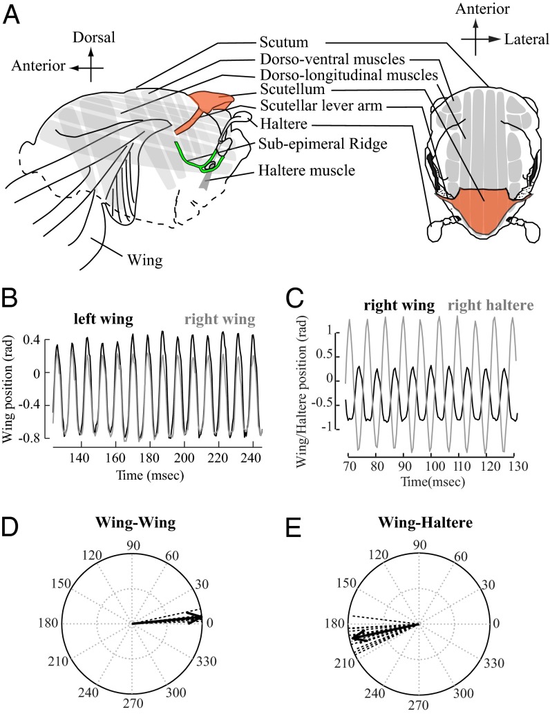
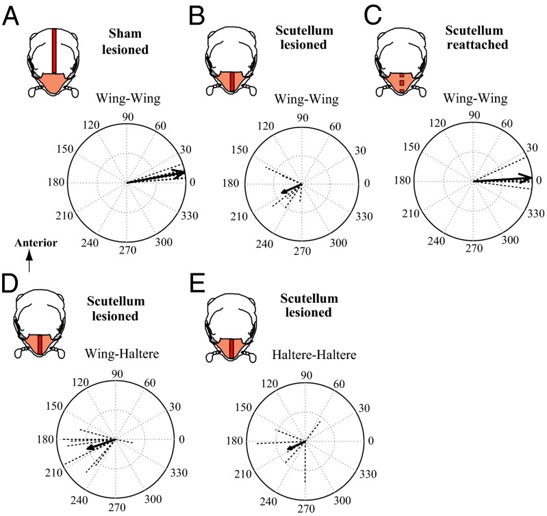
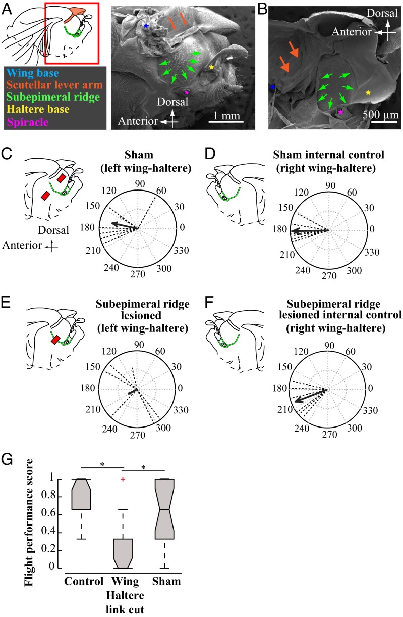
The spectacular success and diversification of insects rests critically on two major evolutionary adaptations. First, the evolution of flight, which enhanced the ability of insects to colonize novel ecological habitats, evade predators, or hunt prey; and second, the miniaturization of their body size, which profoundly influenced all aspects of their biology from development to behavior. However, miniaturization imposes steep demands on the flight system because smaller insects must flap their wings at higher frequencies to generate sufficient aerodynamic forces to stay aloft; it also poses challenges to the sensorimotor system because precise control of wing kinematics and body trajectories requires fast sensory feedback. These tradeoffs are best studied in Dipteran flies in which rapid mechanosensory feedback to wing motor system is provided by halteres, reduced hind wings that evolved into gyroscopic sensors. Halteres oscillate at the same frequency as and precisely antiphase to the wings; they detect body rotations during flight, thus providing feedback that is essential for controlling wing motion during aerial maneuvers. Although tight phase synchrony between halteres and wings is essential for providing proper timing cues, the mechanisms underlying this coordination are not well understood. Here, we identify specific mechanical linkages within the thorax that passively mediate both wing-wing and wing-haltere phase synchronization. We demonstrate that the wing hinge must possess a clutch system that enables flies to independently engage or disengage each wing from the mechanically linked thorax. In concert with a previously described gearbox located within the wing hinge, the clutch system enables independent control of each wing. These biomechanical features are essential for flight control in flies.

Keywords: halteres; insect thorax; insect wings; wing clutch; wing hinge.


Conflict of interest statement

The authors declare no conflict of interest.

Figures

Fig. 1.
Fig. 1.
Wings and halteres are precisely coordinated at high frequencies ≥100 Hz. (A) Diagram of Dipteran thorax in lateral and dorsal view. (B) The right (gray) and the left (black) wings move in phase with each other. (C) The wing (black) and the ipsilateral haltere (gray) move antiphase to each other. (D) Vector strength representation of control data for wing–wing phase (tethered flies; n = 6; mean phase angle ϕ = 5.63°, vector length r = 0.9965; P < 0.001; nonparametric Moore’s test for uniformity). Dotted lines indicate the mean vector (∼20 wing strokes) for individual insects, and solid line indicates mean for the treatment. (E) Vector strength representation of control data of wing–haltere phase (tethered flies; n = 12; ϕ = 192.14°; r = 0.9572; P < 0.001; right and left side data pooled).
Fig. 2.
Fig. 2.
Wing–wing coordination is mediated by a passive mechanical linkage within the scutellum. Vector strength data for the phase between wing–wing (A–C), wing–ipsilateral haltere (D), and haltere–haltere (E) pairs. (Insets) Treatment type as a dorsal view of thorax with scutum (white) and scutellum (brown), and a line on thorax (red) indicating the surgical lesion. (A) Similar to controls (Fig. 1C), scutum-lesioned flies show well-coordinated wing movements (ϕ = 10.35°, r = 0.9932, n = 6, P < 0.001). (B) Scutellum-lesioned flies have disrupted wing–wing coordination and randomly distributed phase angles (ϕ = 203.63°, r = 0.3822, n = 6, P > 0.01). (C) In flies with reattached scutellum, wing coordination is restored (ϕ = 3.60°, r = 0.9769, n = 6, P < 0.001). (D) In scutellum-lesioned flies, halteres move antiphase to ipsilateral wings (ϕ = 198.79°, r = 0.5033, n = 12, P < 0.001; left and right data pooled) similar to control (Fig. 1E). (E) Haltere–haltere coordination is disrupted in scutellum-lesioned flies (ϕ = 203.89, r = 0.3217, n = 6, P > 0.1).
Fig. 3.
Fig. 3.
Each wing–haltere pair is coordinated via a separate mechanical linkage running through the subepimeral ridge. (A, Inset) Red box around the fly wing base highlights the approximate region of the thorax imaged using a SEM. (A and B) SEM images of H. illucens thorax with wing (blue asterisk) and haltere (yellow asterisk) base, scutellar lever arm (brown arrows), epimeral ridge (green arrows), and spiracle (purple asterisk) in lateral (A) and hemisectional (B) views. (C–F) Phase relationships between ipsilateral wing–haltere pair. (C–F, Left) Lateral thorax with red bar indicating lesioned area relative to the supepimeral ridge (green). All treatments were performed on the left side, leaving the right intact as an internal control. (C) In sham-treated flies, the left wing–haltere pair, which is perforated above and below the ridge, continues to move antiphase to each other (ϕ = 167.84°; r = 0.6780; n = 7; P < 0.01; Moore’s test), similar to D, the right side untreated wing–haltere pair that acts as internal control (ϕ = 181.14°; r = 0.9420; P < 0.001, n = 7). (E) Lesioning the subepimeral ridge disrupts antiphase coordination in left wing–haltere pair (ϕ = 207.72°, r = 0.2485, n = 6, P > 0.5), whereas (F) the unlesioned wing–haltere pair on the right side, which acts as internal control, maintains antiphase coordination (ϕ = 201.05°; r = 0.8938, P < 0.005; n = 6). (G) Lesioning the subepimeral ridge and disrupting wing–haltere coordination on both sides impairs flight. We measured flight performance (shown as notched box plots) using the drop test [SI Materials and Methods, Free Flight (Drop Test) Assay for Flies with Severed Subepimeral Ridge; n = 20 per treatment], which measured the ability of each fly to recover from free fall in a vertical cylinder in three separate trials. Flies scored 0 if did not recover flight in all three trials, or 1/3, 2/3, or 1 if it recovered flight, respectively, in one, two, or three of three trials. Significant differences in groups (P < 0.001, asterisk) were identified using nonparametric Kruskal–Wallis ANOVA, followed by the Tukey–Kramer post hoc multicomparison analysis.
Fig. 4.
Fig. 4.
Passive mechanisms for wing–wing and wing–haltere coordination in flies. (A) Wing (blue) and right haltere (green) frequency as a function of wing length for a representative fly (for more data, see Fig. S5). Individual box plots show distribution of ∼20 wing (and haltere) stroke frequency. (B) SEM image of the wing hinge in P. dux in lateral view showing radial stop (RS), pleural wing process (PWP), PteraleC (PtC), and wing (W). (C–F) Simultaneous visualization and quantification of 3D trajectories of the wing tip and wing hinge configurations. (C–E) As shown for a representative tethered P. dux, during flight onset, the wing beat amplitude as a function of time increases from small (black traces, corresponding to mode 0 gearbox configuration) to large (blue corresponding to mode 1, 2, or 3) in a single stroke (magenta, gearbox in transition), and at flight offset the drop in amplitude reverses from high (blue; mode 1, 2, or 3) to low (black; mode 0) amplitude also occurs in one wing stroke (pink; gearbox in transition). The corresponding trajectories for the whole sequence are shown in D (gray silhouette represents fly body and black points are wing base, which were also digitized) and wing hinge dynamics and corresponding wing amplitudes for several flies is shown in E. Here, stroke amplitudes (mean ± SD, n = 6; five animals) at various hinge configurations were compared using Friedman ANOVA with Tukey’s LSD post hoc test (*P < 0.001). (F) Wing hinge configurations during flight initiation. (Inset) Approximate region around the fly thorax that was filmed at macro magnification. (i and ii) Video stills show the wing hinge configuration (traced as in Movie S8; radial stop, red; pleural wing process, yellow; PteraleC, light blue) for the (i) wing transitioning from mode 0 to higher modes and (ii) engaged wing in mode 2 where the wing is flapping and RS touches the second grove of the PWP. (G) Schematic summary of the Dipteran thoracic mechanics. The A-IFM’s (gray box) drive the wings, which are synchronized via the scutellum (brown spring). Each wing is, however, independently and actively engaged and disengaged by the clutch (black switch) and its amplitude modulated by the gearbox (yellow and red). The halteres are driven by their own asynchronous muscles (gray box) and are mechanically linked to the ipsilateral wing by the wing haltere linkage, the subepimeral ridge (green spring). (G, Insets) The clutch in a disengaged state (a) and engaged state (b). (a) Disengaged state: when the clutch is disengaged, only mode 0 is possible. RS sits posteriorly to the PWP. (b) Engaged state: when the clutch is engaged, modes 1, 2, and 3 are possible. RS either contacts a groove in PWP (modes 1 and 2) or moves in front and around the PWP (mode 3).

References

    1. Polilov AA. The smallest insects evolve anucleate neurons. Arthropod Struct Dev. 2012;41(1):29–34. - PubMed
    1. Dudley R. The Biomechanics of Insect Flight. Princeton Univ Press; Princeton, NJ: 2000.
    1. Josephson RK, Malamud JG, Stokes DR. Asynchronous muscle: A primer. J Exp Biol. 2000;203(18):2713–2722. - PubMed
    1. Dickinson MH, Tu MS. The function of dipteran flight muscle. Comp Biochem Physiol A. 1997;116(3):223–238.
    1. Bastian J, Esch H. Nervous control of indirect flight muscles of honey bee. Z Vgl Physiol. 1970;67(3):307.

Publication types