. 2015 Feb 3;112(5):E420-9.
doi: 10.1073/pnas.1424576112. Epub 2015 Jan 20.

MITF drives endolysosomal biogenesis and potentiates Wnt signaling in melanoma cells

Affiliations

MITF drives endolysosomal biogenesis and potentiates Wnt signaling in melanoma cells

Diego Ploper et al. Proc Natl Acad Sci U S A. .

Abstract

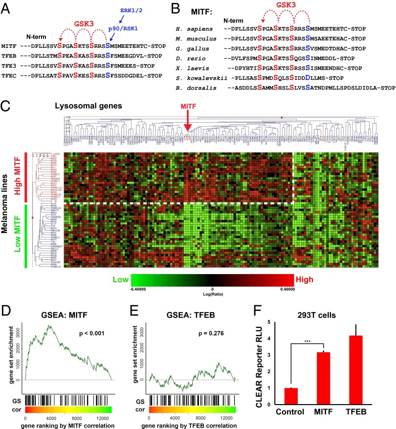
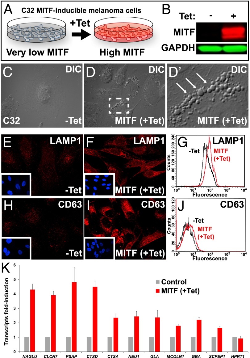
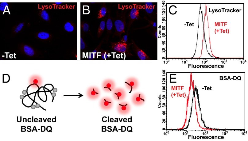
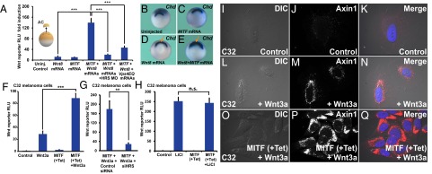
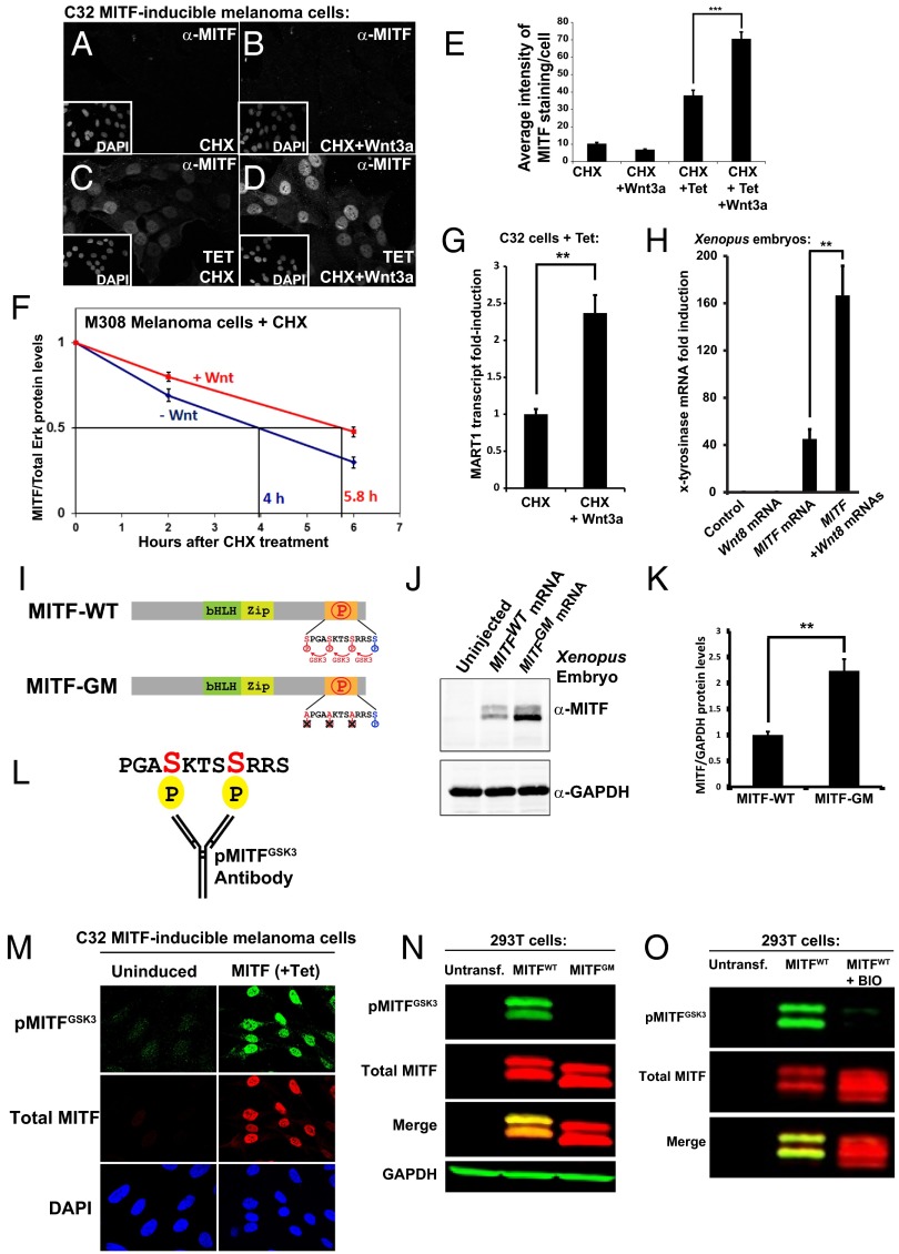
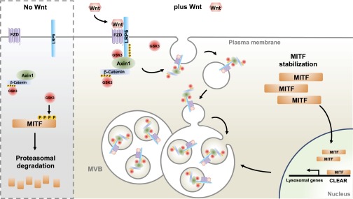
Canonical Wnt signaling plays an important role in development and disease, regulating transcription of target genes and stabilizing many proteins phosphorylated by glycogen synthase kinase 3 (GSK3). We observed that the MiT family of transcription factors, which includes the melanoma oncogene MITF (micropthalmia-associated transcription factor) and the lysosomal master regulator TFEB, had the highest phylogenetic conservation of three consecutive putative GSK3 phosphorylation sites in animal proteomes. This finding prompted us to examine the relationship between MITF, endolysosomal biogenesis, and Wnt signaling. Here we report that MITF expression levels correlated with the expression of a large subset of lysosomal genes in melanoma cell lines. MITF expression in the tetracycline-inducible C32 melanoma model caused a marked increase in vesicular structures, and increased expression of late endosomal proteins, such as Rab7, LAMP1, and CD63. These late endosomes were not functional lysosomes as they were less active in proteolysis, yet were able to concentrate Axin1, phospho-LRP6, phospho-β-catenin, and GSK3 in the presence of Wnt ligands. This relocalization significantly enhanced Wnt signaling by increasing the number of multivesicular bodies into which the Wnt signalosome/destruction complex becomes localized upon Wnt signaling. We also show that the MITF protein was stabilized by Wnt signaling, through the novel C-terminal GSK3 phosphorylations identified here. MITF stabilization caused an increase in multivesicular body biosynthesis, which in turn increased Wnt signaling, generating a positive-feedback loop that may function during the proliferative stages of melanoma. The results underscore the importance of misregulated endolysosomal biogenesis in Wnt signaling and cancer.

Keywords: MITF; Wnt-STOP; lysosome; melanoma; multivesicular body.


Conflict of interest statement

The authors declare no conflict of interest.

Figures

Fig. 1.
Fig. 1.
MITF mRNA correlates with lysosomal gene expression in melanoma cell lines. (A) MITF and the lysosomal master gene regulator TFEB have three putative C-terminal GSK3 phosphorylation sites, with a previously validated priming site. (B) The C-terminal GSK3 sites on MITF have been highly conserved throughout evolution, including in the oriental fruit fly Bactrocera dorsalis. (C) Heat map obtained from a RNA microarray panel of 51 melanoma cell lines, which cluster into two distinct groups, one with high MITF and the other with low MITF expression, when queried for a panel of 89 lysosomal genes. The group with high MITF expression (which includes all cell lines with MITF genomic amplifications) up-regulates many, but not all, lysosomal genes (dashed line). (D and E) GSEA of an expression dataset consisting of a panel of 83 additional, different, melanoma lines confirms that MITF, but not TFEB, significantly correlates with the lysosomal gene set in melanoma. Microarray data for melanoma cell lines was obtained from Hoek et al. (30). Genes were ranked by their correlation (cor) with MITF or TFEB (red to green = high to low correlation). The positions of lyosomal genes (22) among over 12,000 genes per cell line are marked as vertical lines (GS). Enrichment of the lysosomal gene set at the top of the ranked lists was assessed with a permutation based Kolmogorov–Smirnoff nonparametric rank test. A significant correlation for MITF, but not for TFEB, was found. (F) Transfection of MITF activated a CLEAR element-Firefly luciferase reporter (22), in transient transfections of HEK 293T cells. Renilla luciferase driven by the CMV promoter was used for normalization purposes. ***P < 0.001.
Fig. 2.
Fig. 2.
Tet-inducible MITF expression increases late endolysosomal vesicles in the C32 melanoma cell line. (A) Schematic diagram of the C32 Tet-inducible MITF melanoma cell line (20). (B) Strong induction of MITF after Tet treatment was detected in C32 cells by Western blot with anti-MITF antibody. (C–D′) Increase in the number of vesicular structures upon MITF induction observed by differential interference contrast light microscopy. Note vesicular structures seen at high power (arrows). (E and F) MITF induction increases immunostaining of the late endosomal marker LAMP1. (G) Quantification by flow cytometry of the increase in LAMP1 levels upon MITF induction. (H and I) MITF induction increases immunostaining of the MVB marker CD63. (J) Quantification by flow cytometry of the increase in CD63 upon MITF induction. (K) MITF induction increased the transcripts of many lysosomal genes containing CLEAR elements in C32 melanoma cells, as validated by RT-qPCR. MITF induction in C32 melanoma cells up-regulated transcripts for the CLEAR element lysosomal genes NAGLU, CLCN7, PSAP, CTSD, CTSA, NEU1, GLA, MCOLN1, GBA, and SCPEP1. Error bars indicate the SEM from three independent experiments.
Fig. 3.
Fig. 3.
MITF expands acidic organelle compartments but does not increase lysosomal activity. (A and B) Acidic organelles visualized by treatment of living cells with LysoTracker dye. (C) Quantification by flow cytometry of the increase in acidic organelles observed upon MITF induction. (D) Schematic representation of the BSA-DQ reagent used for detecting lysosomal proteolytic activity. BSA-DQ added to the culture medium is endocytosed, but only fluoresces when cleaved by proteases inside lysosomes. (E) MITF induction decreases lysosomal activity, as quantified by flow cytometry.
Fig. 4.
Fig. 4.
MITF enhances Wnt signaling in Xenopus and in melanoma cells in an ESCRT-dependent manner, and causes increased MVB localization of the destruction complex component Axin1. (A) MITF enhances Wnt signaling in Xenopus ectodermal explants. This enhancement is ESCRT-dependent, as it is blocked by HRS/Vps27 morpholino or a dominant-negative form of the Vps4 ATPase (HRS MO and VPS4EQ) (***P < 0.001). (B–E) MITF cooperates with a low dose of Wnt8 mRNA, expanding the Spemann organizer (arrows), the region that expresses chordin mRNA in Xenopus whole-mount in situ hybridization. (F) MITF induction increases Wnt signaling in the C32 MITF-inducible melanoma cell line. The Wnt BAR firefly luciferase reporter and EF1α-driven Renilla luciferase were permanently introduced with lentivectors into the C32 cell line. (G) HRS/Vps27 knockdown by HRS siRNA decreased Wnt signaling in Tet-induced C32 cells (**P < 0.01). (H) MITF induction did not affect Lithium chloride-induced β-catenin signaling. (IQ) Immunostaining for Axin1, the key scaffold of the β-catenin destruction complex. Note that Wnt signaling relocalizes Axin1 to vesicular structures, and that this effect is strongly enhanced by MITF induction with Tet. For relocalization of other Wnt components (pLRP6, GSK3, and p-β-catenin) after MITF induction and Wnt3a protein treatment, see Figs. S6–S8.
Fig. 5.
Fig. 5.
MITF protein stabilization by Wnt via novel C-terminal GSK3 phosphorylation sites. (A–D) MITF immunostainings in C32 cells using a 40× objective. Wnt3a protein treatment for 5 h stabilized MITF protein in the presence of CHX. (E) Quantification of MITF staining per nucleus from a previous experiment (***P < 0.001). (F) MITF protein levels (normalized to total Erk1/2) from three independent Western blot experiments upon treatment of M308 melanoma cells with Wnt3a; Wnt prolongs the half-life of endogenous MITF protein. (G) RT-qPCR of the MITF target gene MART1 in Tet-induced C32 cells treated with CHX and Wnt3a (**P < 0.01). (H) RT-qPCR for the MITF target gene x-tyrosinase obtained from Xenopus laevis embryos microinjected with Wnt8, MITF, or MITF + Wnt8 mRNAs. Wnt8 markedly increased MITF activity (**P < 0.01). (I) Diagram depicting MITF wild-type (MITF-WT) and a MITF GSK3 phosphorylation-resistant mutant (MITF-GM). (J) Western blot of Xenopus laevis embryos injected with equal amounts of mRNA for MITF-WT or MITF-GM and blotted for MITF. GAPDH was used as a loading control. (K) Quantification of Western blots from three independent Xenopus experiments showing that MITF-GM is more stable than MITF-WT (**P < 0.01). (L) Diagram of a pMITFGSK3 antibody raised against the indicated peptide corresponding to the C-terminal region of MITF with two phosphorylations. (M) pMITFGSK3 antiserum mirrors the total MITF immunostaining pattern detected with an anti-MITF mAb in Tet-induced C32 cells. This indicates that the phospho-antiserum is specific for MITF. (N) Western blot of HEK 293T cells transiently transfected with MITF-WT or MITF-GM and blotted for pMITFGSK3, total MITF, and GAPDH as a loading control. Note that MITF-GM is not recognized by the phospho-specific MITF antibody. (O) Western blot of HEK 293T cells transiently transfected with MITF-WT treated with or without BIO, a specific GSK3 inhibitor. Note that pMITFGSK3, but not total MITF (anti-MITF mAb), is inhibited by BIO.
Fig. 6.
Fig. 6.
Model portraying a positive feedback loop involving MITF, MVBs, and Wnt signaling in proliferative stages of melanoma. Without Wnt signaling, GSK3 phosphorylates MITF on novel C-terminal phosphorylation sites, targeting MITF for proteasomal degradation. Upon Wnt signaling, destruction complex components are sequestered into MVBs, inhibiting GSK3 and stabilizing MITF. In turn, MITF induces late endolysosomes that further sequester destruction complex components upon Wnt signaling, enhancing overall Wnt responsiveness. This positive-feedback loop is proposed to function in the proliferative stages of melanoma, in which MITF and Wnt signaling peak.

Comment in

  • MITF regulation - more hints from Wnt.
    Ogmundsdottir MH, Steingrimsson E. Ogmundsdottir MH, et al. Pigment Cell Melanoma Res. 2015 Jul;28(4):372-3. doi: 10.1111/pcmr.12366. Epub 2015 Apr 13. Pigment Cell Melanoma Res. 2015. PMID: 25786516 No abstract available.

References

    1. MacDonald BT, Tamai K, He X. Wnt/β-catenin signaling: Components, mechanisms, and diseases. Dev Cell. 2009;17(1):9–26. - PMC - PubMed
    1. Angers S, Moon RT. Proximal events in Wnt signal transduction. Nat Rev Mol Cell Biol. 2009;10(7):468–477. - PubMed
    1. Clevers H, Nusse R. Wnt/β-catenin signaling and disease. Cell. 2012;149(6):1192–1205. - PubMed
    1. Cadigan KM, Peifer M. Wnt signaling from development to disease: Insights from model systems. Cold Spring Harb Perspect Biol. 2009;1(2):a002881. - PMC - PubMed
    1. Peifer M, Sweeton D, Casey M, Wieschaus E. Wingless signal and Zeste-white 3 kinase trigger opposing changes in the intracellular distribution of Armadillo. Development. 1994;120(2):369–380. - PubMed

Publication types

Substances

Associated data