. 2015 Jan;19(1):35-42.
doi: 10.4196/kjpp.2015.19.1.35. Epub 2014 Dec 31.

Isolation and in vitro culture of vascular endothelial cells from mice

Affiliations

Isolation and in vitro culture of vascular endothelial cells from mice

Shinkyu Choi et al. Korean J Physiol Pharmacol. 2015 Jan.

Abstract

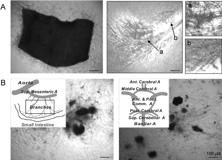
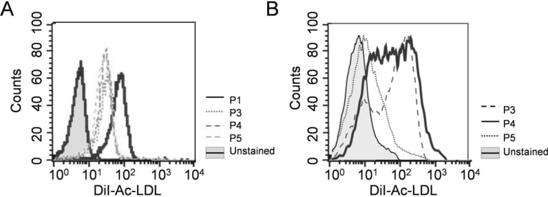
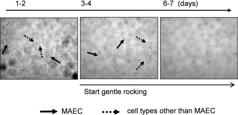
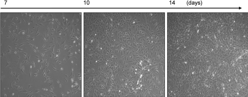
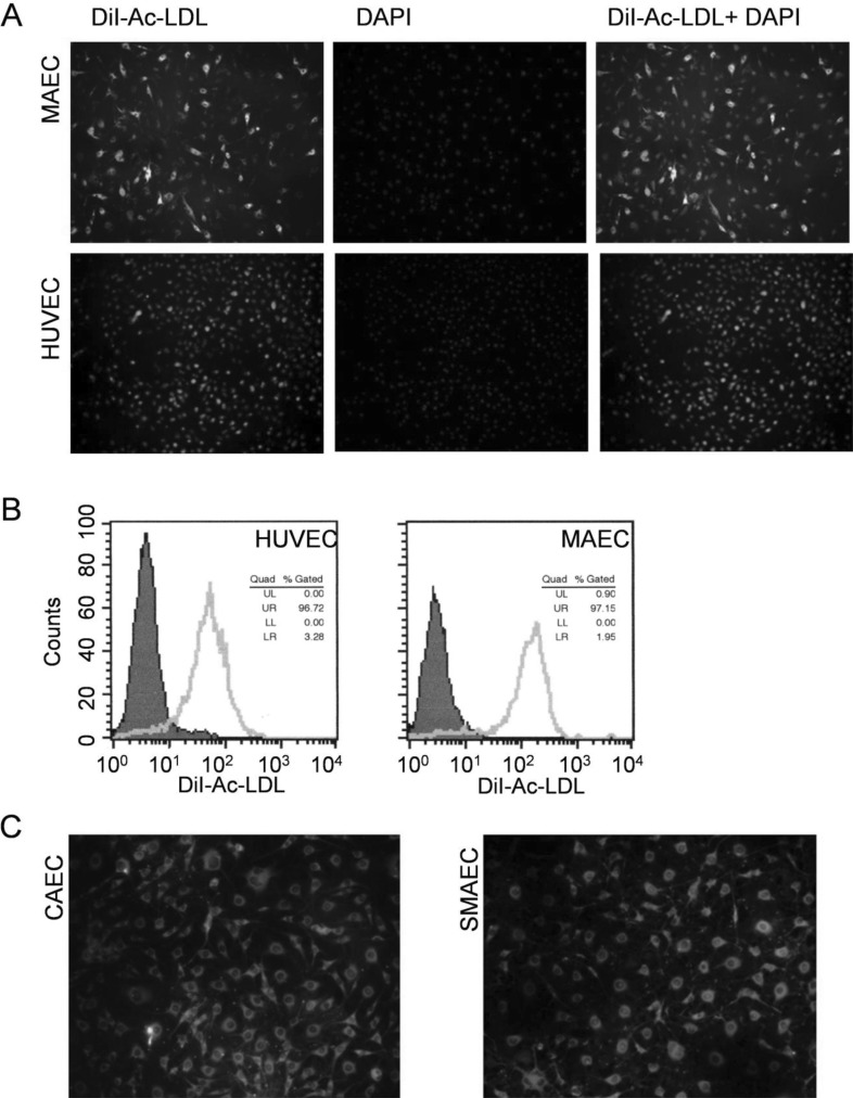
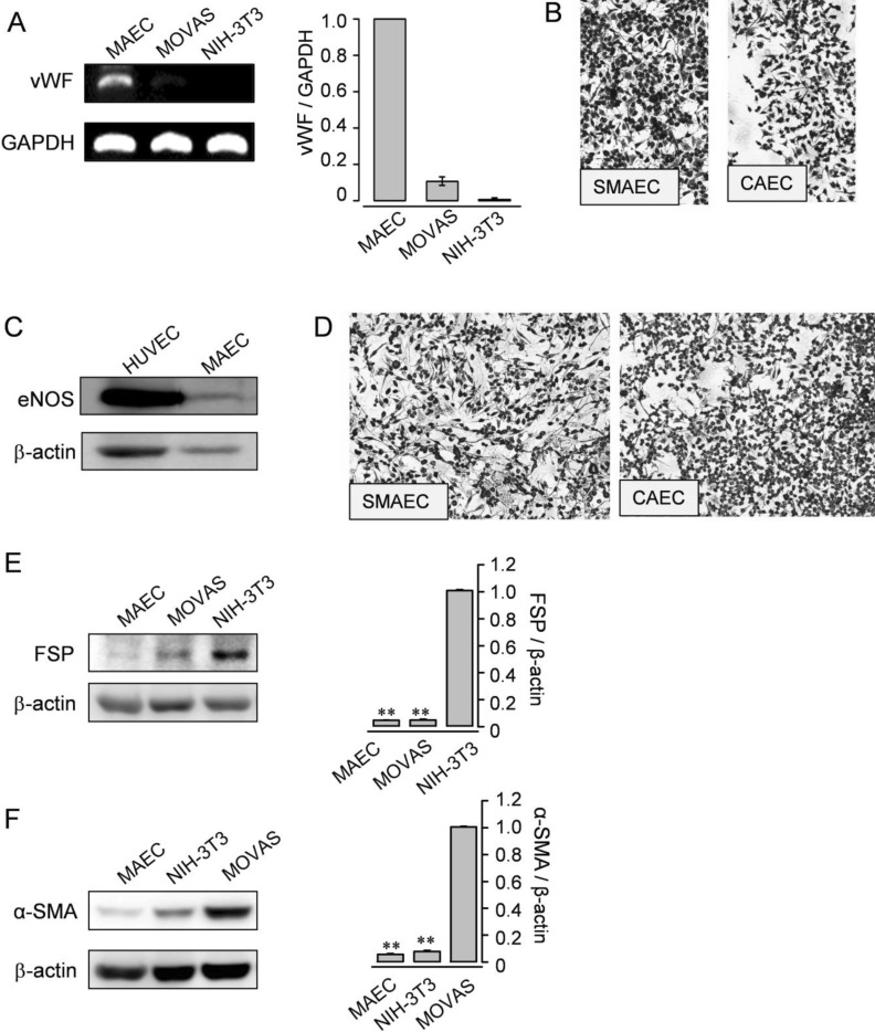
In cardiovascular disorders, understanding of endothelial cell (EC) function is essential to elucidate the disease mechanism. Although the mouse model has many advantages for in vivo and in vitro research, efficient procedures for the isolation and propagation of primary mouse EC have been problematic. We describe a high yield process for isolation and in vitro culture of primary EC from mouse arteries (aorta, braches of superior mesenteric artery, and cerebral arteries from the circle of Willis). Mouse arteries were carefully dissected without damage under a light microscope, and small pieces of the vessels were transferred on/in a Matrigel matrix enriched with endothelial growth supplement. Primary cells that proliferated in Matrigel were propagated in advanced DMEM with fetal calf serum or platelet-derived serum, EC growth supplement, and heparin. To improve the purity of the cell culture, we applied shearing stress and anti-fibroblast antibody. EC were characterized by a monolayer cobble stone appearance, positive staining with acetylated low density lipoprotein labeled with 1,1'-dioctadecyl-3,3,3',3'-tetramethyl-indocarbocyanine perchlorate, RT-PCR using primers for von-Willebrand factor, and determination of the protein level endothelial nitric oxide synthase. Our simple, efficient method would facilitate in vitro functional investigations of EC from mouse vessels.

Keywords: Endothelial cells; In vitro culture; Mouse vessels.


Figures

Fig. 1
Fig. 1
Pieces of mouse vessels explanted on/in a Matrigel support. Cells started to migrate from the edges of explants and reached confluence. (A) Aortic pieces were placed with the intima side down on Matrigel (left panel) and removed after 5~7 days (middle panel). Higher magnification images of the boxed areas, designated as a and b, are displayed in the right upper and lower sides, respectively. Cells proliferated to form a tube-like structure. (B) Schematic representations of branches of the SMA (left panel) and the CA from the circle of Willis (right panel). Branches of the SMA and the CA from the circle of Willis were explanted in Matrigel, and cells migrated from small segments of the vessels. Images were obtained by phase-contrast microscopy at 50× magnification.
Fig. 2
Fig. 2
Labeling of MAEC with DiI-Ac-LDL. (A, B) Active DiI-Ac-LDL uptake was examined by flow cytometry in MAEC at passages 1-5. MAEC at passage 1 without DiI-Ac-LDL were used as a negative control.
Fig. 3
Fig. 3
Elimination of cells in the multilayer. Cells growing in the multilayer were markedly increased after passaging (left panel). When exposed to anti-fibroblast antibody and gentle rocking, cells in the multilayer gradually disappeared. Four or five days after the exposure, only cells growing in a monolayer remained. Lined arrows represent cells in a single layer, and dotted arrows are cells in a multilayer.
Fig. 4
Fig. 4
Purified MAECs by elimination of cells in the multilayer. Cells from aortic explants were exposed to anti-fibroblast antibody and gentle rocking, eliminating cells in the multilayer. A confluent monolayer of MAECs was demonstrated. Note the presence of contact inhibition and cobblestone morphology in all panels. Images were obtained by phase-contrast microscopy at 100× magnification.
Fig. 5
Fig. 5
Typical EC characteristics of isolated cells. (A) Cells were stained with DiI-Ac-LDL, and HUVECs were employed as a positive control (left panel). DAPI was used for nucleus staining (center panel), and merged images are displayed (right panel). DiI-Ac-LDL uptake occurred in nearly all of the cells. (B) Flow cytometry analysis was performed in MAECs (passage 4) and HUVEC. DiI-Ac-LDL-labeled ECs showed a dominant fluorescence shift compared to unlabeled negative control cells. (C) Active DiI-Ac-LDL uptake in EC isolated from the CA (CAEC, left panel) and from branches of the SMA (SMAEC, right panel).
Fig. 6
Fig. 6
Expression of typical genes and proteins from ECs. (A) RT-PCR analysis for vWF was performed (passage 5) in MAEC. SMC (MOVAS) and fibroblasts (NIH-3T3) were employed as negative controls. GAPDH was used as an internal standard. (B) Immunocytochemistry of the isolated EC from branches of the SMA or the CA for vWF. (C) Western blotting for eNOS was examined in MAEC (passage 2) and HUVEC. HUVEC were used as a positive control. (D) Immunocytochemistry of the isolated EC from branches of the SMA or the CA for eNOS. (E and F) Western blotting for FSP (E) or α-SMA (F) was examined in MAEC (passage 5), MOVAS, and NIH-3T3. NIH-3T3 or MOVAS was used as a positive control. β-actin was used as an internal standard. (A, E, and F) Results are representative of three independent experiments. The data shown are mean±SEM of three independent experiments.

References

    1. Carmeliet P. Angiogenesis in health and disease. Nat Med. 2003;9:653–660. - PubMed
    1. Choi S, Kim JA, Na HY, Kim JE, Park S, Han KH, Kim YJ, Suh SH. NADPH oxidase 2-derived superoxide downregulates endothelial KCa3.1 in preeclampsia. Free Radic Biol Med. 2013;57:10–21. - PubMed
    1. Choi S, Kim JA, Na HY, Cho SE, Park S, Jung SC, Suh SH. Globotriaosylceramide induces lysosomal degradation of endothelial KCa3.1 in fabry disease. Arterioscler Thromb Vasc Biol. 2014;34:81–89. - PubMed
    1. Park S, Kim JA, Joo KY, Choi S, Choi EN, Shin JA, Han KH, Jung SC, Suh SH. Globotriaosylceramide leads to K(Ca)3.1 channel dysfunction: a new insight into endothelial dysfunction in Fabry disease. Cardiovasc Res. 2011;89:290–299. - PubMed
    1. Ni CW, Kumar S, Ankeny CJ, Jo H. Development of immortalized mouse aortic endothelial cell lines. Vasc Cell. 2014;6:7. - PMC - PubMed