Osteochondritis dissecans of the knee
- PMID: 25606539
- PMCID: PMC4295664
Osteochondritis dissecans of the knee
Abstract
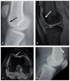
Osteochondritis dissecans (OCD) of the knee is a common cause of knee pain and dysfunction among skeletally immature and young adult patients. OCD is increasingly frequently seen in pediatric, adolescent and young adult athletes. If it is not recognized and treated appropriately, it can lead to secondary osteoarthritis with pain and functional limitation. Stable lesions in skeletally immature patients should initially be managed non-operatively. Unstable juvenile lesions and stable juvenile lesions that fail to heal with non-operative treatment require a surgical treatment. By contrast, adult OCD of the knee rarely responds to conservative measures because of limited healing potential. Operative treatment depends on the lesion stage, and there exist several surgical options.
Keywords: cartilage; knee; osteochonditis dissecans; osteochondral.
Figures


References
-
- Cahill BR. Osteochondritis dissecans of the knee: treatment of juvenile and adult forms. J Am Acad Orthop Surg. 1995;3:237–247. - PubMed
-
- Clanton TO, DeLee JC. Osteochondritis dissecans. History, pathophysiology and current treatment concepts. Clin Orthop Relat Res. 1982;(167):50–64. - PubMed
-
- Glancy GL. Juvenile osteochondritis dissecans. Am J Knee Surg. 1999;12:120–124. - PubMed
-
- Kocher MS, Micheli LJ. The pediatric knee: evaluation and treatment. In: Insall JN, Scott WN, editors. Surgery of the Knee. 3rd ed. New York, NY: Churchill-Livingstone; 2001. pp. 1356–1397.
-
- Pappas A. Osteochondrosis dissecans. Clin Orthop Relat Res. 1981;(158):59–69. - PubMed
LinkOut - more resources
Full Text Sources
