. 2015 Jan 20;108(2):279-90.
doi: 10.1016/j.bpj.2014.11.3464.

Molecular model for the solubilization of membranes into nanodisks by styrene maleic Acid copolymers

Affiliations

Molecular model for the solubilization of membranes into nanodisks by styrene maleic Acid copolymers

Stefan Scheidelaar et al. Biophys J. .

Abstract

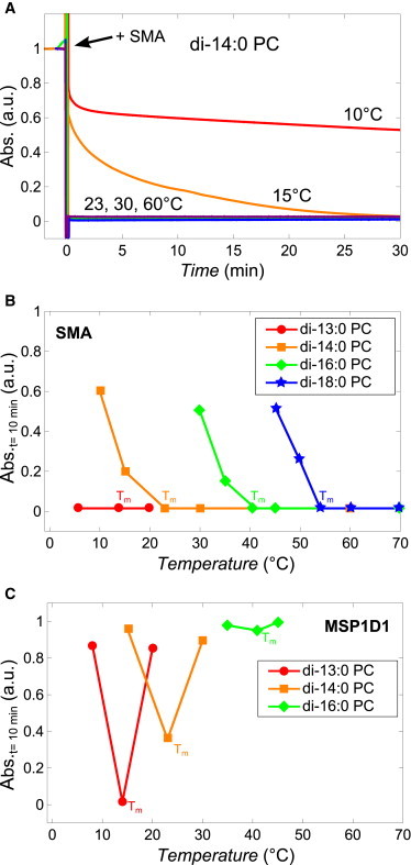
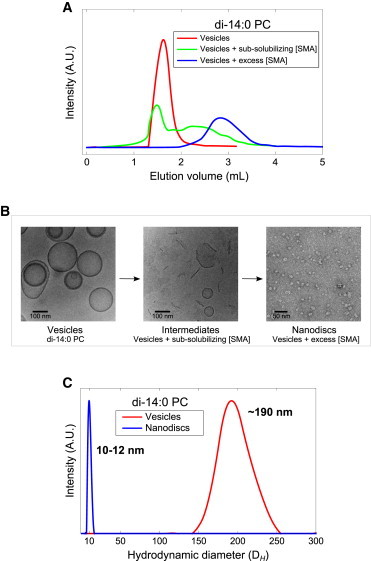
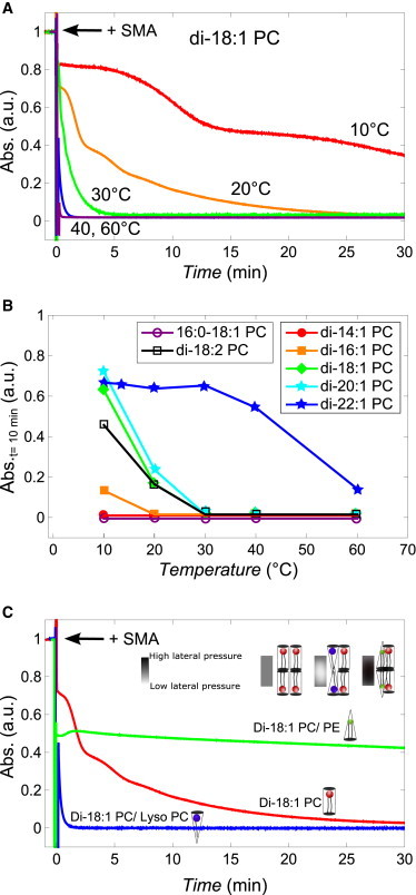
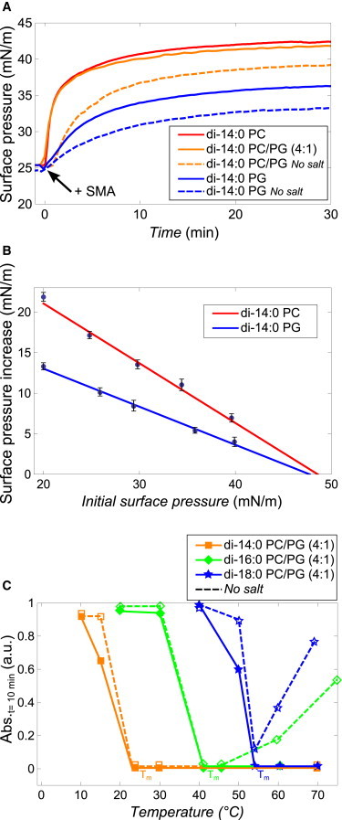
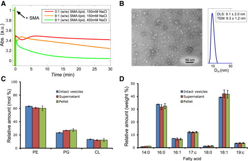
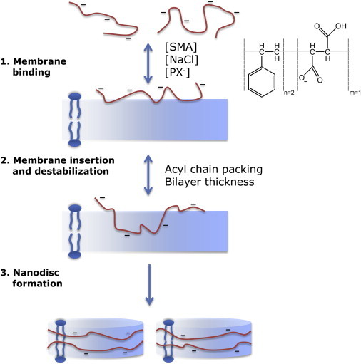
A recent discovery in membrane research is the ability of styrene-maleic acid (SMA) copolymers to solubilize membranes in the form of nanodisks allowing extraction and purification of membrane proteins from their native environment in a single detergent-free step. This has important implications for membrane research because it allows isolation as well as characterization of proteins and lipids in a near-native environment. Here, we aimed to unravel the molecular mode of action of SMA copolymers by performing systematic studies using model membranes of varying compositions and employing complementary biophysical approaches. We found that the SMA copolymer is a highly efficient membrane-solubilizing agent and that lipid bilayer properties such as fluidity, thickness, lateral pressure profile, and charge density all play distinct roles in the kinetics of solubilization. More specifically, relatively thin membranes, decreased lateral chain pressure, low charge density at the membrane surface, and increased salt concentration promote the speed and yield of vesicle solubilization. Experiments using a native membrane lipid extract showed that the SMA copolymer does not discriminate between different lipids and thus retains the native lipid composition in the solubilized particles. A model is proposed for the mode of action of SMA copolymers in which membrane solubilization is mainly driven by the hydrophobic effect and is further favored by physical properties of the polymer such as its relatively small cross-sectional area and rigid pendant groups. These results may be helpful for development of novel applications for this new type of solubilizing agent, and for optimization of the SMA technology for solubilization of the wide variety of cell membranes found in nature.


Figures

Figure 1
Figure 1
Kinetics of solubilization of lipid vesicles (400 nm) induced by the SMA copolymer and membrane scaffold protein MSP1D1. (A) Normalized time trace of absorbance at 350 nm showing the kinetics of solubilization of di-14:0 PC vesicles induced by the SMA copolymer at different temperatures. (B) Normalized absorbance values at 350 nm of saturated lipid vesicle dispersions after 10 min of incubation with SMA copolymer (3:1 w/w SMA/lipid) at different temperatures (solid lines are added to guide the eye). Tm values of the different lipids are indicated. (C) Normalized absorbance values at 350 nm of saturated lipid vesicle dispersions after 10 min of incubation with the membrane scaffold protein MSP1D1 (1:1 w/w MSP1D1/lipid) at different temperatures (solid lines are added to guide the eye). To see this figure in color, go online.
Figure 2
Figure 2
Structural characterization of di-14:0 PC lipid vesicle (200 nm) solubilization by the SMA copolymer. (A) Size-exclusion chromatograms of di-14:0 PC lipid vesicles (red), di-14:0 PC lipid vesicles incubated with a subsolubilizing amount of SMA corresponding to a 1:4 (w/w) SMA/phospholipid (green), and di-14:0 PC lipid vesicles incubated with excess SMA, which corresponds to a 3:1 (w/w) SMA/phospholipid (blue). (B) Cryo- (left, middle) and negative staining (right) transmission electron microscopy images of particles obtained from the fractions corresponding to the three chromatograms from Fig. 2A showing vesicles (left), intermediate vesicular structures and open bilayer fragments (middle), and nanodisks (right). (C) Size distributions from DLS experiments on vesicles (red) and nanodisks obtained by incubating vesicles with excess SMA (blue). To see this figure in color, go online.
Figure 3
Figure 3
(A) Kinetics of solubilization of unsaturated lipid vesicles (400 nm) induced by the SMA copolymer and the effect of lateral chain pressure. In all cases, a 3:1 (w/w) SMA/lipid solubilization kinetics of di-18:1 PC vesicles is induced by the SMA copolymer at different temperatures. (B) Normalized absorbance values at 350 nm of unsaturated lipid vesicle dispersions after 10 min of incubation with SMA copolymer at different temperatures (solid lines are added to guide the eye). (C) Normalized absorbance time trace of di-18:1 PC vesicles (red), di-18:1 PC/di-18:1 PE (1:1 molar ratio, green), and di-18:1-PC/lyso-18:1-PC (4:1 molar ratio, blue) after the addition of SMA copolymer, all at 20°C. (Inset) Schematic representation of the expected changes in the lateral chain pressure. To see this figure in color, go online.
Figure 4
Figure 4
Effect of electrostatic interactions on membrane insertion and solubilization by SMA copolymers. (A) Surface pressure increase in time upon the addition of excess SMA to monolayers of di-14:0 PC (red), di-14:0 PC/PG (4:1) (orange), and di-14:0 PG (blue) in 50 mM Tris-HCl pH 8.0 with 150 mM NaCl (solid) or without NaCl (dashed). (B) Surface pressure increase as function of initial surface pressure for di-14:0 PC and di-14:0 PG lipid monolayers in 50 mM Tris-HCl pH 8.0 with 150 mM NaCl as subphase. (Solid lines) Linear fit with the maximum insertion pressure extrapolated to be ∼48 mN/m. Error bars are standard errors based on at least two independent measurements. (C) Normalized absorbance values at 350 nm of lipid vesicle dispersions after 10 min of incubation with the SMA copolymer at different temperatures. The diameter of the vesicles was 400 nm and a 3:1 (w/w) SMA/lipid was used. (Solid lines) Data recorded in Tris-HCl pH 8.0 buffer with 150 mM NaCl; (dashed lines) without NaCl. To see this figure in color, go online.
Figure 5
Figure 5
Characterization of the solubilization of vesicles (400 nm) of an E. coli native lipid extract by the SMA copolymer. (A) Time traces of absorbance at 350 nm of E. coli vesicle solubilization at different SMA copolymer and salt concentrations, all at 20°C. (B) Negative staining TEM image and DLS size distribution of E.coli nanodisks as prepared by the addition of SMA in a 9:1 (w/w) SMA/lipid in the presence of 450 mM NaCl. (Inset) Average diameters and standard deviations that were found. Analysis by TEM was based on 20 individual nanodisk particles and analysis by DLS was based on multiple measurements on a single independent sample as described in Materials and Methods. (C) Lipid composition (mol %) and (D) fatty acid composition (weight %) of intact vesicles of E. coli lipids, and of pellet and supernatant obtained after partial solubilization by SMA. Error bars represent the standard deviation of three independent experiments. To see this figure in color, go online.
Figure 6
Figure 6
Schematic illustration of membrane solubilization by the SMA copolymer. (Inset) Schematic representation of the SMA copolymer used in this study. Step 1: anionic SMA copolymers bind to the lipid membrane, a process that is driven mainly by hydrophobic interactions and modulated by electrostatic interactions. Binding increases when the salt concentration increases and the number of anionic lipids (PX) decreases. Step 2: insertion of the SMA copolymer in the membrane hydrophobic core, a process mainly determined by acyl-chain packing. A high lateral pressure in the acyl-chain region will decrease the efficiency of insertion. Step 3: insertion into the hydrophobic core leads to membrane destabilization and to the formation of vesicular intermediates. Further solubilization will lead to the formation of nanodisks, which are stabilized by a SMA copolymer belt around the nanodisk. The kinetics and efficiency of membrane destabilization and nanodisk formation is determined by acyl-chain packing properties and bilayer thickness. To see this figure in color, go online.

References

    1. Knowles T.J., Finka R., Overduin M. Membrane proteins solubilized intact in lipid containing nanoparticles bounded by styrene maleic acid copolymer. J. Am. Chem. Soc. 2009;131:7484–7485. - PubMed
    1. Jamshad M., Lin Y.-P., Dafforn T.R. Surfactant-free purification of membrane proteins with intact native membrane environment. Biochem. Soc. Trans. 2011;39:813–818. - PubMed
    1. Orwick M.C., Judge P.J., Watts A. Detergent-free formation and physicochemical characterization of nanosized lipid-polymer complexes: Lipodisq. Angew. Chem. Int. Ed. Engl. 2012;51:4653–4657. - PubMed
    1. Orwick-Rydmark M., Lovett J.E., Watts A. Detergent-free incorporation of a seven-transmembrane receptor protein into nanosized bilayer Lipodisq particles for functional and biophysical studies. Nano Lett. 2012;12:4687–4692. - PubMed
    1. Long A.R., O’Brien C.C., Alder N.N. A detergent-free strategy for the reconstitution of active enzyme complexes from native biological membranes into nanoscale discs. BMC Biotechnol. 2013;13:41. - PMC - PubMed

Publication types