. 2015 Jan 20;108(2):360-7.
doi: 10.1016/j.bpj.2014.11.3474.

Small peptide binding stiffens the ubiquitin-like protein SUMO1

Affiliations

Small peptide binding stiffens the ubiquitin-like protein SUMO1

Hema Chandra Kotamarthi et al. Biophys J. .

Abstract

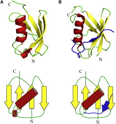
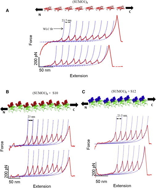
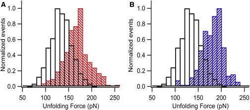
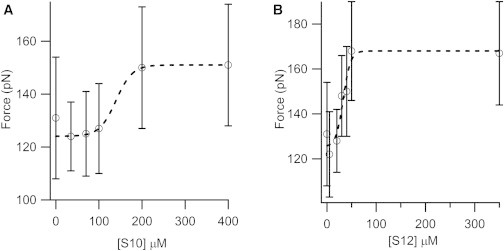
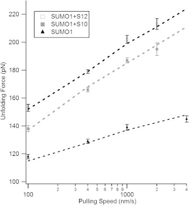
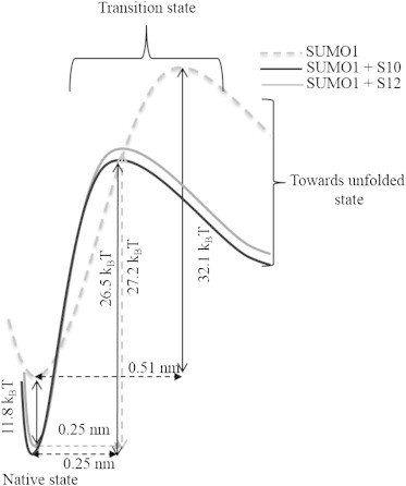
Posttranslational modification by small ubiquitin-like modifiers (SUMOs), known as SUMOylation, is a key regulatory event in many eukaryotic cellular processes in which SUMOs interact with a large number of target proteins. SUMO binding motifs (SBMs) are small peptides derived from these target proteins that interact noncovalently with SUMOs and induce conformational changes. To determine the effect of SBMs on the mechanical properties of SUMO1 (the first member of the human SUMO family), we performed single-molecule force spectroscopy experiments on SUMO1/SBM complexes. The unfolding force of SUMO1 (at a pulling speed of 400 nm/s) increased from ∼ 130 pN to ∼ 170 pN upon binding to SBMs, indicating mechanical stabilization upon complexation. Pulling-speed-dependent experiments and Monte Carlo simulations measured a large decrease in distance to the unfolding transition state for SUMO1 upon SBM binding, which is by far the largest change measured for any ligand binding protein. The stiffness of SUMO1 (measured as a spring constant for the deformation response along the line joining the N- and C-termini) increased upon SBM binding from ∼ 1 N/m to ∼ 3.5 N/m. The relatively higher flexibility of ligand-free SUMO1 might play a role in accessing various conformations before binding to a target.


Figures

Figure 1
Figure 1
Structure and topology map of SUMO1 in ligand-free (A) and ligand-bound (B) forms. N- and C-termini, along which the stretching force is applied in the pulling experiments, are labeled in the figure. Protein Data Bank IDs of the structures of SUMO1 and S12-bound SUMO1 are 1A5R and 2ASQ, respectively. Another SBM peptide, S10, also binds in the same region of SUMO1 but in the reverse orientation. The unstructured N-terminus of SUMO1 (i.e., 21 residues) is not shown in the figure. To see this figure in color, go online.
Figure 2
Figure 2
SUMO binding peptides (SBMs) alter the mechanical properties of SUMO1. Force-extension (FX) traces of (SUMO1)8 (A), (SUMO1)8 in the presence of S10 (B), and (SUMO1)8 in presence of S12 (C) are shown. Cartoons of octameric SUMO1 and its complex with the SBM peptides appear above the corresponding FX traces. All force peaks in the FX traces are fitted to the WLC model (dotted lines) to obtain ΔLC. SUMO1 has a ΔLC of ∼24 nm and is not altered upon binding to the peptides. The unfolding force of SUMO1 increases upon binding to the SBMs, as can be seen from the FX traces and the force scale. SUMO1 is being pulled along the line joining the N- and C-termini in the experiment, as indicated by the arrows. To see this figure in color, go online.
Figure 3
Figure 3
The unfolding force of SUMO1 increases upon binding the SBM peptides. The overlay of unfolding force histograms of SUMO1 (open bars) and SUMO1 bound to S10 (red-hatched bars) (A) and S12 (blue-hatched bars) (B) are shown. The histograms clearly show that the average unfolding force of SUMO1 increased from 132 pN to 166 pN upon binding to S10 and to 179 pN upon binding to S12. To see this figure in color, go online.
Figure 4
Figure 4
Dependence of the unfolding force of SUMO1 on SBM peptide concentration. The unfolding force of SUMO1 increases with increasing concentrations of S10 (A) and S12 (B), with a sudden change at concentrations close to the Kd of the complex of the protein and the SBM peptide. Sigmoidal fits are shown by dotted lines (see text for more details). The unfolding force histograms of SUMO1 at various concentrations of S10 and S12 are shown in Fig. S3.
Figure 5
Figure 5
The distance to the unfolding transition state (Δxu) for SUMO1 becomes smaller upon binding to the SBM peptides. Shown are the pulling-speed-dependent unfolding forces of SUMO1 alone (triangles) and in the presence of S10 (solid squares) and S12 (open squares). The graph shows that SUMO1 is more dependent on the pulling speed in the presence of the SBM peptides. Dashed lines are Monte Carlo fits to the experimental data. SUMO1 in the presence of the SBM peptides has a Δxu of 0.25 nm, which is smaller than that of the ligand-free SUMO1 (0.51 nm). Values are expressed as the mean ± SE. The ku0and Δxu values are given in Table 2. The speed dependence data for ligand-free SUMO1 is taken from our earlier report (19).
Figure 6
Figure 6
Schematic energy landscape of SUMO1 bound to SBM peptides S10 and S12. SUMO1 in its native state is stabilized by ligand binding. The distance to the unfolding transition state (Δxu) decreases from 0.51 nm to 0.25 nm upon ligand binding. The activation energies of unfolding are similar for SUMO1 in the presence of S10 and S12 but lower than the ligand-free SUMO1. The values of Δxu and ku0 used for this calculation are obtained from Monte Carlo simulations. The ΔG value for the native-state stabilization of SUMO1 upon binding S12 is an estimate from earlier studies (16), and S10 is assumed to have the same value in the above representation.

References

    1. Sanchez-Ruiz J.M. Ligand effects on protein thermodynamic stability. Biophys. Chem. 2007;126:43–49. - PubMed
    1. Hu X., Li H. Force spectroscopy studies on protein-ligand interactions: a single protein mechanics perspective. FEBS Lett. 2014;588:3613–3620. - PubMed
    1. Ramanujam V., Kotamarthi H.C., Ainavarapu S.R. Ca2+ binding enhanced mechanical stability of an archaeal crystallin. PLoS ONE. 2014;9:e94513. - PMC - PubMed
    1. Kotamarthi H.C., Sharma R., Ainavarapu S.R. Multiple unfolding pathways of leucine binding protein (LBP) probed by single-molecule force spectroscopy (SMFS) J. Am. Chem. Soc. 2013;135:14768–14774. - PubMed
    1. Ainavarapu S.R., Li L., Fernandez J.M. Ligand binding modulates the mechanical stability of dihydrofolate reductase. Biophys. J. 2005;89:3337–3344. - PMC - PubMed

LinkOut - more resources