Review
. 2015 Feb 3;54(4):962-73.
doi: 10.1021/bi501520w. Epub 2015 Jan 21.

DNA charge transport within the cell

Affiliations
Review

DNA charge transport within the cell

Michael A Grodick et al. Biochemistry. .

Abstract

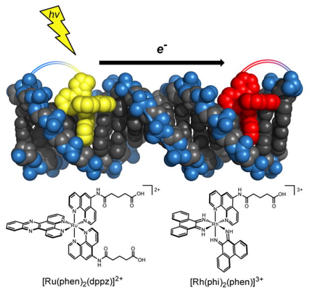
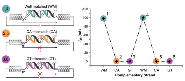
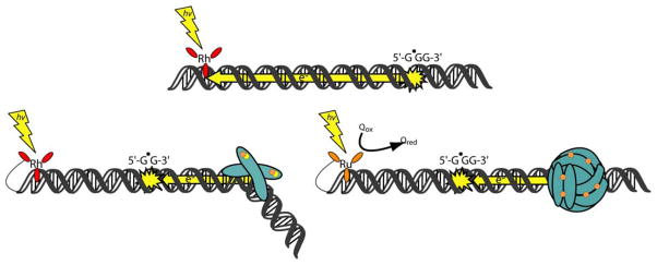
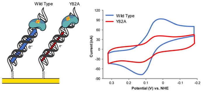
The unique characteristics of DNA charge transport (CT) have prompted an examination of roles for this chemistry within a biological context. Not only can DNA CT facilitate long-range oxidative damage of DNA, but redox-active proteins can couple to the DNA base stack and participate in long-range redox reactions using DNA CT. DNA transcription factors with redox-active moieties such as SoxR and p53 can use DNA CT as a form of redox sensing. DNA CT chemistry also provides a means to monitor the integrity of the DNA, given the sensitivity of DNA CT to perturbations in base stacking as arise with mismatches and lesions. Enzymes that utilize this chemistry include an interesting and ever-growing class of DNA-processing enzymes involved in DNA repair, replication, and transcription that have been found to contain 4Fe-4S clusters. DNA repair enzymes containing 4Fe-4S clusters, that include endonuclease III (EndoIII), MutY, and DinG from bacteria, as well as XPD from archaea, have been shown to be redox-active when bound to DNA, share a DNA-bound redox potential, and can be reduced and oxidized at long-range via DNA CT. Interactions between DNA and these proteins in solution, in addition to genetics experiments within Escherichia coli, suggest that DNA-mediated CT can be used as a means of cooperative signaling among DNA repair proteins that contain 4Fe-4S clusters as a first step in finding DNA damage, even within cells. On the basis of these data, we can consider also how DNA-mediated CT may be used as a means of signaling to coordinate DNA processing across the genome.


Figures

Figure 1
Figure 1
Measuring DNA CT with metal complexes. Shown is a characteristic DNA assembly used to monitor DNA-mediated redox chemistry in solution, using metallointercalators to monitor luminescence quenching by electron transfer through the DNA base stack. The chemical structures of the covalent donor and acceptor, Ru(phen)2dppz2+ (phen = 1,10-phenanthroline, dppz = dipyrido[3,2-a:2′,3′-c]phenazine) (left) and Rh(phi)2phen3+ (phi = 9,10-phenanthrenequinone diimine) (right), respectively, are shown.
Figure 2
Figure 2
Measuring DNA CT in single molecules. A single DNA duplex is made to covalently bridge a gap in an electronically wired, carbon nanotube device such that the measured current flow through the device reflects DNA CT efficiency. To confirm that charge flow through the device is DNA-mediated and to illustrate the sensitivity of DNA CT to single base mismatches, an experiment was designed to allow the introduction of a single base mismatch through thermal dehybridization and rehybridization of the bridging duplex (left). One strand of the DNA duplex is covalently attached at either side of the gap (black), while the other, noncovalent strand is cycled between a well matched strand (turquoise) and strands with a single base mismatch (orange, purple) by sequential dehybridization, rinsing, and rehybridization. During this cycling between well matched and mismatched duplexes, the source-drain current (ISD) for the device was measured at a constant gating voltage (VG = −3V) and plotted for each bridging duplex (right plot, where the colors and numbers of the duplexes in the left illustration correspond to those on the plot). This experiment clearly illustrates the high sensitivity of DNA CT to single base mismatches; DNA CT is inhibited when the device is bridged with a mismatched duplex and restored when the device is rehybridized with a well matched duplex.
Figure 3
Figure 3
Measuring DNA CT with DNA-modified electrodes. Schematic of a multiplex chip with four different DNAs in the four quadrants of the chip. Left side: mismatched (front) and well matched (back) 17-mers with DNA-bound, redox-active protein containing a [4Fe-4S] cluster signified by the cluster of two orange and two yellow spheres. The protein binds the DNA, which is covalently attached by one end to the gold surface, and reduction of the cluster proceeds via DNA CT. Right side: mismatched (front) and well matched (back) 100-mers with a covalent, small molecule redox probe. The location of the mismatch in the 17-mer and 100-mer is circled in red. DNA CT to the redox-active protein or small molecule probe is significantly attenuated (red X) in the presence of a single base mismatch for both the 17-mer and 100-mer.,
Figure 4
Figure 4
Initial experiments to probe DNA CT in biology. Illustrated are some examples of experimental constructs used to evaluate the capabilities of different cellular players to participate in DNA CT including cellular DNA (top) and proteins with redox-active cofactors (bottom left and right). Top: the capacity of cellular DNA to funnel damage over long distances in the genome via DNA CT was studied within its native environment inside a variety of organelles. Damage is induced by a photoexcited, intercalated metal complex and the appearance of damage at the 5′-guanine of distant guanine repeat sites supports that DNA CT facilitates this transport of electrons over such long distances. Bottom Left: SoxR, an oxidative stress response transcription factor binds DNA as a dimer, with each monomer containing a [2Fe-2S] cluster signified by a cluster of one orange and one yellow sphere. SoxR, is activated by oxidative DNA damage from a distance, induced by a covalent, photoexcited metal complex. Bottom Right: Dps, a 12-subunit protein that binds DNA as a spherical dodecamer, contains 12 intersubunit ferroxidase sites that, in the depicted experiment, are occupied by 1 Fe2+ each (12 Fe2+/Dps; each bound Fe2+ is represented by a single orange sphere). Dps uses ferroxidase activity to protect DNA from reactive oxygen species and can neutralize guanine radicals from a distance when they are formed by a photoexcited metal complex.
Figure 5
Figure 5
Electrochemistry of DNA glycosylases on DNA-modified electrodes. Both single and multiplexed DNA-modified gold electrodes have been used to study the electrochemical characteristics of DNA glycosylases containing [4Fe-4S] clusters. In these experiments, the protein is bound to the DNA, which is attached to the gold electrode surface, and the [4Fe-4S] cluster is reduced via DNA CT (left). Cyclic voltammetry has been used to determine that the midpoint redox potential of these enzymes is around 80 mV vs. NHE.,, and to establish that EndoIII Y82A is deficient in its ability to perform DNA-mediated CT (right). The intensity of the electrochemical signal of EndoIII Y82A is much lower than that of WT EndoIII, depicted by the red and blue arrows, respectively, in the illustration (left). The redox-active [4Fe-4S] clusters in the proteins are signified here by clusters of two orange and two yellow spheres.
Figure 6
Figure 6
Model for the enhanced DNA lesion search efficiency of DNA repair proteins with 4Fe-4S clusters by DNA CT signaling. Repair proteins with 4Fe-4S clusters, such as MutY and EndoIII, may use DNA CT to effectively scan long stretches of genomic DNA for damage as illustrated (from bottom to top).,,, In the cytoplasm, the 4Fe-4S cluster of repair proteins is in the 2+ oxidation state (purple), but the cluster is oxidized to the 3+ state (turquoise) upon DNA binding if an electron can be transferred to a distally bound protein (recepient) via DNA CT (yellow arrows). Importantly, the intervening DNA must be free of damage for this transfer to occur. Successful electron transfer reduces the cluster of the recepient protein from the 3+ to 2+ oxidation state which decreases its binding affinity for DNA and promotes its dissociation. This protein, diffusing freely in the cytoplasm, can then bind at a different DNA location and repeat this use of DNA CT to scan another segment of the genome. If, however, the protein binds a location where there is damage intervening it and a potential recepient protein, electron transfer cannot occur and the potential recepient protein remains tightly bound to the DNA with its cluster in the 3+ oxidation state. The bound proteins (either in the 3+ or 2+ state) which are now localized in the vicinity of the damage, can then process along the DNA (dashed black arrow) to efficiently find and repair the damage. The redox-active [4Fe-4S] clusters in the proteins are signified here by clusters of two orange and two yellow spheres.
Figure 7
Figure 7
AFM reveals that CT proficiency determines whether proteins localize near DNA mismatches. When wild-type, CT-proficient EndoIII, SaXPD, or DinG (blue in left diagram and cyclic voltammogram (CV) on right) are independently incubated with mixtures of long strands of DNA (3.8 kbps) containing a mismatch (yellow X) and well-matched short strands of DNA (1.9 kbps), a redistribution of the enzymes to the mismatched strand of DNA is observed (right).,, This is consistent with DNA-mediated charge transport promoting redistribution to the site that attenuates DNA CT. This redistribution to the damaged strand is not observed for the CT-deficient EndoIII Y82A or SaXPD L325V mutants (red in diagram and in CV on right), nor is it observed when CT-deficient mutants are mixed with either SaXPD or DinG.
Figure 8
Figure 8
ATP hydrolysis by helicases with 4Fe-4S clusters measured by CV. The helicases SaXPD and DinG both have a redox potential of ~80 mV vs. NHE., When ATP is added to each enzyme on DNA-modified electrodes with 5′ to 3′ single-stranded overhangs, substrates that each enzyme is competent to unwind, an increase in the intensity of the electrochemical signal is observed. As the protein incubates on the electrode, the intensity of the signal slowly grows in over time. After the addition of ATP to the solution, the change in the growth of the signal increases by 10–20% for XPD and 40–50% for DinG. Shown above, 5 mM ATP was added to DinG that been allowed to equilibrate on a DNA-modified electrode overnight (black). The overlaid traces show the signal at several time points during the incubation of ATP including 25 minutes (grey), 50 minutes (light blue), and 80 minutes (blue). ATP hydrolysis leads to a significant increase in the intensity of the electrochemical signal.
Figure 9
Figure 9
Cultures of InvA strains transformed with complementation and rescue plasmids. Shown is a photograph of cultures of InvA, InvA Δnth, and InvA ΔpurR in addition to InvA Δnth transformed with plasmids that express RNaseH, EndoIII Y82A, EndoIII D138A, or WT EndoIII, all grown in minimal media to stationary phaseIn minimal media, it is clear that silencing the nth gene in InvA leads to a severe growth defect that cannot be rescued by expression of the CT-deficient EndoIII Y82A but can be rescued by expression of WT EndoIII, EndoIII D138A, or RNaseH. InvA ΔpurR was included as a control to show that the mutation in purR that was linked to the Δnth deletion when constructing the InvA Δnth strain did not cause the observed growth defect. An empty plasmid was used as a control to show that the presence of a plasmid does not affect growth.

References

    1. Genereux JG, Barton JK. Mechanisms for DNA Charge Transport. Chem Rev. 2010;110:1642–1662. - PMC - PubMed
    1. Muren NB, Olmon ED, Barton JK. Solution, surface, and single molecule platforms for the study of DNA-mediated charge transport. Phys Chem Chem Phys. 2012;14:13754–13771. - PMC - PubMed
    1. Sontz PA, Muren NB, Barton JK. DNA charge transport for sensing and signaling. Acc Chem Res. 2012;45:1792–1800. - PMC - PubMed
    1. Genereux JC, Boal AK, Barton JK. DNA-mediated charge transport in redox sensing and signaling. J Am Chem Soc. 2010;132:891–905. - PMC - PubMed
    1. Eley DD, Spivey DI. Semiconductivity of organic substances. Part 9. Nucleic acid in the dry state. Trans Faraday Soc. 1962;58:411–415.

Publication types