. 2015 Jan 21;10(1):e0115701.
doi: 10.1371/journal.pone.0115701. eCollection 2015.

Isoproterenol acts as a biased agonist of the alpha-1A-adrenoceptor that selectively activates the MAPK/ERK pathway

Affiliations

Isoproterenol acts as a biased agonist of the alpha-1A-adrenoceptor that selectively activates the MAPK/ERK pathway

Alicja J Copik et al. PLoS One. .

Abstract

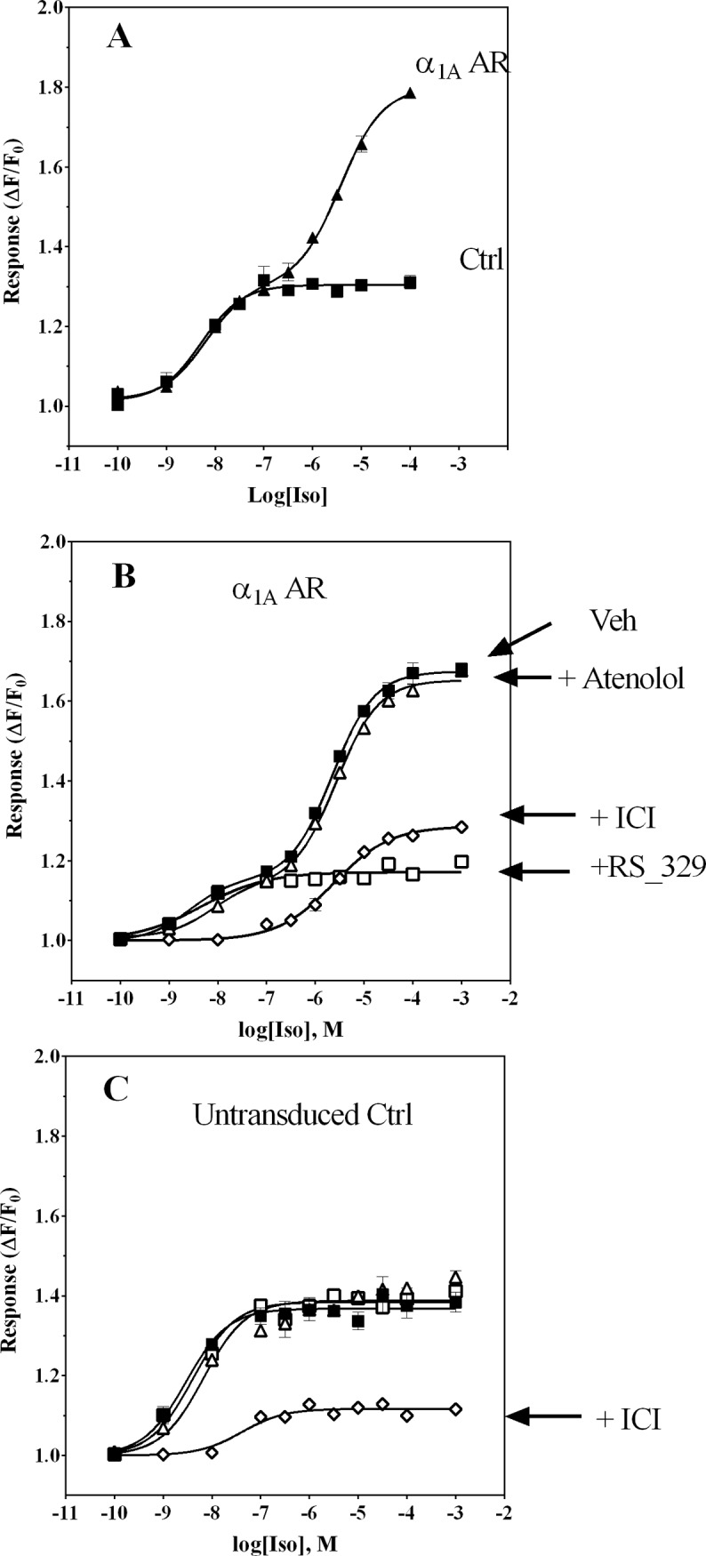
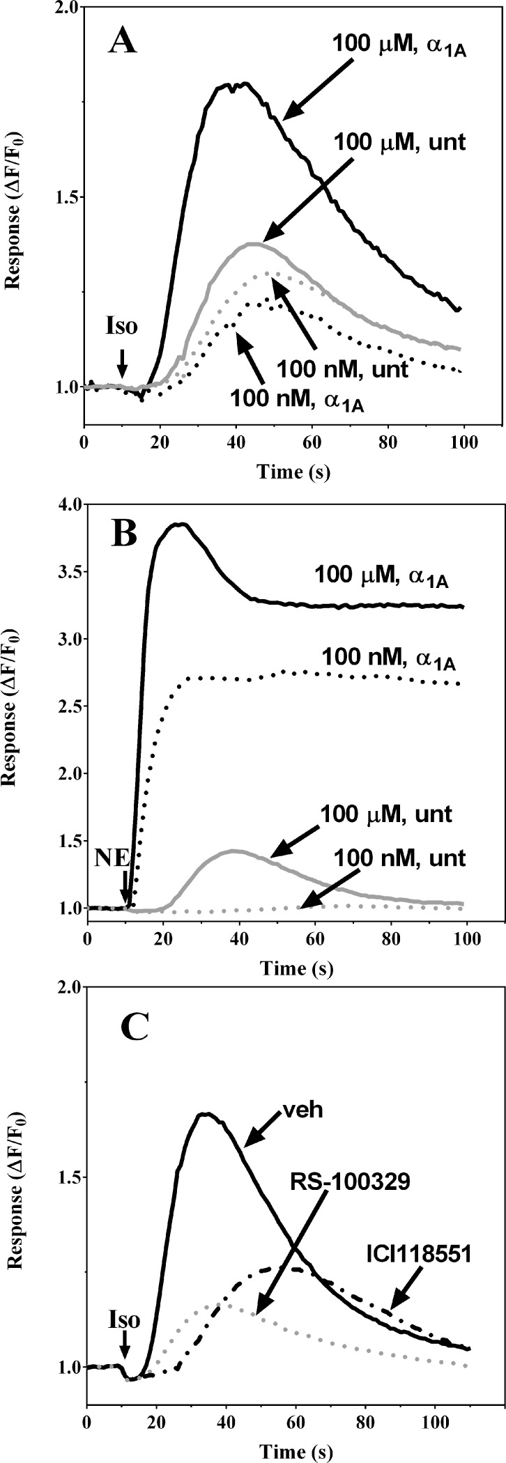
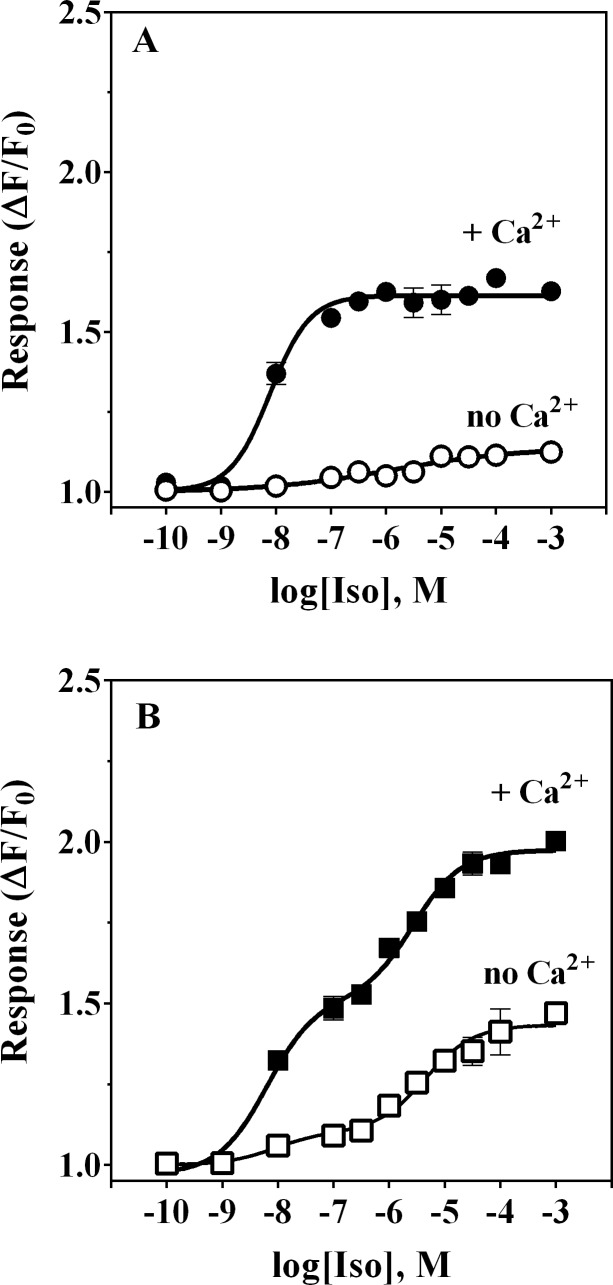
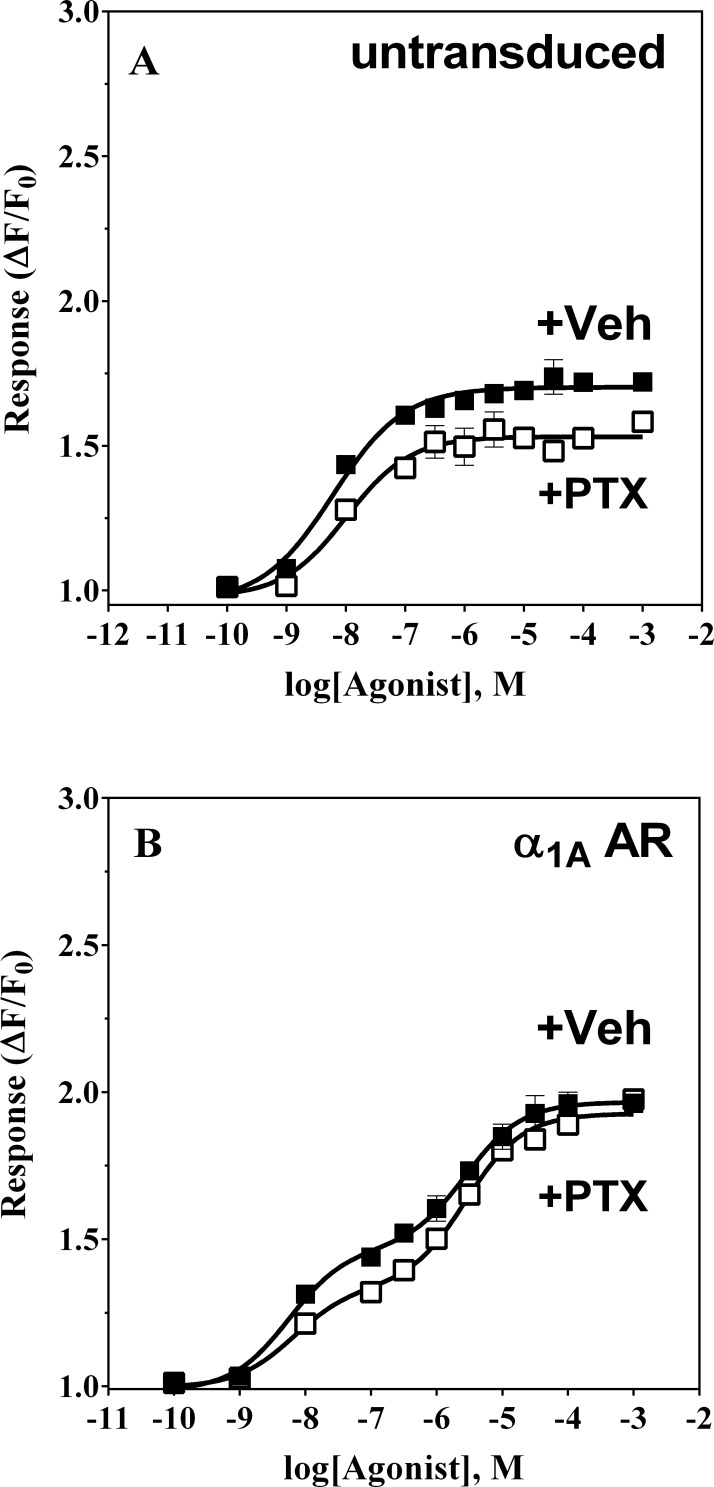
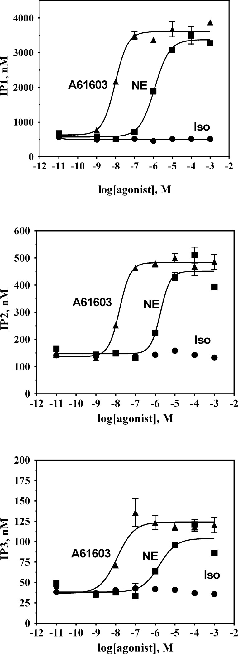
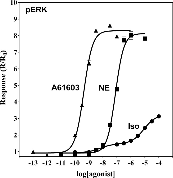
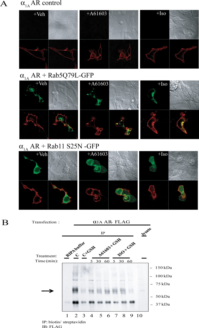
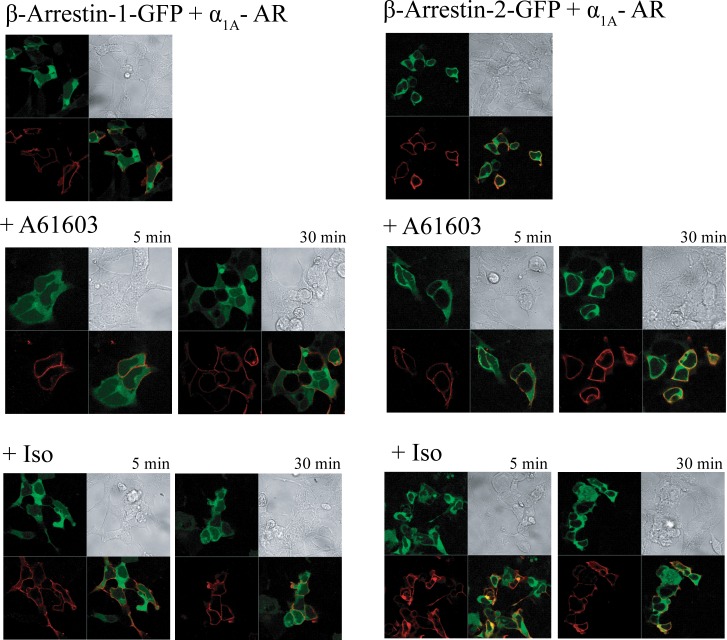
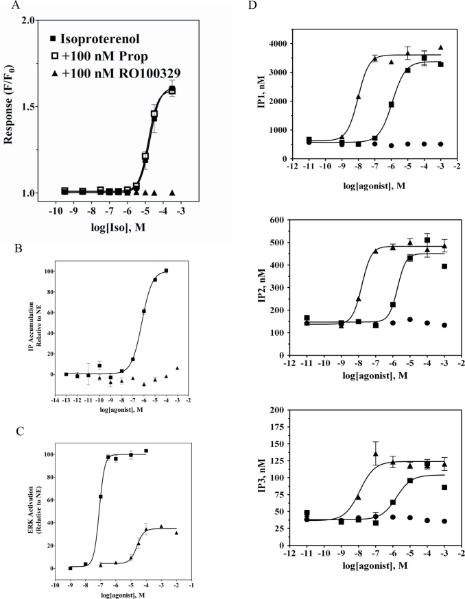
The α1A-AR is thought to couple predominantly to the Gαq/PLC pathway and lead to phosphoinositide hydrolysis and calcium mobilization, although certain agonists acting at this receptor have been reported to trigger activation of arachidonic acid formation and MAPK pathways. For several G protein-coupled receptors (GPCRs) agonists can manifest a bias for activation of particular effector signaling output, i.e., not all agonists of a given GPCR generate responses through utilization of the same signaling cascade(s). Previous work with Gαq coupling-defective variants of α1A-AR, as well as a combination of Ca2+ channel blockers, uncovered cross-talk between α1A-AR and β2-AR that leads to potentiation of a Gαq-independent signaling cascade in response to α1A-AR activation. We hypothesized that molecules exist that act as biased agonists to selectively activate this pathway. In this report, isoproterenol (Iso), typically viewed as β-AR-selective agonist, was examined with respect to activation of α1A-AR. α1A-AR selective antagonists were used to specifically block Iso evoked signaling in different cellular backgrounds and confirm its action at α1A-AR. Iso induced signaling at α1A-AR was further interrogated by probing steps along the Gαq /PLC, Gαs and MAPK/ERK pathways. In HEK-293/EBNA cells transiently transduced with α1A-AR, and CHO_α1A-AR stable cells, Iso evoked low potency ERK activity as well as Ca2+ mobilization that could be blocked by α1A-AR selective antagonists. The kinetics of Iso induced Ca2+ transients differed from typical Gαq- mediated Ca2+ mobilization, lacking both the fast IP3R mediated response and the sustained phase of Ca2+ re-entry. Moreover, no inositol phosphate (IP) accumulation could be detected in either cell line after stimulation with Iso, but activation was accompanied by receptor internalization. Data are presented that indicate that Iso represents a novel type of α1A-AR partial agonist with signaling bias toward MAPK/ERK signaling cascade that is likely independent of coupling to Gαq.


Conflict of interest statement

Competing Interests: With regard to competing interests, please note the affiliation of authors AJC, KN, SS, HH, AK, PJD, BF, APDWF, DB and MEM to Roche Palo Alto LLC. Again, this does not alter the authors’ adherence to PLOS ONE policies on sharing data and materials.

Figures

Figure 1
Figure 1. Biphasic concentration-response relationship for Iso-mediated Ca2+ responses in α1A-AR transduced HEK-293/EBNA cells.
Ca2+ transient responses (expressed as ΔF/F0) were measured as a function of Iso concentration in fluo3-loaded cells by fluorometric plate imaging (FLIPR). HEK-293/EBNA cells were first exposed for 3–4 h to baculovirus encoding α1A-AR or an irrelevant negative control protein (aldehyde oxidase), then cultured in fresh medium containing 4 mM NaBu for 18 h prior to use in experiments as described in Materials and Methods. Panel A: Control cell (■) Ca2+ transient responses exhibited a monophasic increase in amplitude, whereas responses in cells transiently expressing α1A-AR (▲) revealed a biphasic concentration-response relationship, with large response amplitude at high Iso concentrations. Panel B: Blockade of Iso responses by pre-treatment of cells with antagonists selective for β-ARs or α1A-ARs reveals roles for both receptors in mediating Ca2+ transient responses in HEK-293/EBNA cells transiently expressing α1A-AR. Although the β2-AR-selective antagonist ICI 118551 at 10 nM (◇) abolished responses to low Iso concentrations, responses to high Iso concentrations were attenuated by only ∼ 50%. The α1A-AR-selective antagonist RS100329 (□) blocked only responses to high Iso concentrations. Neither phase of the concentration-response profile was affected by the β1-AR-selective antagonist atenolol (1 μM, Δ). Vehicle-treated cell responses represented by ■. Panel C: In non-transduced control cells, only the β2-AR-selective antagonist ICI 118551 at 10 nM (◇) was effective at blocking responses to Iso: both α1A-AR-selective RS100329 (10 nM, □) and β1-AR-selective atenolol (1 μM, Δ) were ineffective. Each data point represents an average of duplicate determinations; results shown are representative of experiments repeated at least 3 times.
Figure 2
Figure 2. The Iso-induced Ca2+ mobilization response in α1A-AR transduced HEK-293/EBNA cells is slower in onset and shorter in duration than the response to NE.
Ca2+ transient response kinetics (expressed as ΔFt/F0) are shown for fluo3-loaded cells monitored fluorometrically following addition of Iso or NE. HEK-293/EBNA cells were first exposed to recombinant baculoviral strains (3–4 h) encoding either α1A-AR or an irrelevant negative control protein (aldehyde oxidase), then cultured in fresh culture medium containing 4 mM NaBu for 18 h prior to use in experiments as described in Materials and Methods. Panel A: Slow onset of the Iso agonist response which returns to baseline within a 2 min interval is evident in representative traces from α1A-AR transduced HEK-293/EBNA cells (black lines) and negative control cells (gray lines) during responses elicited by addition of Iso (↓) at 100 nM (dashed lines) or 100 μM (solid lines). Panel B: Representative Ca2+ transients in α1A-AR transduced HEK-293/EBNA cells showing rapid onset and sustained NE response following application of NE (↓) at 100 nM (dashed lines) or 100 μM (solid lines) to α1A-AR transduced (black lines) or negative control-transduced cells (gray lines). Panel C: α1A-AR transduced HEK-293/EBNA cells exhibit distinct kinetics for Iso-mediated responses via occupancy of β-AR vs α1A-AR transduced, as revealed by monitoring of responses following pre-treatment of cells for 20 min with vehicle (solid black line), 10 nM RS-100329 (dashed gray line) or 10 nM ICI118551 (dashed black line). Representative traces are shown for responses to 100 μM Iso (↓).
Figure 3
Figure 3. Iso-induced Ca2+ mobilization in α1A-AR transduced HEK-293/EBNA cells is partially dependent on the presence of extracellular Ca2+.
Ca2+ transient responses (expressed as ΔF/F0) were measured as a function of Iso concentration in untransduced control cells (Panel A) or α1A-AR transduced HEK-293/EBNA cells (Panel B), either in the presence (filled symbols) or absence (open symbols) of 2 mM Ca2+ in the assay buffer. Responses in untransduced cells were virtually abolished in the absence of extracellular Ca2+ (Panel A). In α1A-AR transduced cells, responses to low concentrations of Iso in the absence of extracellular Ca2+ were also essentially abolished whereas the low potency phase of response was diminished by approximately 50% (Panel B). Each data point is an average of duplicate determinations; this experiment was repeated twice.
Figure 4
Figure 4. Pertussis toxin pretreatment does not impair Ca2+ responses to Iso.
HEK-293/EBNA cells were exposed for 3 h to a baculoviral strain carrying α1A-AR or to viral medium. Cells were treated with 100 ng/mL of pertussis toxin (PTX) or vehicle for 18 h prior to experiments. Panel A: untransduced HEK-293/EBNA cells exposed to PTX (filled symbols) or vehicle (open symbols) were treated with Iso (■,□) during fluorometric imaging of the Ca2+-tracking dye. Panel B: untreated (open symbols) or PTX-pretreated (filled symbols) α1A-AR HEK-293/EBNA cells were stimulated with Iso (■,□) during fluorometric imaging of the Ca2+-tracking dye. Each experiment was performed in duplicate two independent times.
Figure 5
Figure 5. Inositol phosphates production in α1A-AR transduced HEK-293/EBNA cells occurs in response to A-61603 and NE, but not in response to Iso.
HEK-293/EBNA cells were exposed to baculovirus encoding α1A-AR for 3–4 h, then cultured in fresh medium containing 4 mM NaBu for 18 h prior to use in experiments as described in Materials and Methods. IP1 (top), IP2 (middle) and IP3 (bottom) formation was measured in α1A-AR transduced HEK-293/EBNA cells stimulated with increasing concentrations of A-61603 (▲), NE (■) or Iso(●). IP1, IP2, and IP3 levels were determined via LC-MS. Plots are representative of three independent experiments with each data point being the average of triplicates.
Figure 6
Figure 6. MAPK activation in α1A-AR transduced HEK-293/EBNA cells treated with A-61603, NE or Iso.
α1A-AR transduced HEK-293/EBNA cells were pre-treated with NaBu for 18 h to induce receptor expression. Cells were stimulated with A-61603 (▲), NE (■) or Iso(●) for 5 min. Agonist treatment was terminated by addition of of SureFire lysis solution. Samples were analyzed for levels of phospho-ERK using an AlphaScreen SureFire p-ERK assay kit. Plots are representative of four independent experiments, with each data point being the average of triplicates.
Figure 7
Figure 7. Stimulation of α1A-AR transduced HEK-293/EBNA with A-61603 and Iso leads to an increase in intracellular α1A – AR.
A. HEK293 cells were transiently transfected with α1A – AR only (top panels), or co-transfected with α1A – AR and Rab5 variant Q79L (middle panels) or Rab11 variant S25N (bottom panels). Following serum-deprivation, cells were stimulated with vehicle, 1μM A-61603 or 1mM ISO for 2h. Cells were then fixed and analyzed by confocal microscopy. B. HEK293 cells were transiently transfected with α1A – AR. After serum deprivation for 24h, cells were pre-treated with a membrane impermeable, disulfide-cleavable biotin reagent to label plasma membrane α1A – AR. Cells were then left untreated, or stimulated 1 μM A-61603 or 1mM ISO for 5, 30, or 60 min. After treatment, one dish of control cells was harvested without any further manipulations (C: total α1A – AR). The remaining seven dishes were divided into one control (C+GSH), three treated with A-61603 (A-61603+GSH) and three treated with ISO (ISO+GSH). They were stripped of surface biotin label using a reducing agent, in order to reveal internalized, labeled α1A – AR. Samples were then analyzed by immunoprecipitation (IP) with streptavidin followed by immunoblotting (IB) with an anti-FLAG antibody.
Figure 8
Figure 8. Treatment of α1A-AR transduced HEK-293/EBNA with A-61603 and Iso does not trigger intracellular redistribution of arrestins.
HEK293 cells were co-transfected with FLAG-tagged α1A – AR and GFP- tagged β-arrestin-1 (left panels) or β-arrestin-2 (right panels). Following serum-deprivation for 24h, cells were left untreated (top panels), or stimulated with 1μM A-61603 (middle panels) or 1mM Iso (bottom panels) for the indicated amount of time. Cells were then fixed, permeabilized, stained with Alexa Fluor-568 conjugated anti-FLAG antibodies, and analyzed employing confocal microscopy.
Figure 9
Figure 9. Concentration-response behavior to Iso in Chinese hamster ovary cells stably expressing recombinant α1A-AR.
A. Ca2+ transients (expressed as ΔF/F0) were measured as a function of Iso concentration in fluo3-loaded cells by fluorometric plate imaging (FLIPR). Responses to Iso were monitored following pre-treatment with either vehicle (■), 100 nM propranolol (□) or 100 nM RO100329 (▲). Inositol phosphate accumulation (B) and levels of phospho-ERK (C) were measured as a function of increasing concentrations of A-61603 (■) or Iso (▲) and are represented relative to NE. D. IP1 (top), IP2 (middle) and IP3 (bottom) formation was measured in CHO cells stably expressing α1A-AR, stimulated with increasing concentrations of A-61603 (▲), NE (■) or Iso(●). IP1, IP2, and IP3 levels were determined via LC-MS. Plots are representative of three independent experiments with each data point being the average of triplicates.

References

    1. Ahlquist R (1948) A study of the adrenotropic receptors. American Journal of Physiology 153: 586–600. - PubMed
    1. Furchgott RF (1959) The receptors for epinephrine and norepinephrine (adrenergic receptors). Pharmacol Rev 11: 429–441; discussion 441–422 - PubMed
    1. Lands AM, Arnold A, McAuliff JP, Luduena FP, Brown TG Jr. (1967) Differentiation of receptor systems activated by sympathomimetic amines. Nature 214: 597–598. 10.1038/214597a0 - DOI - PubMed
    1. Arch JR, Ainsworth AT, Cawthorne MA, Piercy V, Sennitt MV, et al. (1984) Atypical beta-adrenoceptor on brown adipocytes as target for anti-obesity drugs. Nature 309: 163–165. 10.1038/309163a0 - DOI - PubMed
    1. Berthelsen S, Pettinger WA (1977) A functional basis for classification of alpha-adrenergic receptors. Life Sci 21: 595–606. 10.1016/0024-3205(77)90066-2 - DOI - PubMed

Publication types

MeSH terms