. 2015 Feb 19;518(7539):431-4.
doi: 10.1038/nature14160. Epub 2015 Jan 21.

Structure of the key species in the enzymatic oxidation of methane to methanol

Affiliations

Structure of the key species in the enzymatic oxidation of methane to methanol

Rahul Banerjee et al. Nature. .

Abstract

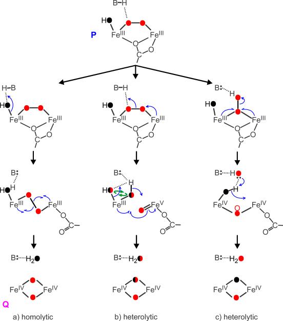
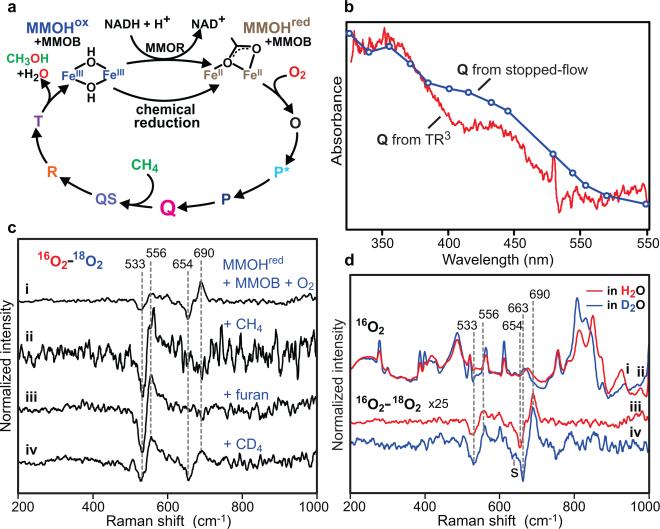
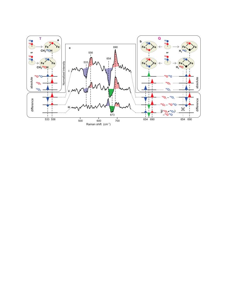
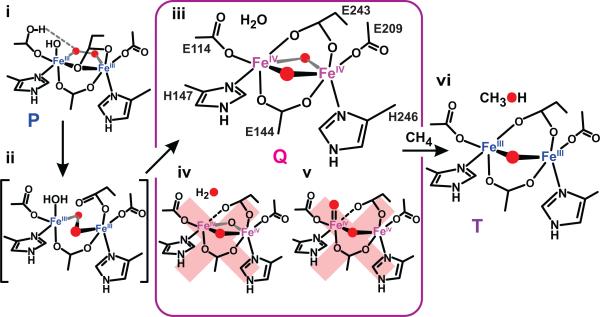
Methane monooxygenase (MMO) catalyses the O2-dependent conversion of methane to methanol in methanotrophic bacteria, thereby preventing the atmospheric egress of approximately one billion tons of this potent greenhouse gas annually. The key reaction cycle intermediate of the soluble form of MMO (sMMO) is termed compound Q (Q). Q contains a unique dinuclear Fe(IV) cluster that reacts with methane to break an exceptionally strong 105 kcal mol(-1) C-H bond and insert one oxygen atom. No other biological oxidant, except that found in the particulate form of MMO, is capable of such catalysis. The structure of Q remains controversial despite numerous spectroscopic, computational and synthetic model studies. A definitive structural assignment can be made from resonance Raman vibrational spectroscopy but, despite efforts over the past two decades, no vibrational spectrum of Q has yet been obtained. Here we report the core structures of Q and the following product complex, compound T, using time-resolved resonance Raman spectroscopy (TR(3)). TR(3) permits fingerprinting of intermediates by their unique vibrational signatures through extended signal averaging for short-lived species. We report unambiguous evidence that Q possesses a bis-μ-oxo diamond core structure and show that both bridging oxygens originate from O2. This observation strongly supports a homolytic mechanism for O-O bond cleavage. We also show that T retains a single oxygen atom from O2 as a bridging ligand, while the other oxygen atom is incorporated into the product. Capture of the extreme oxidizing potential of Q is of great contemporary interest for bioremediation and the development of synthetic approaches to methane-based alternative fuels and chemical industry feedstocks. Insight into the formation and reactivity of Q from the structure reported here is an important step towards harnessing this potential.


Figures

Extended Data Figure 1
Extended Data Figure 1. Speciation plots of the sMMO reaction with O2
Plots were computed using known rate constants of the catalytic steps,,, for the following conditions. a, No added substrate, b, presence of 0.45 mM CH4, c, presence of 3.5 mM furan and d, presence of 0.45 mM CD4 at pH 7.0, 4 °C. Rate constants used in simulation of individual conditions are shown for each step. All rate constants are first order (s−1) except for the Q to T step, which is first order in both Q and substrate (M−1 s−1) but is given as a pseudo first order constant for the current substrate concentration. The rate constant for the formation of intermediate O (oxygen binding) is unknown, but it is assumed to be fast based upon typical rates for metalloenzymes of the MMO type. It is irreversible because the rate constant of the next step (O → P*) is independent of O2 concentration.
Extended Data Figure 2
Extended Data Figure 2. Absolute TR3 spectra of the sMMO reaction with O2
Top, reaction using 16O2-containing buffer (blue), 18O2-containing buffer (red) or buffer background in the absence of MMOH/MMOB (black). Middle, 16O218O2 difference spectra of the MMOHred/MMOB reaction with O2 at pH 7.0, 4 °C and Δt ≈ 3.0 s. Bottom, 16O218O2 difference spectra of the oxygenated buffers in the absence of MMOH and MMOB proteins. Intensities of sMMO spectra were normalized to protein vibration. In the absence of protein, relative intensity was normalized using buffer vibrations.
Extended Data Figure 3
Extended Data Figure 3. Power dependence of TR3 spectra of sMMO
16O218O2 difference spectra obtained using 65 mW (i) and 15 mW (ii) excitation laser power show the same normalized intensity of oxygen vibrations in Q, indicating that no detectable photodecomposition is taking place under current conditions.
Extended Data Figure 4
Extended Data Figure 4. A comparison of the electronic absorption spectra of compound T (red trace) and MMOHred (blue trace)
T exhibits an absorption band in the near-ultraviolet region, giving rise to its resonance Raman enhancement. Single wavelength time courses of the reaction of 25 μM MMOHred/MMOB with a 450 μM solution of CH4 and 450 μM O2 at 4 °C, pH 7 were recorded throughout the visible region (concentrations after mixing). The absorbance at each wavelength at the time of maximal T formation given by the speciation plot shown in Extended Data Fig. 1b was extracted and used to make the red trace shown.
Extended Data Figure 5
Extended Data Figure 5. Potential O-O bond cleavage mechanisms in the dinuclear centre of MMO
The most divergent mechanisms are shown along with expected isotopic composition of oxygen derived from O2 (red) and solvent (black). All mechanisms are triggered by proton-dependent rearrangement of P,. The monodentate carboxylate bridge (E243) found in the diferrous enzyme is likely to maintain this position in P, but return to the non-bridging position in Q, as found in the resting enzyme, to accommodate the diamond core structure. The catalytic base B, which mediates proton dependency, has not been definitively identified. Based on structural similarity to other di-iron O2-activating enzymes and DFT computations for P-analogues in those systems,,, we have proposed that E114 (a ligand to solvent-coordinated iron in P), is this base. Other ligands not directly involved in cleavage are omitted for clarity (see Fig. 3). Equal intensities of Q-16O2 and Q-18O2 modes, and the absence of Q-16O18O mode in Fig. 2c, (i) argue against isotope scrambling in Q formation. This and all other experimental results reported to date are in full accord with the nominally concerted homolytic cleavage mechanism. We postulate that the loss of E243 bridge facilitates the conversion of cis-μ-peroxo adduct in P to the trans-μ-peroxo conformation and the ensuing O-O bond cleavage (a) to form the diamond core structure detected here. This transition is supported by DFT computations. In contrast, the stepwise, end-on heterolytic cleavage mechanism (c) (analogous to formation of compound I in cytochrome P450) leads to the mixed isotope cluster in Q-18O2 and can be ruled out. Heterolytic cleavage of trans-μ-peroxo is essentially isoelectronic to and experimentally indistinguishable from the homolytic mechanism (a). The proton-assisted heterolytic cleavage of cis-μ-peroxo bridge (b) cannot be ruled out yet, but several observations argue against it. (1) We did not observe isotope scrambling (curved green arrows), which is expected upon formation of two terminal oxygenic ligands on the same iron. While scrambling may not occur if ligands are highly stabilized, structural basis for such putative stabilization is not apparent. Scrambling may also not be observed if formation of diamond core is fast following bond cleavage, in which case mechanism (b) becomes, in essence, a stepwise, proton-assisted homolytic cleavage, also indistinguishable from (a). (2) Two iron atoms in di-ferric P and di-ferryl Q are in the same oxidation states and in indistinguishable electronic environments,. Such symmetry is unfavourable for O-O bond polarization and charge separation in the FeIII/FeV state during heterolytic cleavage. The deprotonated state of the peroxo bridge in P also argues against overall polarity of the site that would aid heterolytic cleavage.
Figure 1
Figure 1. Reaction of sMMO with O2
a, The catalytic cycle includes stable MMOHox and MMOHred and detectable transient species O, P*, P, Q and T; transient states QS and R were predicted from kinetic, spectroscopic, and chemical studies. b, An in situ transient difference electronic absorption spectrum of the reaction mixture (vs anaerobic MMOHred) in the TR3 instrument at Δt ≈ 3.0 s (red, 1.0 × 0.12 mm probe volume) in comparison with spectrum of Q reconstructed from stopped-flow kinetic traces (markers, blue). c, Transient 16O218O2 difference resonance Raman spectra of sMMO reveal vibrations of iron-bound oxygen atoms. Measurement conditions: pH 7.0, 4 °C, Δt ≈ 3.0 s. Spectra recorded without the substrate (i) show marked changes when 0.45 mM CH4 (ii), 3.5 mM furan (iii) or 0.45 mM CD4 (iv) is added. d, Solvent vibrations in the absolute resonance Raman spectra are sensitive to H2O (i)/D2O (ii) substitution. 16O218O2 difference spectra of sMMO recorded in H2O (iii) show little sensitivity to D2O (iv) substitution. The upshift of 18O vibration of Q and the appearance of a low-frequency shoulder (marked with S) in D2O is attributed to Fermi resonance with a protein-derived metal ligand.
Figure 2
Figure 2. Fingerprinting cluster structure using 16O18O mixed oxygen isotope
a, b, Asymmetrically labelled 16O18O can initially bind in two equiprobable orientations, yielding an even mixture of two isotopomers in T or Q (panels a and b, top) exhibiting characteristic vibrations (panels a and b, bottom). Two scenarios are possible for Q, depending on whether two (left) or one (right) O2-derived atoms (red and blue) are incorporated into the cluster. If only one atom is incorporated, then the second oxygen atom in the core structure would derive from solvent (black). Water and methanol exemplify a departing oxygen atom while other ligands are omitted for simplicity. Since the two isotopomers of T (a) and those of singly labelled Q (b, right) have identical composition as corresponding 16O2 and 18O2 derivatives, they will exhibit both vibrations simultaneously at half the intensity. Doubly labelled Q (panel b, left) will be different from both symmetrically labelled derivatives and thus, c, will exhibit a new vibration (green), as illustrated by isotope difference spectra (c). The 16O216O18O difference (ii) in singly labelled cluster (a, b right) will appear as the 16O218O2 difference (i) with reduced intensity. The 16O18O derivative will be identical to the average of symmetrical isotopomers, yielding no signal in trace iii, as observed experimentally for T. The new frequency in doubly labelled Q should appear in both difference (ii) and (iii), and can, indeed, be seen in experimental data at 673 cm−1. Spectral superimposition inflates the apparent isotopic shift when frequencies of isotopomers are close (traces (ii) and (iii), right), but not when bands are well separated (trace (i)).
Figure 3
Figure 3. Formation of compound Q and its reaction with methane
Homolytic mechanism of O-O bond cleavage (ii) upon formation of Q from P (i) follows from the structure of Q (iii) presented here. Alternative structures of Q (iv, v) discounted by current results are also shown. The iron shown on the left in the Q structure (iii) may retain the solvent found in P. In this case, E243 would not bind to this iron, but would be likely to hydrogen bond with the bound solvent,. T (vi) contains a single atom from O2 while another is incorporated in the product.

Comment in

  • Biochemistry: Breaking methane.
    Rosenzweig AC. Rosenzweig AC. Nature. 2015 Feb 19;518(7539):309-10. doi: 10.1038/nature14199. Epub 2015 Jan 21. Nature. 2015. PMID: 25607367 Free PMC article.

Similar articles

Cited by

References

    1. Lee SK, Nesheim JC, Lipscomb JD. Transient intermediates of the methane monooxygenase catalytic cycle. J. Biol. Chem. 1993;268:21569–21577. - PubMed
    1. Lee SK, Fox BG, Froland WA, Lipscomb JD, Münck E. A transient intermediate of the methane monooxygenase catalytic cycle containing a FeIVFeIV cluster. J. Am. Chem. Soc. 1993;115:6450–6451.
    1. Shu L, et al. An FeIV2O2 diamond core structure for the key intermediate Q of methane monooxygenase. Science. 1997;275:515–518. - PubMed
    1. Dunietz BD, et al. Large scale ab initio quantum chemical calculation of the intermediates in the soluble methane monooxygenase catalytic cycle. J. Am. Chem. Soc. 2000;122:2828–2839.
    1. Han WG, Noodleman L. Structural model studies for the high-valent intermediate Q of methane monooxygenase from broken-symmetry density functional calculations. Inorganica Chim. Acta. 2008;361:973–986. - PMC - PubMed

Publication types