. 2015 Feb 5;518(7537):55-60.
doi: 10.1038/nature14121. Epub 2015 Jan 21.

Biocontainment of genetically modified organisms by synthetic protein design

Affiliations

Biocontainment of genetically modified organisms by synthetic protein design

Daniel J Mandell et al. Nature. .

Erratum in

Abstract

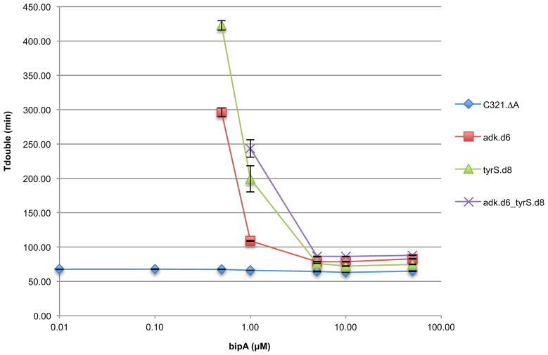
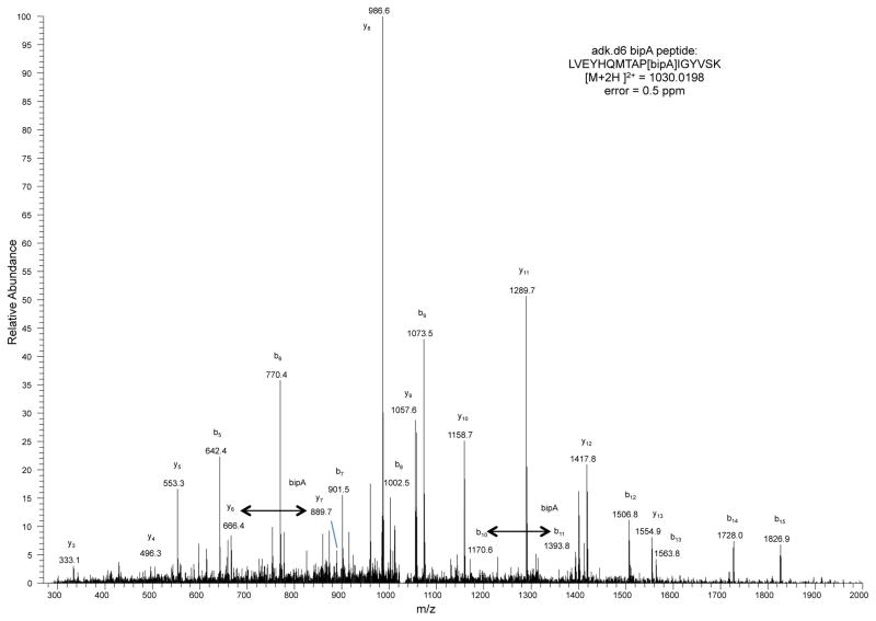
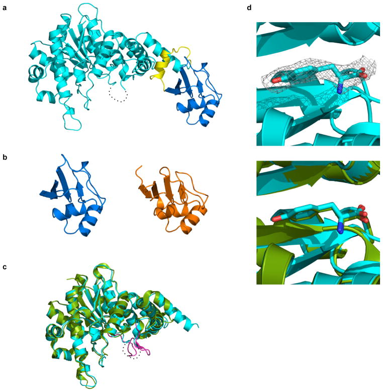
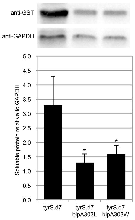
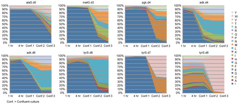
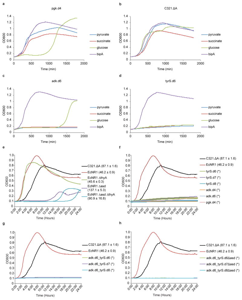
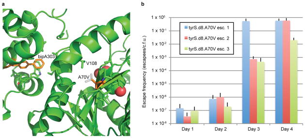
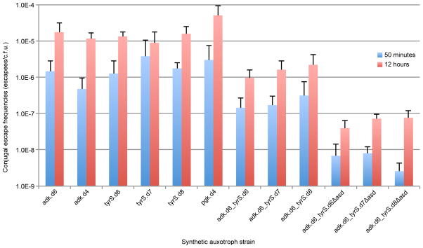
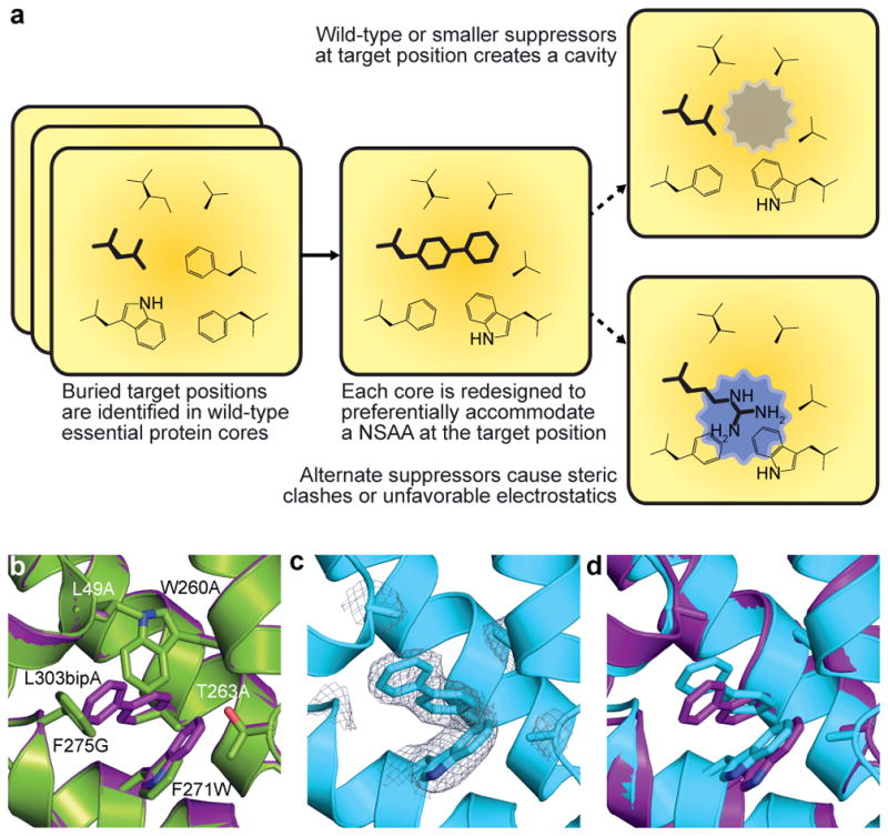
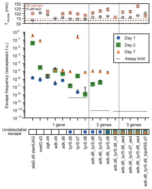
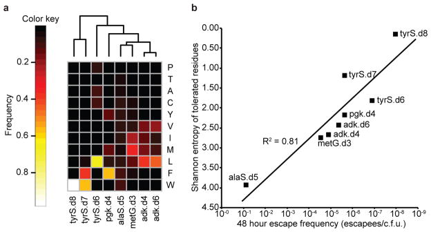
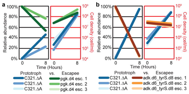
Genetically modified organisms (GMOs) are increasingly deployed at large scales and in open environments. Genetic biocontainment strategies are needed to prevent unintended proliferation of GMOs in natural ecosystems. Existing biocontainment methods are insufficient because they impose evolutionary pressure on the organism to eject the safeguard by spontaneous mutagenesis or horizontal gene transfer, or because they can be circumvented by environmentally available compounds. Here we computationally redesign essential enzymes in the first organism possessing an altered genetic code (Escherichia coli strain C321.ΔA) to confer metabolic dependence on non-standard amino acids for survival. The resulting GMOs cannot metabolically bypass their biocontainment mechanisms using known environmental compounds, and they exhibit unprecedented resistance to evolutionary escape through mutagenesis and horizontal gene transfer. This work provides a foundation for safer GMOs that are isolated from natural ecosystems by a reliance on synthetic metabolites.


Conflict of interest statement

The authors declare no competing financial interests.

Figures

Extended Data Figure 1
bipA dependence in synthetic auxotrophs. Prototrophic and synthetic auxotrophic strains were grown in titrations of bipA and monitored in a microplate reader (Methods). Media for all bipA concentrations contained SDS, chloramphenicol, and arabinose. Doubling times for three technical replicates are shown. Positive and negative error bars are s.e.m. Growth was undetectable for synthetic auxotrophs at 0.00 μM, 0.01 μM and 0.10 μM bipA, as well as 0.50 μM bipA for adk.d6_tyrS.d8.
Extended Data Figure 2
Mass spectrometry of NSAA-dependent enzymes. Mass spectrometry was performed and peptide-spectrum matches (PSMs) were obtained as described in the Methods. Datasets were culled of minor contaminant PSMs and researched with SEQUEST against adk.d6 (X at position 178) or tyrS.d7 and tyrS.d8 (X at 303) sequences without taking into account enzyme specificity. To interrogate the sequences for bipA, tryptophan and leucine, the amino acid X was given the mass of leucine and searches were performed with differential modifications of +110.01565 and +72.99525 to account for the masses of bipA and tryptophan, respectively. In all samples, only bipA, and not leucine or tryptophan, was detected at these positions. The peptide spectrum match for adk.d6 is shown. Peptides observed to contain bipA are LVEYHQMTAP[bipA]IGYVSK (adk.d6), AQYV[bipA]AEQVTR (tyrS.d7) and AQYV[bipA]AEQATR (tyrS.d8).
Extended Data Figure 3
Crystal structure of tyrS.d7. a, Overall structure of the redesigned enzyme. The N-terminal domain (residues 4-330) that catalyzes tyrosine activation, the C-terminal tRNA binding domain (residues 350-424) and their connecting region are colored cyan, blue and yellow, respectively. The residues 232-241 are disordered (dash line). b, Comparison between the C-terminal tRNA recognition domains of tyrS.d7 (blue) and of T. thermophilus TyrS (orange; PDB code 1H3E). The residues 352-442 of the hyperthermophilic TyrS are shown. c, The N-terminal domain of the engineered protein is superposed on the crystal structure of its parental enzyme (green; PDB code 1X8X). The KMSKS loop of the parental enzyme is highlighted in magenta. d, Tyrosine molecule bound to tyrS.d7. An electron density map of L-tyrosine is shown as a gray mesh (2Fo-Fc contoured at 1.2 σ; upper panel). A tyrosine and the surrounding protein fold of tyrS.d7 (cyan) are very similar to those of the wild type TyrS structure (green; lower panel).
Extended Data Figure 4
Western blot analysis of tyrS.d7 variants. Variants of tyrS.d7 with leucine or tryptophan at the bipA position were expressed as GST fusions under identical conditions and analyzed by Western blot (Methods). Soluble protein content was quantified by densitometry and normalized to GAPDH. Mutating bipA to leucine or tryptophan reduced soluble TyrS levels by 2.5- or 2.1-fold, respectively (p < 0.05 by two-tailed unpaired student’s t-test with unequal variances). Three technical replicates were performed; a representative image is shown.
Extended Data Figure 5
Population selection dynamics for canonical amino acid substitutions at designed UAG positions. For each plot, degenerate MAGE oligos were used to create a population of cells in which the UAG codon was mutated to all 64 codons. Codon substitutions leading to survival in the absence of bipA were selected by growth in LBL media without bipA and arabinose supplementation. Aliquots of the culture population were taken at 1 hour, 4 hours, confluence 1 (once the culture reached confluence), confluence 2 (after regrowth of a 100-fold dilution of confluence 1), and confluence 3 (after regrowth of a 100-fold dilution of confluence 2). The amino acid identity at the bipA position was probed by targeted Illumina sequencing. Residual bipA-containing proteins were expected to remain active until intracellular protein turnover cleared them from the cell, making the 1 hour time point a reasonable representation of initial diversity present in the population. This data shows the relative fitness of amino acid substitutions in a given protein variant; relative fitness across multiple protein variants cannot be accurately assessed from this data.
Extended Data Figure 6
Natural metabolites can circumvent auxotrophies. (a–d) Synthetic auxotrophs of pgk can be complemented by pyruvate or succinate. Strains were cultured in LBL in the presence of pyruvate, succinate, glucose or bipA (10 μM) and monitored by kinetic growth. a, The single-enzyme synthetic auxotroph pgk.d4 grows similarly to prototrophic C321.ΔA (b) in the presence of pyruvate and succinate, but not glucose. Synthetic auxotrophs of adk (c) and tyrS (d) grow robustly in bipA but cannot be complemented by pyruvate or succinate. Growth of pgk.d4 and adk.d6 in glucose after 1000 minutes is due to mutational escape (loss of bipA dependence). e, The synthetic auxotroph parental strain (C321.ΔA), a second prototrophic MG1655-derived strain (EcNR1), and three natural auxotroph derivatives of EcNR1 were grown in LBL supplemented with 166.66 ml/L bacterial lysate (Teknova). Growth curves are shown with doubling times ± one standard deviation of three technical replicates next to the labels. The conditions fully complement the metabolic auxotrophy of EcNR1.ΔthyA, which doubles as robustly as prototrophic EcNR1. Strains lacking the asd gene (EcNR1.Δasd and the EcNR1.ΔasdΔthyA double knockout) show more impairment but enter exponential growth with doubling times of 91 to 137 minutes, respectively. f, (single-) and g, (double-)enzyme synthetic auxotrophies are not complemented by natural products in rich media or bacterial lysate. h, When the Δasd auxotrophy is combined with double-enzyme synthetic auxotrophies the natural products are no longer sufficient to support growth. No growth is indicated by * in f–h.
Extended Data Figure 7
Analysis of the A70V mutation as an escape mechanism for tyrS.d8. a, The X-ray structure of tyrS.d7 is shown; tyrS.d8 varies by the single mutation V307A. BipA303, A70 and their neighboring side chains are shown in sticks, with bipA303 and A70 colored orange. The bound tyrosine substrate is shown in space fill. The A70V mutation (white sticks) may stabilize the catalytic domain when bipA is replaced by natural amino acids by tightly packing with neighboring side chains including V108. b, Escape frequencies on nonpermissive media for three separately constructed tyrS.d8 A70V strains are shown for days 1 through 4. Although escapees are growth impaired in the absence of bipA (Supplementary Table 10), all cells form colonies after 5 days, suggesting that A70V confers 100% survival on nonpermissive media.
Extended Data Figure 8
Conjugal escape frequencies of synthetic auxotrophs. Single, double, and triple-enzyme auxotrophs were assayed to determine the frequency of escape by horizontal genetic transfer and recombination from a prototrophic donor as described in the methods. These results highlight the benefit of having multiple auxotrophies distributed throughout the genome. Notably, scaling from a single synthetic auxotroph to three distributed auxotrophies results in a reduction of conjugal escape by at least two orders of magnitude.
Figure 1
Figure 1. Computational design of NSAA-dependent essential proteins
a, Overview of the computational second-site suppressor strategy. b, Computational design of a NSAA-dependent tyrosyl-tRNA synthetase (purple) overlaid on the wild-type structure (green; PDB code 2YXN). Six substituted residues are shown as sticks. c, X-ray crystallography of the redesigned synthetase with an electron density map (2Fo-Fc contoured at 1.0 σ) for substituted residues; substitution F236A is on a disordered loop and is not observed. d, The crystal structure of the redesigned enzyme (cyan) superimposed onto the computationally predicted model (purple).
Figure 2
Figure 2. Escape frequencies and doubling times of auxotrophic strains
Escape frequencies for engineered auxotrophic strains calculated as colonies observed per colony forming unit (c.f.u.) plated over 3 technical replicates on solid media lacking arabinose and bipA. Assay limit is calculated as 1/(total c.f.u. plated) for the most conservative detection limit of a cohort, with a single-enzyme auxotroph limit of 3.5 × 10⁻⁹ escapees/c.f.u., a double-enzyme auxotroph limit of 8.3 × 10⁻¹¹ escapees/c.f.u. and a triple-enzyme auxotroph limit of 6.41 × 10⁻¹¹ escapees/c.f.u. Positive error bars are standard error of the mean (s.e.m.) of the escape frequency over three technical replicates (Methods). The top panel presents the doubling times for each strain in the presence of 10 μM or 100 μM bipA, with the parental strain doubling times represented by the dashed horizontal lines. MetG.d3 growth was undetectable in 100 μM bipA. Positive and negative error bars are s.e.m.
Figure 3
Figure 3. Structural specificity at designed UAG positions in eight NSAA-dependent enzymes correlates with escape frequencies
a, Amino acid preferences at UAG positions in eight synthetic auxotrophs were determined by replacing the UAG codon with full NNN degeneracy and then sequencing the resulting populations with an Illumina MiSeq. Frequencies of each amino acid as a fraction of total sequences observed after three 1:100 passages to confluence are shown (top 11 most frequent amino acids only). Samples are clustered by Euclidean distance between amino acid frequencies. The frequency of an amino acid reports on the fitness conferred by the corresponding natural amino acid suppressor at the UAG position relative to all other amino acids. b, Shannon entropy was computed over the distributions of amino acids preferred at the UAG positions of the eight single-enzyme auxotrophs and plotted against the 48 hour escape frequency for each strain. Entropy correlates log-linearly with escape frequency, suggesting that enzyme cores with high structural specificity for bipA at the UAG position have less fit evolutionary routes to escape. Strains alaS.d5 and metG.d3 have a deactivated mutS gene.
Figure 4
Figure 4. Competition between synthetic auxotroph escapees and prototrophic E. coli
C321.ΔA was competed in the absence of bipA against escapees from a single-enzyme bipA auxotroph (pgk.d4, moderate NSAA dependence), or from a double-enzyme bipA auxotroph (adk.d6_tyrS.d8, strong bipA-dependence). Populations were seeded with 100-fold excess escapees and grown for 8 hours in nonpermissive conditions. The populations were evaluated using flow cytometry for episomally expressed fluorescent proteins at t = 0 and t = 8 hours. Results from separate competition experiments against 3 different escapees are shown for each synthetic auxotroph. a, Pgk.d4 escapees continue to expand in a mixed population with C321.ΔA after 8 hours. b, Adk.d6_tyrS.d8 escapees are rapidly outcompeted by C321.ΔA, which overtakes the population after 8 hours.
Figure 5
Figure 5. Synthetic auxotrophy and genomic recoding reduce HGT-mediated escape
a, The positions of key alleles are plotted to scale on the genome schematic. Red lines indicate auxotrophies used in the multi-enzyme auxotrophs and gray lines indicate other auxotrophies that were not included in this assay. Asterisks indicate important alleles associated with the reassignment of UAG translation function (blue are essential genes and green are potentially important genes). Conjugation-mediated reversion of the UAA codons back to the wild-type UAG is expected to be deleterious unless the natural UAG translational termination function is reverted. b, Combining multiple synthetic auxotrophies in a single genome requires a large portion of the genome to be overwritten by wild-type donor DNA, reducing the frequency of conjugal escape (top panel) and increasing the likelihood of overwriting the portions of the genome (bottom panel) that provide expanded biological function (e.g., prfA encodes RF1, which mediates translational termination at UAG codons). Positive error bars indicate standard deviation.

Comment in

  • Synthetic biology: GMOs in lockdown.
    Nunes-Alves C. Nunes-Alves C. Nat Rev Microbiol. 2015 Mar;13(3):125. doi: 10.1038/nrmicro3443. Epub 2015 Feb 9. Nat Rev Microbiol. 2015. PMID: 25659321 No abstract available.
  • Synthetic biology: GMOs in lockdown.
    Nunes-Alves C. Nunes-Alves C. Nat Rev Genet. 2015 Mar;16(3):127. doi: 10.1038/nrg3909. Nat Rev Genet. 2015. PMID: 25690387 No abstract available.

References

    1. Moe-Behrens GH, Davis R, Haynes KA. Preparing synthetic biology for the world. Frontiers in microbiology. 2013;4:5. doi: 10.3389/fmicb.2013.00005. - DOI - PMC - PubMed
    1. Molin S, et al. Conditional Suicide System for Containment of Bacteria and Plasmids. Nat Biotech. 1987;5:1315–1318.
    1. Li Q, Wu Y-J. A fluorescent, genetically engineered microorganism that degrades organophosphates and commits suicide when required. Appl Microbiol Biotechnol. 2009;82:749–756. doi: 10.1007/s00253-009-1857-3. - DOI - PubMed
    1. Curtiss R., 3rd Biological containment and cloning vector transmissibility. The Journal of infectious diseases. 1978;137:668–675. - PubMed
    1. Ronchel MC, Ramos JL. Dual system to reinforce biological containment of recombinant bacteria designed for rhizoremediation. Appl Environ Microbiol. 2001;67:2649–2656. doi: 10.1128/AEM.67.6.2649-2656.2001. - DOI - PMC - PubMed

Publication types

MeSH terms

Associated data