. 2015 Jan 21;10(1):e0117076.
doi: 10.1371/journal.pone.0117076. eCollection 2015.

Regulation of neuronal morphogenesis and positioning by ubiquitin-specific proteases in the cerebellum

Affiliations

Regulation of neuronal morphogenesis and positioning by ubiquitin-specific proteases in the cerebellum

Julius Anckar et al. PLoS One. .

Erratum in

Abstract

Ubiquitin signaling mechanisms play fundamental roles in the cell-intrinsic control of neuronal morphogenesis and connectivity in the brain. However, whereas specific ubiquitin ligases have been implicated in key steps of neural circuit assembly, the roles of ubiquitin-specific proteases (USPs) in the establishment of neuronal connectivity have remained unexplored. Here, we report a comprehensive analysis of USP family members in granule neuron morphogenesis and positioning in the rodent cerebellum. We identify a set of 32 USPs that are expressed in granule neurons. We also characterize the subcellular localization of the 32 USPs in granule neurons using a library of expression plasmids encoding GFP-USPs. In RNAi screens of the 32 neuronally expressed USPs, we uncover novel functions for USP1, USP4, and USP20 in the morphogenesis of granule neuron dendrites and axons and we identify a requirement for USP30 and USP33 in granule neuron migration in the rodent cerebellar cortex in vivo. These studies reveal that specific USPs with distinct spatial localizations harbor key functions in the control of neuronal morphogenesis and positioning in the mammalian cerebellum, with important implications for our understanding of the cell-intrinsic mechanisms that govern neural circuit assembly in the brain.


Conflict of interest statement

Competing Interests: The authors have declared that no competing interests exist.

Figures

Figure 1
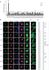
Figure 1. Expression and subcellular locale of USPs in granule neurons.
A. Analysis of USP gene expression in primary granule neurons. Neurons were isolated from P6 rat pups. After one or five days in vitro (DIV), total neuronal mRNA was isolated, reverse transcribed to cDNA, and analyzed by quantitative RT-PCR using gapdh as a control gene. Asterisks indicate significant changes in expression between DIV1 and DIV5 (P<0.05, t-test). B. Subcellular localization of neuronally expressed USPs in primary granule neurons. Cells isolated from P6 rat pups were cultured in vitro and at DIV2 transfected with expression plasmids encoding the indicated GFP-tagged USP. To visualize the entirety of the neuron, cells were cotransfected with plasmids encoding mCherry. Two days after transfection, cells were fixed and subjected to immunocytochemical analyses using the GFP and dsRED antibodies. Staining with the DNA dye bisbenzimide (Hoechst) was used to visualize the cell nucleus. An enlarged view of the localization of each USP in neurons is shown in the indicated panel. Bar = 10μm. C. Subcellular localization of neuronally expressed USPs in 293T cells. D. Summary of the subcellular localization of USPs in neurons. Abbreviations: Cent—Centrosome; Cyt—cytoplasm; ER—endoplasmic reticulum; Mito—mitochondria; Mt—microtubules; Nuc—nucleus; Nos—nucleolus; Ves—vesicles.
Figure 2
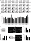
Figure 2. USP RNAi screen reveals functions for USP4 and USP20 in granule neuron axon development.
A. Generation of a plasmid-based shRNA library targeting neuronally expressed USPs. Target sequences of 32 candidate USPs were inserted into a pBS/U6 backbone vector, and the efficiency of the shRNA constructs was analyzed by cotransfecting 293T cells with the indicated GFP-USP expression constructs together with empty vector (U6) or shRNA-encoding plasmids. Whole cell lysates were analyzed by immunoblotting using the GFP and ERK antibodies, the latter to serve as loading control. B. Effect of knockdown of 32 USP genes on axon growth. Cerebellar granule neurons prepared from P6 rat pups were transfected with a GFP-expression plasmid and the indicated RNAi plasmid. Four days later, neurons were fixed and subjected to immunocytochemistry using the GFP antibody, and total axon length in transfected neurons was determined. Total axon length in granule neurons transfected with each RNAi plasmid was normalized to the total length in control U6-transfected granule neurons. A total of 3665 cells were counted. Asterisks indicate statistically significant effects (P<0.005, t-test) C. Lysates of 293T cells transfected with the GFP-USP20 expression plasmid together with the indicated USP20 RNAi or control U6 RNAi plasmid were immunoblotted with the indicated antibodies. D-G. USP20 knockdown significantly decreased the total axon length (P<0.001, ANOVA, n = 3, 279 neurons) and led to aberrant axon branching (P<0.001, ANOVA, n = 3, 212 neurons) in granule neurons. Granule neurons were transfected with the USP20 RNAi plasmids or the U6 control plasmid as in panel 2B. Panel E shows an enlarged view of the boxed area in D. H. Lysates of 293T cells transfected with GFP-USP4 together with the U6 or indicated USP4 RNAi plasmid were immunoblotted with the indicated antibodies. I-J. USP4 RNAi significantly decreased total axon length in granule neurons (P<0.001, ANOVA, n = 3, 234 neurons counted). Granule neurons were transfected with the USP4 RNAi plasmids or the U6 control plasmid as in Fig. 2B. The size of all scale bars is 50μm.
Figure 3
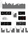
Figure 3. Regulation of granule neuron dendrite morphogenesis by USP1 and USP4.
A. Cerebellar granule neurons were prepared as in Fig. 2B, transfected at DIV2 and analyzed at DIV5. Total dendrite length in granule neurons transfected with each RNAi plasmid was normalized to the total dendrite length of control U6 transfected neurons. Asterisks indicate statistically significant effects (P<0.005, t-test). B, C. USP4 knockdown stimulates dendrite growth. Granule neurons were transfected as in panel A, and analyzed for dendrite length. Arrows and arrowheads indicate dendrites and axons, respectively. USP4 knockdown significantly increased total dendrite length (P<0.001, ANOVA, n = 3; 209 cells counted). D-E. USP1 knockdown simplifies granule neuron dendrite arbors. Neurons transfected with GFP and USP1 RNAi or U6 control plasmid were subjected to immunocytochemistry using the GFP and the MAP2 antibodies. Arrows and arrowheads indicate dendrites and axons, respectively. F. Lysates of 293T cells transfected with GFP-USP1 together with U6 or the indicated USP1 RNAi plasmid were immunoblotted with the indicated antibodies. G-K. Granule neurons were transfected as in A with the USP1 RNAi plasmid or control U6 plasmid. USP1 knockdown significantly reduced primary and secondary/tertiary dendrite numbers and increased length of longest dendrite (P<0.001 for 3H; P<0.001 for 3I; P<0.005 for 3J, ANOVA, n = 3; 206 cells), but had little or no effect on total dendrite length. Arrows and arrowheads indicate dendrites and axons, respectively. L-N. USP1 and USP4 RNAi regulate dendrite development in vivo. P4 rat pups were injected with plasmids encoding GFP together with the control U6 RNAi plasmid or the indicated RNAi plasmid targeting USP1 or USP4. Dendrite length and primary dendrite number of transfected cells were determined following immunohistochemistry of coronal sections of GFP-positive cerebella. For panel 3M, 81 cells and for panel 3N, 131 cells were counted. USP4 knockdown significantly increased total granule neuron dendrite length (P<0.005, t-test) and reduced primary granule neuron dendrite number (P<0.001, t-test) in vivo. The size of all scale bars is 20μm. Arrows and arrowheads indicate dendrites and axons, respectively.
Figure 4
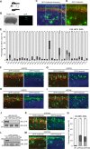
Figure 4. USP30 and USP33 are required for granule neuron migration in the rodent cerebellar cortex in vivo.
A-D. Cerebella of P4 rat pups were injected with expression plasmids encoding synapsin-mCitrin and the U6 control and subjected to electroporation. Pups were sacrificed five days later, at P9, and coronal sections of cerebella were analyzed by immunohistochemistry. Transfected neurons and Purkinje cells were visualized with the GFP (green) and calbindin antibodies (red), respectively. Staining with the DNA dye bisbenzimide (Hoechst) was used to label cell nuclei. Panel D shows an enlarged view of the boxed area in panel C. Bar in panel B = 500μm. E. An in vivo RNAi screen targeting USPs in the intact mammalian brain. P4 rat cerebella were subjected to in vivo electroporation as in panels A-D, and neuronal migration was analyzed by counting the number of neurons residing in the indicated layers. For each RNAi condition, at least three different brains were used for quantification. In total, 21536 cells were counted. F-I. Representative images showing impaired migration of neurons in rat pups transfected with RNAi targeting USP22, USP30, USP33 or USP36. J. Lysates of 293T cells transfected with USP30-GFP together with U6 or the indicated USP30 RNAi plasmid were immunoblotted with the indicated antibodies. K, N. USP30 knockdown impairs neuronal migration in the cerebellum. Cerebella of P4 rat pups were subjected to in vivo electroporation as in panels A-D. L. Lysates of 293T cells transfected with GFP-USP33 together with U6 or the indicated USP33 RNAi plasmid were immunoblotted with the indicated antibodies. M, N. USP33 knockdown impairs neuronal migration in the cerebellum. Cerebella of P4 rat pups were subjected to in vivo electroporation as in panels A-D. EGL—external granule layer; PCL—Purkinje cell layer; IGL—internal granule layer. The size of all scale bars is 20μm, unless otherwise noted.

References

    1. Altman J, Bayer SA (1997) Development of the cerebellar system: in relation to its evolution, structure, and functions (Boca Raton: CRC Press; ).
    1. Hatten ME, Heintz N (1995) Mechanisms of neural patterning and specification in the developing cerebellum. Annual Rev Neurosci 18:385–408. - PubMed
    1. Chedotal A (2010) Should I stay or should I go? Becoming a granule cell. Trends Neurosci 33:163–172. 10.1016/j.tins.2010.01.004 - DOI - PubMed
    1. Komuro H, Rakic P (1998) Distinct modes of neuronal migration in different domains of developing cerebellar cortex. J Neurosci 18:1478–1490. - PMC - PubMed
    1. Hamori J, Somogyi J (1983) Differentiation of cerebellar mossy fiber synapses in the rat: a quantitative electron microscope study. Journal Comp Neurol 220: 365–377. - PubMed

Publication types

LinkOut - more resources