Comparative Study
. 2014 May 6;14(1):21.
doi: 10.1186/1470-7330-14-21.

Muscle metastases: comparison of features in different primary tumours

Comparative Study

Muscle metastases: comparison of features in different primary tumours

Alexey Surov et al. Cancer Imaging. .

Abstract

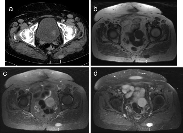
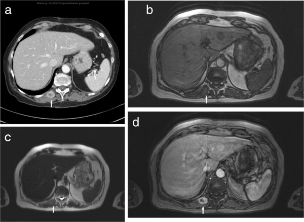
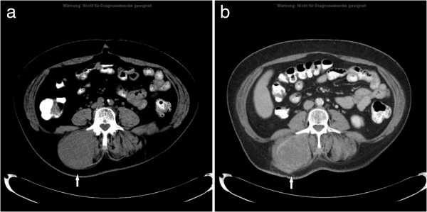
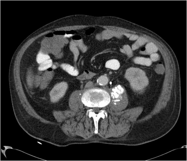
Background: Muscle metastases (MM) from solid tumours are rare. The aim of this study was to describe radiological features of MM, and to compare their patterns in different malignancies.

Methods: A retrospective search in the statistical database of our institution revealed 61 cases of MM. Additionally, a retrospective search in Pubmed database was performed. Together with our cases the present analysis comprises 461 patients (682 MM).

Results: MM derived from the following malignancies: lung cancer (25.1%), gastrointestinal tumours (21.0%), and urological tumours (13.2%). Other neoplasias with MM were rare. MM were localised most frequently in the thigh muscles, the extraocular musculature, and the gluteal and paravertebral muscles. The localisation of MM was different in several primary malignancies.

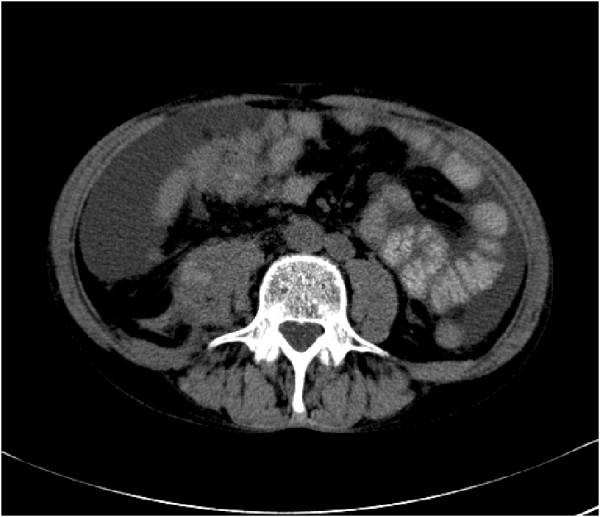
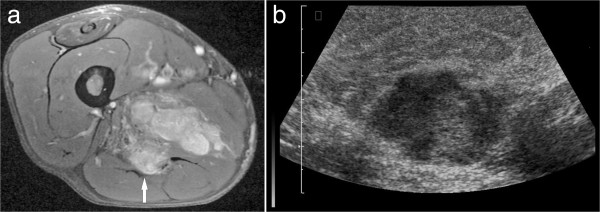
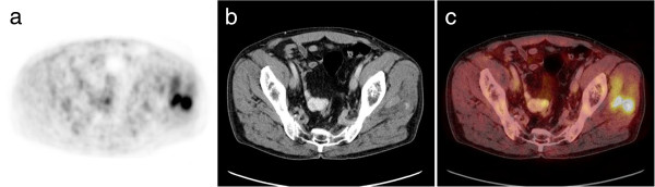
Conclusion: MM present with a broad spectrum of radiological features. Different CT imaging findings of MM were observed in different primary tumours. The localisation of MM also varies with different primary malignancies.


Figures

Figure 1
Figure 1
Imaging findings in a 50 year old woman with known history of melanoma. (a) CT image showing an oval mass with homogeneous contrast enhancement (type I) in the left gluteal musculature. (b) The lesion is hypointense on T1w image (spin echo pulse sequence, TR/TE: 569/11 ms). (c) The lesion is hyperintense on T2w image (short tau inversion recovery (STIR), TR/TE: 5210/80 ms). (d) After intravenous administration of contrast medium the lesion shows marked homogeneous enhancement (T1w spin echo with fat saturation, TR/TE: 610/12 ms).
Figure 2
Figure 2
Type II metastasis in the right paravertebral musculature in a patient with lung carcinoma. (a) On CT, the lesion (arrow) shows central low attenuation and rim enhancement. (b) On the T1w image (T1w flash 2D, TR/TE: 142/2.2 ms) the lesion (arrow) is isointense in comparison to the unaffected musculature. (c) On T2w image (half-Fourier acquisition turbo spin echo pulse sequence, HASTE, TR/TE: 800/120 ms) the lesion is isointense (arrow). (d) On MRI after administration of contrast medium (T1w flash 2D image with fat saturation, TR/TE: 209/2.3 ms) the lesion shows central low attenuation and rim enhancement (arrow).
Figure 3
Figure 3
MM type III in a patient with known metastatic soft tissue sarcoma. CT images before (a) and after administration of contrast medium (b) documenting a massive hypodense enlargement of the right paravertebral musculature with inhomogeneous enhancement (arrow).
Figure 4
Figure 4
MM type IV in a patient with urinary bladder carcinoma. CT image demonstrates multiple calcifications in the left iliopsoas muscle (arrow).
Figure 5
Figure 5
MM type V in a patient with known history of esophageal cancer. CT without intravenous administration of contrast medium shows hyperdense areas (arrow) in the right iliopsoas muscle.
Figure 6
Figure 6
MM in the thigh musculature in a patient with known history of metastatic renal cell carcinoma. (a) T1w image (turbo spin echo sequence with fat saturation, TR/TE: 615/11 ms) after contrast administration shows a lobulated mass (arrow) with inhomogeneous enhancement (arrow). On ultrasound, the mass was mixed hypo- to hyperechoic in comparison to the unaffected muscle (b).
Figure 7
Figure 7
Imaging findings in a patient with lung cancer. PET (a) showing focal uptake in the left gluteal musculature (arrow). CT scan (b) detecting intramuscular masses in this area (arrow). Fusion image (c).

References

    1. Acinas Garcia O, Fernandez FA, Satue EG, Buelta L, Val-Bernal JF. Metastasis of malignant neoplasms to skeletal muscle. Rev Esp Oncol. 1984;31:57–67. - PubMed
    1. Hasegawa S, Sakurai Y, Imazu H, Matsubara T, Ochiai M, Funabiki T, Suzuki K, Mizoguchi Y, Kuroda M, Kasahara M. Metastasis to the forearm skeletal muscle from an adenocarcinoma of the colon: report of a case. Surg Today. 2000;30:1118–1123. doi: 10.1007/s005950070013. - DOI - PubMed
    1. Nabeyama R, Tanaka K, Matsuda S, Iwamoto Y. Multiple intramuscular metastases 15 years after radical nephrectomy in a patient with stage IV renal cell carcinoma. J Orthop Sci. 2001;6:189–192. doi: 10.1007/s007760100070. - DOI - PubMed
    1. Willis RA. The Spread of Tumours in the Human Body. London: Butterworth; 1952.
    1. Glockner DM, White LM, Sundaram M, McDonald DJ. Unsuspected metastases presenting as solitary soft tissue lesions: a fourteen-year review. Skeletal Radiol. 2000;29:270–274. doi: 10.1007/s002560050606. - DOI - PubMed

Publication types

LinkOut - more resources