. 2015 Jan 22:5:7918.
doi: 10.1038/srep07918.

Cloud computing approaches for prediction of ligand binding poses and pathways

Affiliations

Cloud computing approaches for prediction of ligand binding poses and pathways

Morgan Lawrenz et al. Sci Rep. .

Abstract

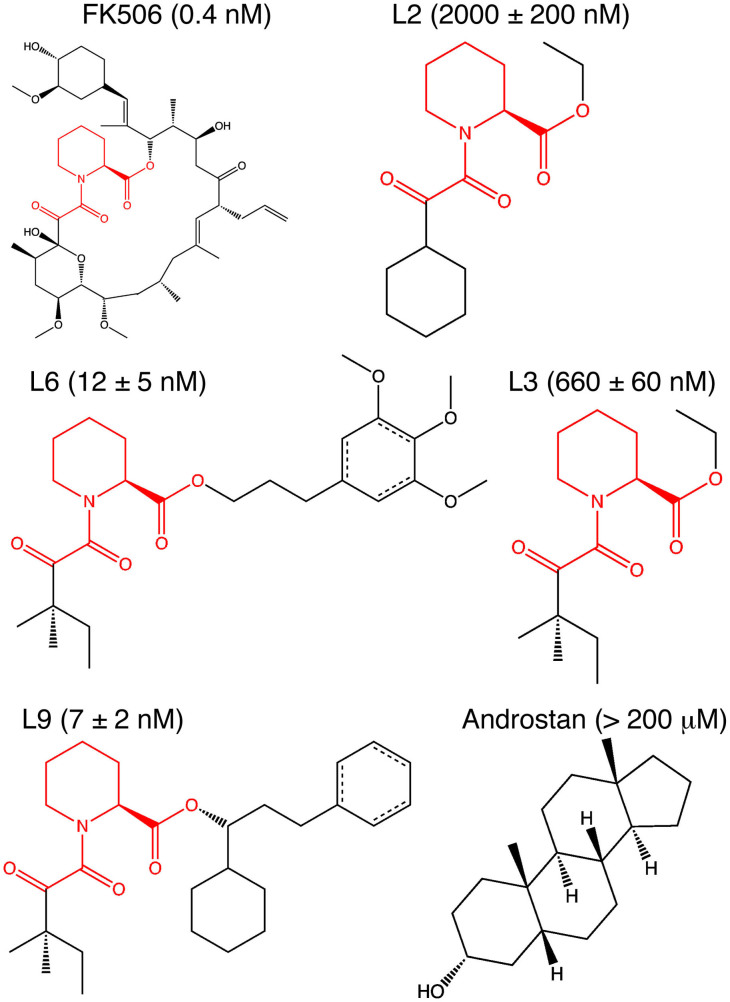
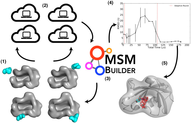
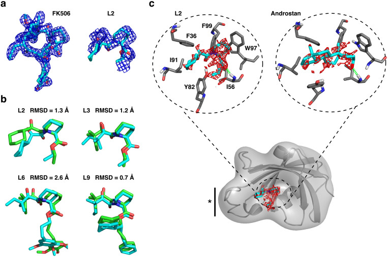
We describe an innovative protocol for ab initio prediction of ligand crystallographic binding poses and highly effective analysis of large datasets generated for protein-ligand dynamics. We include a procedure for setup and performance of distributed molecular dynamics simulations on cloud computing architectures, a model for efficient analysis of simulation data, and a metric for evaluation of model convergence. We give accurate binding pose predictions for five ligands ranging in affinity from 7 nM to > 200 μM for the immunophilin protein FKBP12, for expedited results in cases where experimental structures are difficult to produce. Our approach goes beyond single, low energy ligand poses to give quantitative kinetic information that can inform protein engineering and ligand design.


Figures

Figure 1
Figure 1. FKBP12 ligands used for predictions in this study.
The chemical structures highlight the common core in the ligands L2, L3, L6 and L9 derived from FK506 from Holt, et al. Ki values are also listed. A structure is available for L9, but structure factors for computing electron densities are only available for FK506.
Figure 2
Figure 2. Overview of the scheme used to for FKBP12 ligand predictions.
The scheme for the approach described in this study is illustrated, with steps as follows: 1) select diverse structures for the protein-ligand complex, (2) perform extensive MD simulations on cloud computing architectures, (3) construct a ligand-binding MSM at the end of a simulation period, (4) evaluate the MSM convergence, and repeat steps 1–4 until convergence is reached and reliable predictions are available (5). In step 4, we show the convergence of the lowest free energy state for L2, selected from MSMs built at ≈ 10 μs intervals and plotted as the state RMSD to the reference pose as the rolling mean with standard deviations over 2 data points (≈20 μs).
Figure 3
Figure 3. Comparison of FKBP12 ligand pose predictions with experiment.
(a) The available electron density for FK506 is shown in the left panel, compared with the adjusted density that corresponds to the common scaffold for L2, in the right panel. The density is shown at the 1σ contour level of the 2Fo–Fc difference map computed in PHENIX. The predicted L2 pose is shown in cyan stick representation to illustrate overlap with the available density. (b). Overlap of the predicted pose from the MSM (cyan), determined as the converged, highly populated ligand MSM state, with the validation pose (green). The validation pose is derived from crystallography experiments for L9, or from overlap and minimization of common scaffolds for L2, L3, and L6 with the L9 and FK506 structures, as done previously for accurate binding free energy predictions for these ligands. The RMSD between the structures is listed. (c). The 1.0 kcal/mol contour of the 3-D MSM-weighted free energy map within the active site is shown for L2 and 5 Androstan-3α-ol, with key binding residues labeled and a solid bar with a star denoting the 80's loop region. The free energy minimum surface is shown for all ligands in Supplementary Information Fig. 3. The L2 surface can be closely compared to the electron density contour in (a).

References

    1. Chae P. S. et al. Maltose-neopentyl glycol (MNG) amphiphiles for solubilization, stabilization and crystallization of membrane proteins. Nat. Methods 7, 1003–1008 (2010). - PMC - PubMed
    1. Hoeppner A., Schmitt L. & Smits S. Proteins and their ligands: Their importance and how to crystallize them. Advanced Topics on Crystal Growth Ferreira, S. O. (ed.) (InTech, 2013).
    1. Feixas F., Lindert S., Sinko W. & McCammon J. A. Exploring the role of receptor flexibility in structure-based drug discovery. Biophys. Chem. 186, 31–45 (2014). - PMC - PubMed
    1. Schneider G. Virtual screening: an endless staircase? Nat. Rev. Drug Discovery 9, 273–276 (2010). - PubMed
    1. Dror R. O. et al. Activation mechanism of the 2-adrenergic receptor. Proc. Natl. Acad. Sci. U. S. A. 108, 18684–18689 (2011). - PMC - PubMed

Publication types