. 2015 Jan 22:5:7955.
doi: 10.1038/srep07955.

In vitro expansion of corneal endothelial cells on biomimetic substrates

Affiliations

In vitro expansion of corneal endothelial cells on biomimetic substrates

Rachelle N Palchesko et al. Sci Rep. .

Abstract

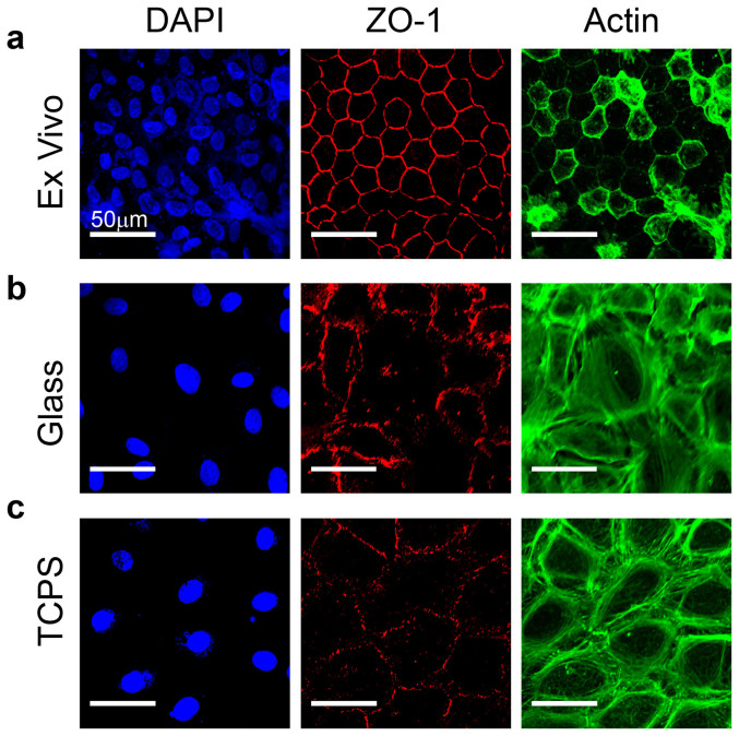
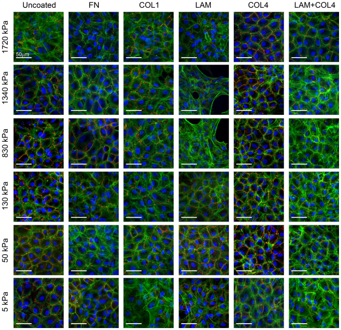
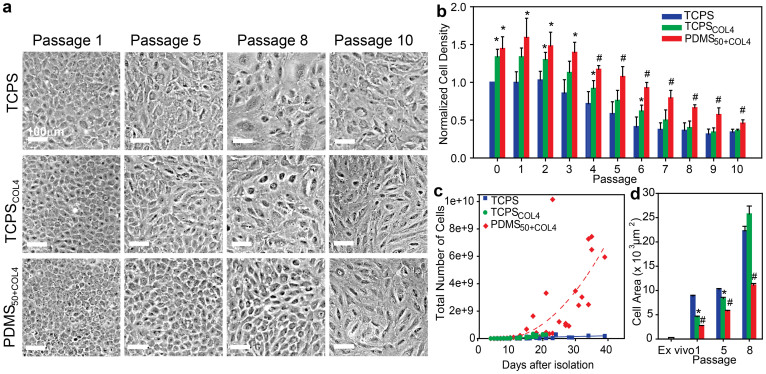
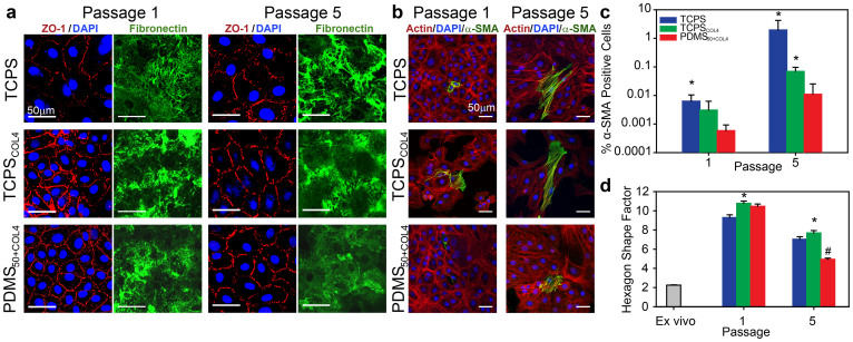
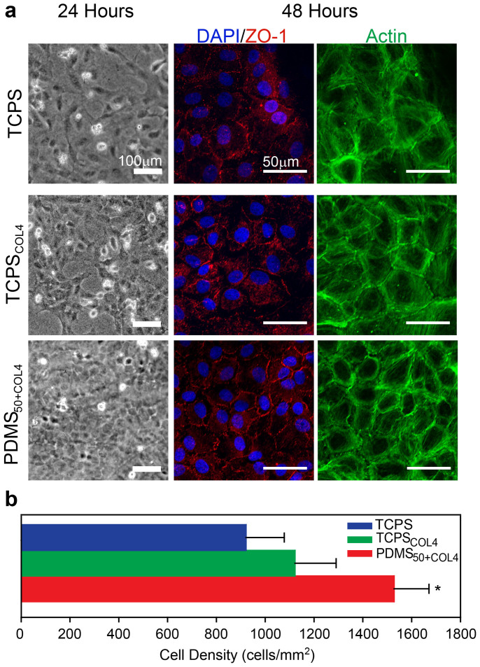
Corneal endothelial (CE) cells do not divide in vivo, leading to edema, corneal clouding and vision loss when the density drops below a critical level. The endothelium can be replaced by transplanting allogeneic tissue; however, access to donated tissue is limited worldwide resulting in critical need for new sources of corneal grafts. In vitro expansion of CE cells is a potential solution, but is challenging due to limited proliferation and loss of phenotype in vitro via endothelial to mesenchymal transformation (EMT) and senescence. We hypothesized that a bioengineered substrate recapitulating chemo-mechanical properties of Descemet's membrane would improve the in vitro expansion of CE cells while maintaining phenotype. Results show that bovine CE cells cultured on a polydimethylsiloxane surface with elastic modulus of 50 kPa and collagen IV coating achieved >3000-fold expansion. Cells grew in higher-density monolayers with polygonal morphology and ZO-1 localization at cell-cell junctions in contrast to control cells on polystyrene that lost these phenotypic markers coupled with increased α-smooth muscle actin expression and fibronectin fibril assembly. In total, these results demonstrate that a biomimetic substrate presenting native basement membrane ECM proteins and mechanical environment may be a key element in bioengineering functional CE layers for potential therapeutic applications.


Conflict of interest statement

Yes, there is potential competing interest. Drs. Palchesko, Funderburgh and Feinberg are co-inventors on a filed patent application related to the expansion technology in this manuscript.

Figures

Figure 1
Figure 1. CE cells lose differentiated morphology when cultured on glass or tissue culture polystyrene.
(a) Example of ex vivo, intact endothelium from the bovine cornea where cells exhibit a polygonal, mostly hexagonal shape and small size with the tight junction protein ZO-1 (red) present at cell borders and F-actin (green) located cortically. (b) CE cells cultured for one passage on a rigid glass substrate have reduced ZO-1 localization at the cell-cell borders and F-actin fibers present cortically as well as throughout the cell body. (c) CE cells cultured on TCPS for one passage appear similar to CE cells on glass, showing a loss of phenotypic shape and ZO-1 and F-actin localization relative to the ex vivo tissue. Cells are stained for nuclei (blue), tight junction protein ZO-1 (red) and F-actin (green) and scale bars are 50 μm.
Figure 2
Figure 2. Screening the response of CE cells to variable substrate elastic modulus and ECM protein coating.
CE cells were cultured on one of 36 different PDMS substrates with elastic modulus of 5, 50, 130, 830, 1340 or 1720 kPa and uncoated or coated with the ECM proteins fibronectin (FN), collagen type I (COLI), laminin (LAM), collagen type IV (COL4) or LAM and COL4. Representative fluorescent images show CE cells stained for nuclei (blue), ZO-1 (red) and F-actin (green), demonstrating distinct differences in morphology based on the substrates properties. In general, CE cells appeared to have a more polygonal morphology, continuous ZO-1 at the cell-cell border and cortical F-actin on substrates with an elastic modulus of 50 kPa for most of the ECM proteins. Similar results were observed for the COL4 coating for most of the elastic modulus formulations. Thus, the combination of elastic modulus and ECM protein that gave the best results in terms of the CE cell morphology most closely resembling that observed in vivo was an elastic modulus of 50 kPa and COL4 coating, which was selected for further cell expansion studies.
Figure 3
Figure 3. CE cells cultured on the biomimetic PDMS50+COL4 substrate maintained a polygonal morphology, higher cell density, increased proliferation rate and smaller cell size.
(a) Representative phase contrast images showing the morphology of CE cells cultured on the three different substrates at four different passages. At P5, CE cells cultured on TCPSCOL4 and TCPS exhibited elongated, irregular cell morphology. By P8 the cells were enlarged and polarized with no resemblance to a hexagonal morphology. In contrast, CE cells cultured on PDMS50+COL4 maintained a hexagonal like morphology up to P8. (b) Normalized cell density for CE cells cultured on TCPS (n = 4), TCPSCOL4 (n = 5) and PDMS50+COL4 (n = 5) (mean ± s.d.). At all passages the density of CE cells on the PDMS50+COL4 was significantly greater than on the TCPS and than on the TCPSCOL4 from P4 to P10. (c) Number of CE cells on the different substrates as a function of time, where each data point represents cell number prior to passaging (up to P5 for TCPS and TCPSCOL4 and P8 for PDMS50+COL4, when CE morphology became non-polygonal). There was a >3000-fold increase in the total cell number on PDMS50+COL4 compared to approximately a 140-fold increase on TCPS and TCPSCOL4 (dashed lines are to guide the eye). (d) Cell area in the ex vivo endothelium and on TCPS, TCPSCOL4 and PDMS50+COL4 at P1, P5 and P8 (mean ± s.e.m.). (*) indicating a statistically significant difference (P < 0.05) between PDMS50+COL4 and TCPS and (#) indicating a statistically significant difference (P < 0.05) between PDMS50+COL4 and both TCPS and TCPSCOL4.
Figure 4
Figure 4. CE cells cultured on PDMS50+COL4 maintained phenotypic ZO-1 staining and hexagonal shape while inhibiting fibronectin fibril formation and α-SMA expression.
(a) Representative images of CE cells on TCPS, TCPSCOL4 and PDMS50+COL4 stained for nuclei (blue) and ZO-1 (red) to show cell morphology and separate images stained for fibronectin (green) to show fibril assembly at the cell-substrate interface at P1 and P5. CE cells on PDMS50+COL4 had more continuous ZO-1 staining at the cell-cell border and decreased fibronectin fibril formation relative to cell on TCPS and TCPSCOL4. (b) Representative images of CE cells on TCPS, TCPSCOL4 and PDMS50+COL4 stained for nuclei (blue), F-actin (red) and α-SMA (green) to show cells that have undergone EMT to a fibroblast-like phenotype at P1 and P5. (c) Percentage of α-SMA positive cells at P1 and P5 showing the increase in EMT on TCPS and TCPSCOL4 relative to PDMS50+COL4 (mean ± s.d., * indicates statistically significant difference relative to PDMS50+COL4, P < 0.05). (d) Hexagon shape factor (HSF) of CE cells in ex vivo, intact endothelium and at P1and P5 TCPS, TCPSCOL4 and PDMS50+COL4 (mean ± s.e.m., * indicates TCPSCOL4 had a statistically significant difference to TCPS and PDMS50+COL4, P < 0.05, and # indicates PDMS50+COL4 had a statistically significant difference to TCPS and TCPSCOL4, P < 0.05).
Figure 5
Figure 5. CE cells expanded on PDMS50+COL4 formed a higher density, tissue engineered endothelium.
(a) Representative phase contrast images and fluorescent confocal images of CE cells expanded on TCPS, TCPSCOL4 and PDMS50+COL4 until P5 and then seeded onto collagen type I gels to form a tissue engineered endothelium. CE cells expanded on PDMS50+COL4 formed a complete monolayer more rapidly and had a polygonal morphology by 24 hours. At 48 hours CE cells stained for nuclei (blue), ZO-1 (red) and F-actin (green) appeared to have more continuous ZO-1 staining at the cell-cell border when expanded first on PDMS50+COL4. (b) Quantification of cell density in the tissue engineered endothelium showed that CE cells expanded on PDMS50+COL4 formed higher density monolayers (mean ± s.d., * indicates PDMS50+COL4 had a statistically significant difference compared to TCPS and TCPSCOL4, P < 0.05).

Similar articles

Cited by

References

    1. Zhu C. & Joyce N. C. Proliferative response of corneal endothelial cells from young and older donors. Invest Ophth Vis Sci. 45, 1743–1751 (2004). - PubMed
    1. Joyce N. C. Proliferative capacity of the corneal endothelium. Prog Retin Eye Res. 22, 359–389 (2003). - PubMed
    1. Joyce N. C., Meklir B., Joyce S. J. & Zieske J. D. Cell cycle protein expression and proliferative status in human corneal cells. Invest Ophth Vis Sci. 37, 645–655 (1996). - PubMed
    1. Joyce N. C., Navon S. E., Roy S. & Zieske J. D. Expression of cell cycle-associated proteins in human and rabbit corneal endothelium in situ. Invest Ophth Vis Sci. 37, 1566–1575 (1996). - PubMed
    1. Murphy C., Alvarado J., Juster R. & Maglio M. Prenatal and Postnatal Cellularity of the Human Corneal Endothelium - a Quantitative Histologic-Study. Invest Ophth Vis Sci. 25, 312–322 (1984). - PubMed

Publication types

LinkOut - more resources