. 2015 Jan 22:15:17.
doi: 10.1186/s12885-015-1023-5.

Nitric oxide donors increase PVR/CD155 DNAM-1 ligand expression in multiple myeloma cells: role of DNA damage response activation

Affiliations

Nitric oxide donors increase PVR/CD155 DNAM-1 ligand expression in multiple myeloma cells: role of DNA damage response activation

Cinzia Fionda et al. BMC Cancer. .

Abstract

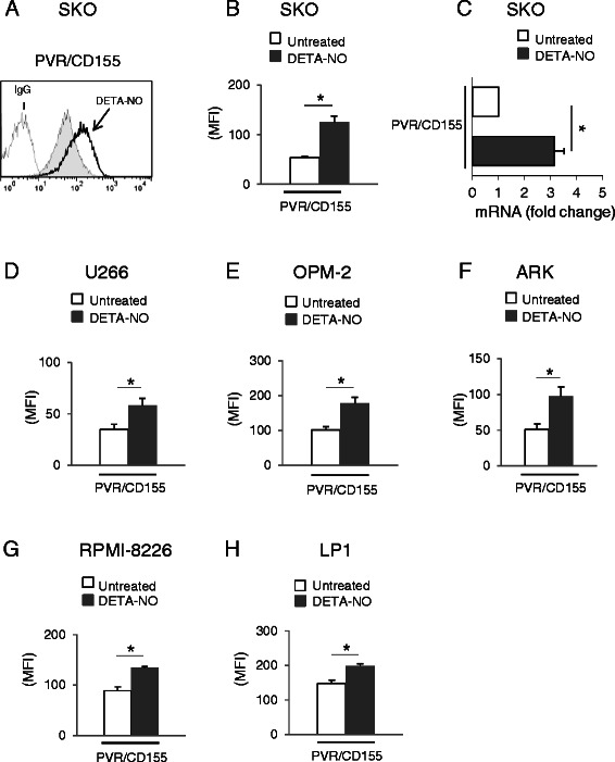
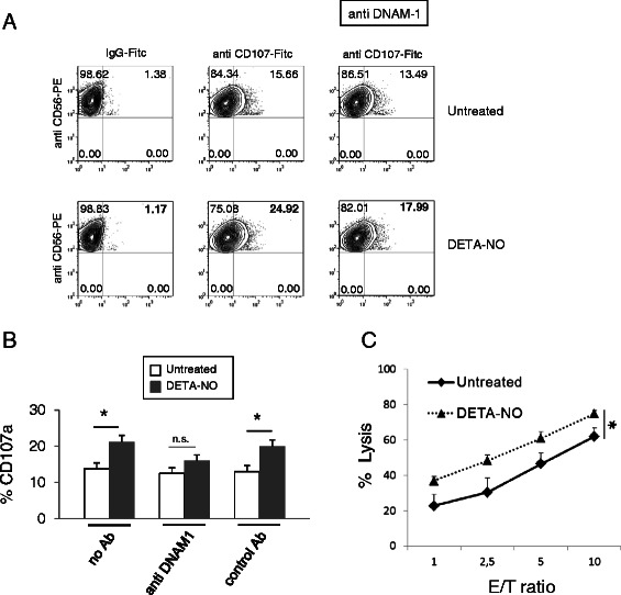
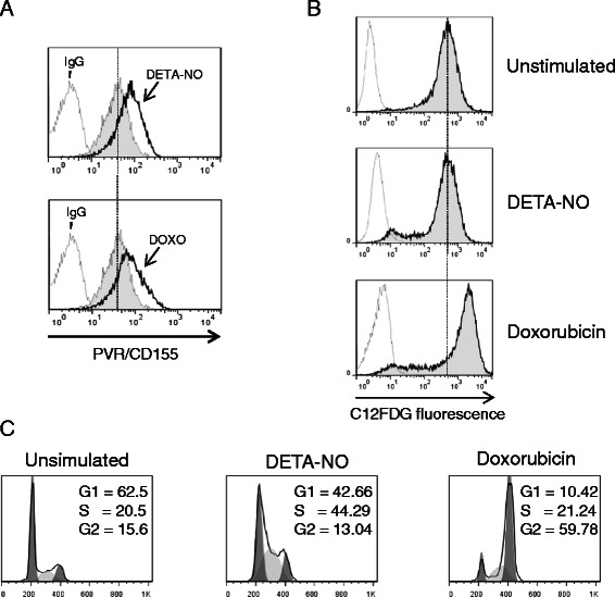
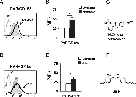
Background: DNAX accessory molecule-1 (DNAM-1) is an activating receptor constitutively expressed by macrophages/dendritic cells and by T lymphocytes and Natural Killer (NK) cells, having an important role in anticancer responses; in this regard, combination therapies able to enhance the expression of DNAM-1 ligands on tumor cells are of therapeutic interest. In this study, we investigated the effect of different nitric oxide (NO) donors on the expression of the DNAM-1 ligand Poliovirus Receptor/CD155 (PVR/CD155) in multiple myeloma (MM) cells.

Methods: Six MM cell lines, SKO-007(J3), U266, OPM-2, RPMI-8226, ARK and LP1 were used to investigate the activity of different nitric oxide donors [DETA-NO and the NO-releasing prodrugs NCX4040 (NO-aspirin) and JS-K] on the expression of PVR/CD155, using Flow Cytometry and Real-Time PCR. Western-blot and specific inhibitors were employed to investigate the role of soluble guanylyl cyclase/cGMP and activation of the DNA damage response (DDR).

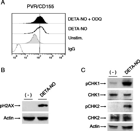
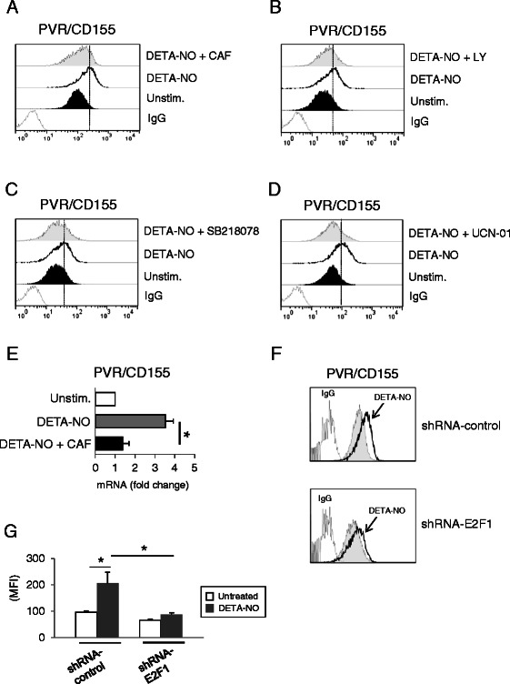
Results: Our results indicate that increased levels of nitric oxide can upregulate PVR/CD155 cell surface and mRNA expression in MM cells; in addition, exposure to nitric oxide donors renders myeloma cells more efficient to activate NK cell degranulation and enhances their ability to trigger NK cell-mediated cytotoxicity. We found that activation of the soluble guanylyl cyclase and increased cGMP concentrations by nitric oxide is not involved in the up-regulation of ligand expression. On the contrary, treatment of MM cells with nitric oxide donors correlated with the activation of a DNA damage response pathway and inhibition of the ATM /ATR/Chk1/2 kinase activities by specific inhibitors significantly abrogates up-regulation.

Conclusions: The present study provides evidence that regulation of the PVR/CD155 DNAM-1 ligand expression by nitric oxide may represent an additional immune-mediated mechanism and supports the anti-myeloma activity of nitric oxide donors.


Figures

Figure 1
Figure 1
Regulation of PVR/CD155 expression on MM cell lines following treatment with NO donor DETA-NO. A) PVR/CD155 surface expression was analyzed by flow cytometry on SKO-007(J3) cells treated with DETA-NO (200 μM) for 48 h. Data are representative of one out of three independent experiments. The grey colored histogram represents basal expression, while thick black colored histogram represents the expression after treatment with DETA-NO. B) The MFI of PVR/CD155 surface expression was calculated based on at least four independent experiments and evaluated by paired Student t test (*P < 0.05). Histograms represent the MFI with specific mAb subtracted from the MFI value of isotype control. These treatments did not affect the cell viability over the time and DETA-NO concentration [200 μM for SKO-007(J3)] chosen for these experiments (as assessed by PI staining, data not shown). C) Real Time PCR analysis of total mRNA obtained from SKO-007(J3) cells, untreated (−) or treated with 200 μM DETA-NO for 24 h as described above. Data, expressed as fold change units, were normalized with β-actin and referred to the untreated cells considered as calibrator and represent the mean of 3 experiments (*P < 0.05). D-H) The MFI of PVR/CD155 surface expression was calculated for U266, OPM-2, ARK, RPMI-8226 and LP1 MM cells, based on at least three independent experiments and evaluated by paired Student t test (*P < 0.05). Histograms represent the MFI with specific mAb subtracted from the MFI value of isotype control. These treatments did not affect the cell viability over the time and DETA-NO concentration [200 μM for U266, 50 μM for OPM-2, 200 μM for ARK, 100 μM for RPMI-8226 and 125 μM for LP1] chosen for these experiments (as assessed by PI staining, data not shown).
Figure 2
Figure 2
NO exposed SKO-007(J3) cells enhances NK cell-mediated cytotoxicity. A) NK cells prepared from PBMCs of healthy donors, were incubated with SKO-007(J3) cells, untreated or treated with DETA-NO for 48 h, and used as target cells in a degranulation assay. The assay was performed at the effector:target (E:T) ratio of 2.5:1. After 2 hours at 37°C, cells were stained with anti-CD56, anti-CD3 and anti-CD107a mAbs. Cell surface expression of CD107a was analyzed on CD56+CD3 cells. In order to evaluate the role of DNAM-1, the assay was performed in parallel treating NK cells with blocking anti-DNAM-1 antibody. Results are representative of one out of three independent experiments. B) The MFI of CD107 were calculated based on at least three independent experiments and evaluated by paired Student t test (*P < 0.05). Histograms represent the MFI with specific mAb subtracted from the MFI value of isotype control. C) NK cells isolated from PBMCs of healthy donors were incubated with SKO-007(J3) cells, untreated or treated with DETA-NO for 48 h as described above, and used as target cells in a standard 4-hour chromium-release assay. The percentage of specific lysis was calculated by counting an aliquot of supernatant and using the formula: 100 x [(sample release - spontaneous release)/total release - spontaneous release)]. All determinations were made in triplicate and E:T ratios ranged from 10:1 to 1:1, as indicated. Data represent the mean (n = 3 experiments, *P < 0.05).
Figure 3
Figure 3
NO enhances PVR/CD155 expression: molecular mechanisms. A) PVR/CD155 surface expression was analyzed by flow cytometry on SKO-007(J3) cells treated with DETA-NO (200 μM) in the presence or absence of the guanylate cyclase inhibitor ODQ (50 μM) for 48 h. Data are representative of one out of three independent experiments. B) Western Blot analysis of total cellular proteins from SKO-007(J3) cells treated with DETA-NO for 18 h. The arrow indicates the expression of the pH2A.X and β-actin, used as loading control. The proteins transferred to nitrocellulose membranes were stained with Ponceau to verify that similar amounts of protein had been loaded in each lane. Data shown are representative of 1 out of 2 independent experiments. C) Western Blot analysis of total cellular proteins from SKO-007(J3) cells treated with DETA-NO for 18 h. Lysates were probed with antibodies to different phosphorylation sites of Chk1 and Chk2, wt Chk1 and Chk2 or β-actin, used as loading control. The proteins transferred to nitrocellulose membranes were stained with Ponceau to verify that similar amounts of protein had been loaded in each lane. Data shown are representative of 1 out of 2 independent experiments.
Figure 4
Figure 4
NO enhances PVR/CD155 expression: role of DDR. A,B) PVR/CD155 surface expression was analyzed by flow cytometry on SKO-007(J3) cells treated with DETA-NO (200 μM) in the presence or absence of caffeine (CAF 1 mM) or LY294002 (LY 20 μM) for 48 h. Data are representative of one out of four independent experiments. C,D) PVR/CD155 surface expression was analyzed by flow cytometry on SKO-007(J3) cells treated with DETA-NO (200 μM) in the presence or absence of the Chk1/2 inhibitors SB218078 and UCN-01 (0.5 μM and 50 nM respectively) for 48 h. Data are representative of one out of four independent experiments. In these experiments, the concentration used for the different inhibitors, did not significantly affect cell viability as assessed by PI staining (data not shown). E) Real Time PCR analysis of total mRNA obtained from SKO-007(J3) cells, treated for 24 h in the presence or absence of caffeine (1 mM) as described above. Data, expressed as fold change units, were normalized with β-actin and referred to the untreated cells considered as calibrator and represent the mean of 3 experiments (*P < 0.05). F) PVR/CD155 surface expression was analyzed by flow cytometry on SKO-007(J3) non-target shRNA (shRNA-control) or pLKO-sh-E2F1 cells, treated with DETA-NO as described above. Data are representative of one out of three independent experiments. G) The MFI of PVR/CD155 surface expression was calculated based on at least three independent experiments and evaluated by paired Student t test (*P < 0.05). Histograms represent the MFI with specific mAb subtracted from the MFI value of isotype control.
Figure 5
Figure 5
NO-induced up-regulation of PVR/CD155 is not related to a senescence-dependent mechanism. A) PVR/CD155 surface expression was analyzed by flow cytometry on SKO-007(J3) cells treated with DETA-NO (200 μM) or with doxorubicin (0.05 μM) for 48 h. Data are representative of one out of three independent experiments. The grey colored histograms represent basal expression of the indicated ligand, while thick black colored histograms represent the expression after treatment with the indicated drug. B) SA-βGal activity of SKO-007(J3) cells treated with DETA-NO or with doxorubicin for 48 h as described above. Data are representative of one out of three independent experiments. The grey colored histograms represent the C12-fluorescein signal. C) SKO-007(J3) cells were treated for 48 hours with the indicated drug as described above. Cells were fixed and stained with PI to analyze cell distribution among the different cell-cycle phases.
Figure 6
Figure 6
Regulation of PVR/CD155 expression on MM cell lines following treatment with the NO-releasing prodrugs NitroAspirin and JS-K. A) PVR/CD155 surface expression was analyzed by flow cytometry on SKO-007(J3) cells treated with NCX4040 (10 μM) for 48 h. Data are representative of one out of three independent experiments. The grey colored histogram represents basal expression of the indicated ligand, while thick black colored histogram represents the expression after treatment with NCX4040. B) The MFI of PVR/CD155 surface expression was calculated based on at least three independent experiments and evaluated by paired Student t test (*P < 0.05). Histograms represent the MFI with specific mAb subtracted from the MFI value of isotype control. C) Molecular structure of NCX4040. D) PVR/CD155 surface expression was analyzed by flow cytometry on SKO-007(J3) cells treated with JS-K (3 μM) for 48 h. Data are representative of one out of three independent experiments. The grey colored histogram represents basal expression of the indicated ligand, while thick black colored histogram represents the expression after treatment with JS-K. E) The MFI of PVR/CD155 surface expression was calculated based on at least three independent experiments and evaluated by paired Student t test (*P < 0.05). Histogram represents the MFI with specific mAb subtracted from the MFI value of isotype control. F) Molecular structure of JS-K. The concentration of the indicated donor used in these experiments, did not significantly affect cell viability as assessed by PI staining (data not shown).

References

    1. Kumar SK, Rajkumar SV, Dispenzieri A, Lacy MQ, Hayman SR, Buadi FK, et al. Improved survival in multiple myeloma and the impact of novel therapies. Blood. 2008;111:2516–20. doi: 10.1182/blood-2007-10-116129. - DOI - PMC - PubMed
    1. Kyle RA, Rajkumar SV. Multiple myeloma. Blood. 2008;111:2962–72. doi: 10.1182/blood-2007-10-078022. - DOI - PMC - PubMed
    1. Mahindra A, Laubach J, Raje N, Munshi N, Richardson PG, Anderson K. Latest advances and current challenges in the treatment of multiple myeloma. Nat Rev Clin Oncol. 2012;9:135–43. doi: 10.1038/nrclinonc.2012.15. - DOI - PubMed
    1. Mohty B, El-Cheikh J, Yakoub-Agha I, Avet-Loiseau H, Moreau P, Mohty M. Treatment strategies in relapsed and refractory multiple myeloma: a focus on drug sequencing and ‘retreatment’ approaches in the era of novel agents. Leukemia. 2012;26:73–85. doi: 10.1038/leu.2011.310. - DOI - PubMed
    1. Ludwig H, Durie BG, McCarthy P, Palumbo A, San MJ, Barlogie B, et al. IMWG consensus on maintenance therapy in multiple myeloma. Blood. 2012;119:3003–15. doi: 10.1182/blood-2011-11-374249. - DOI - PMC - PubMed

Publication types

MeSH terms