. 2015 Mar 15;26(6):1119-28.
doi: 10.1091/mbc.E14-08-1281. Epub 2015 Jan 21.

Rab5-family guanine nucleotide exchange factors bind retromer and promote its recruitment to endosomes

Affiliations

Rab5-family guanine nucleotide exchange factors bind retromer and promote its recruitment to endosomes

Bjorn D M Bean et al. Mol Biol Cell. .

Erratum in

  • Correction.
    [No authors listed] [No authors listed] Mol Biol Cell. 2015 Apr 1;26(7):1411. Mol Biol Cell. 2015. PMID: 25823929 Free PMC article. No abstract available.

Abstract

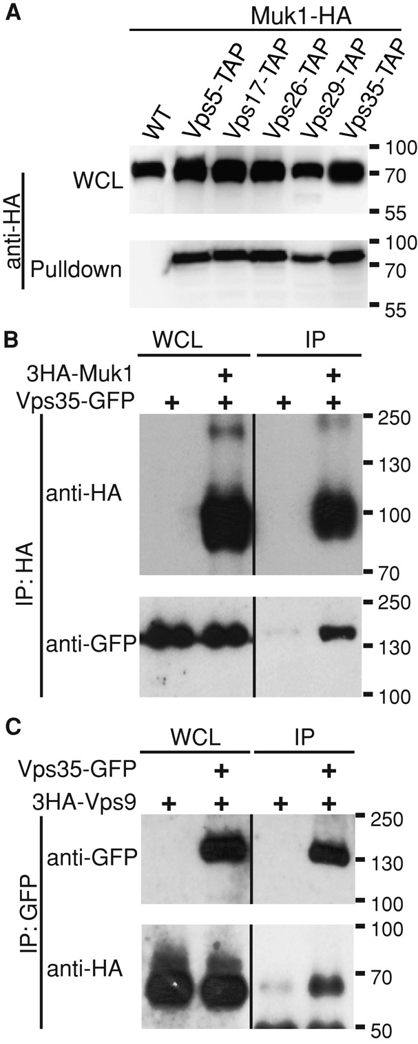
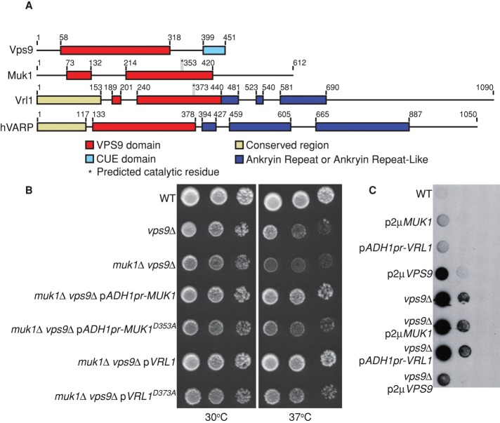
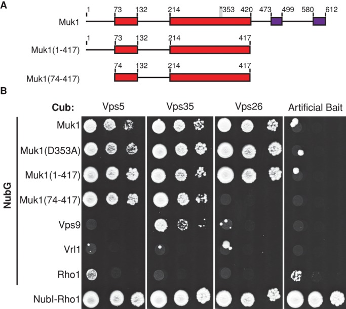
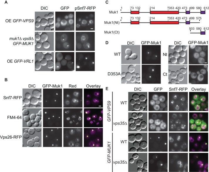
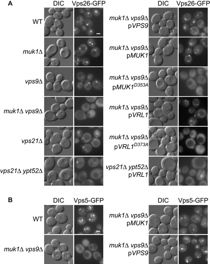
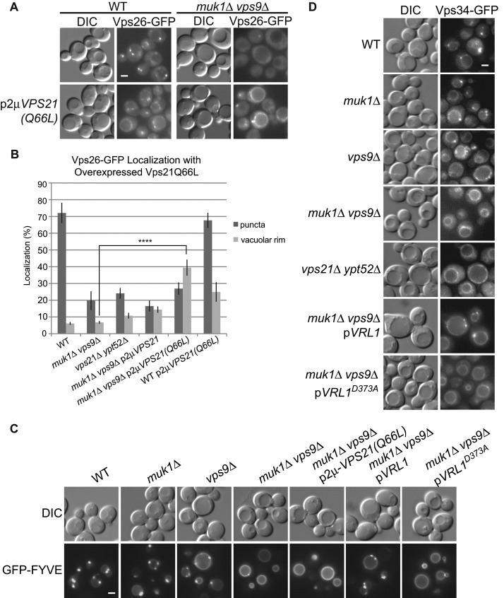
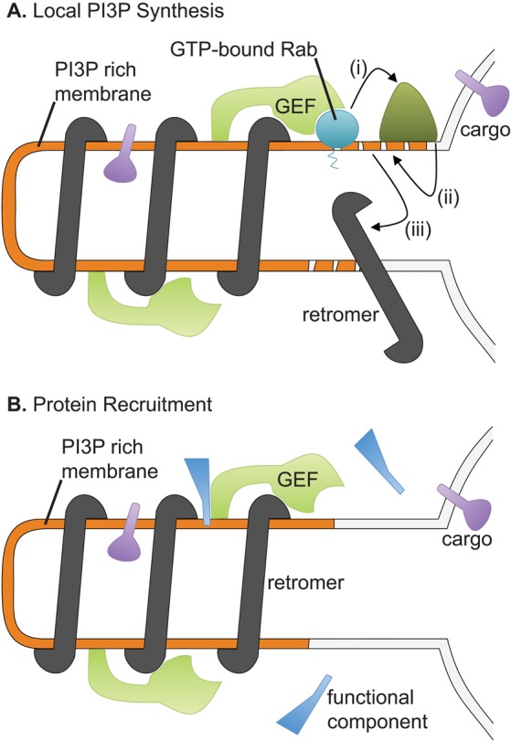
The retromer complex facilitates the sorting of integral membrane proteins from the endosome to the late Golgi. In mammalian cells, the efficient recruitment of retromer to endosomes requires the lipid phosphatidylinositol 3-phosphate (PI3P) as well as Rab5 and Rab7 GTPases. However, in yeast, the role of Rabs in recruiting retromer to endosomes is less clear. We identified novel physical interactions between retromer and the Saccharomyces cerevisiae VPS9-domain Rab5-family guanine nucleotide exchange factors (GEFs) Muk1 and Vps9. Furthermore, we identified a new yeast VPS9 domain-containing protein, VARP-like 1 (Vrl1), which is related to the human VARP protein. All three VPS9 domain-containing proteins show localization to endosomes, and the presence of any one of them is necessary for the endosomal recruitment of retromer. We find that expression of an active VPS9-domain protein is required for correct localization of the phosphatidylinositol 3-kinase Vps34 and the production of endosomal PI3P. These results suggest that VPS9 GEFs promote retromer recruitment by establishing PI3P-enriched domains at the endosomal membrane. The interaction of retromer with distinct VPS9 GEFs could thus link GEF-dependent regulatory inputs to the temporal or spatial coordination of retromer assembly or function.


Figures

FIGURE 1:
FIGURE 1:
Retromer physically interacts with the Rab5-family GEFs Muk1 and Vps9. (A) TAP-tagged retromer subunits were pulled down using calmodulin beads from GAL1pr-MUK1-HA strains. Samples were resolved by 12% SDS–PAGE and detected by immunoblotting. Loading of lysates relative to the pull down was 1:25. (B) Endogenously tagged Vps35-GFP and ADH1pr-3HA-Muk1 were cross-linked with 1.6 mg/ml DSP, 3HA-Muk1 was immunoprecipitated, and copurifying Vps35-GFP was probed by immunoblotting. Loading of lysates relative to immunoprecipitate (IP) was 1:1.6 (anti-HA) and 1:388 (anti-GFP). (C) Cells expressing Vps35-GFP and ADH1pr-3HA-Vps9 were cross-linked and immunoprecipitated with anti-GFP. Copurification of 3HA-Vps9 was detected with anti-HA. Loading of lysates relative to IP was 1:1.6 (anti-GFP) and 1:388 (anti-HA).
FIGURE 2:
FIGURE 2:
Vrl1 is a new yeast VPS9-domain protein. (A) Schematic of yeast VPS9 domain–containing proteins and human VARP based on Superfamily (Gough et al., 2001) and ClustalW alignments. (B) Yeast were spotted in 10× dilution series and grown 2 d at 37°C to assess temperature sensitivity of the indicated strains. VRL1 was expressed from its endogenous promoter. (C) A colony overlay assay was used to assess carboxypeptidase Y (CPY) secretion. Cells spotted in 10× dilution series were overlaid with nitrocellulose and incubated for 16 h. The nitrocellulose was then immunoblotted with anti-CPY antibodies.
FIGURE 3:
FIGURE 3:
The VPS9 domain of Muk1 is sufficient for interaction with retromer subunits in the iMYTH assay. (A) Schematic of Muk1 truncations. The position of the invariant aspartate residue required for GEF activity and conserved C-terminal motifs are indicated. (B) Strains containing Cub-tagged retromer subunits and plasmids encoding Rab5-family GEFs tagged N-terminally with NubG were tested for activation of the HIS3 reporter on selective medium lacking tryptophan, adenine, and histidine. NubG-tagged Rho1 acted as a negative control, whereas the NubI tag, which binds tightly to Cub independently of other interactions, confirmed the expression of Cub fusions.
FIGURE 4:
FIGURE 4:
The yeast VPS9-domain proteins Muk1, Vps9, and Vrl1 localize to endosomes. (A) MUK1pr-GFP-Muk1, ADH1pr-GFP-Vps9, and ADH1pr-GFP-Vrl1 localize to Snf7-RFP–marked late endosomes by fluorescence microscopy. (B) Fluorescence microscopy of ADH1pr-GFP-Muk1 with Snf7-RFP, the lipophilic dye FM4-64, and the tagged retromer subunit RFP-Vps26. (C) Schematic of Muk1 truncations used in D, showing conserved C-terminal motifs identified by alignments (purple). (D) Fluorescence microscopy of full-length or mutated ADH1pr-GFP-Muk1. (E) Deletion of VPS35 does not disrupt the endosomal localization of overexpressed GFP-Vps9 or GFP-Muk1. Scale bars, 2 μm; OE, overexpressed.
FIGURE 5:
FIGURE 5:
Rab5-family GEFs and GTPases are required for endosomal recruitment of retromer. (A) Fluorescence microscopy of Vps26-GFP in strains lacking Rab5-family GEFs and Rab5-family GTPases. Muk1 and Vrl1 are expressed from endogenous promoters. Muk1D353A and Vrl1D373A are predicted to be catalytically inactive. (B) Microscopy of Vps5-GFP in strains with deletions of Rab5-family GEFs. Scale bars, 2 μm.
FIGURE 6:
FIGURE 6:
Rab5-family GEFs are needed for PI3P production at endosomes, and this cannot be bypassed by expressing the constitutively active Rab5-family GTPase Vps21(Q66L). (A) Fluorescence microscopy shows that Vps26-GFP is mislocalized to the vacuolar rim when Vps21(Q66L) is expressed, even in the absence of Rab5-family GEFs. (B) Quantification of Vps26-GFP fluorescence microscopy. Images of Vps26-GFP localization from four independent experiments were manually scored for localization to the vacuolar rim or puncta (N ≥ 140/strain per experiment). Unpaired one-way analysis of variance: p < 0.0001 overall; ****p < 0.0001. (C) Fluorescence microscopy of GFP-FYVE, a biomarker for PI3P, in strains with deletions of Rab5-family GEFs. (D) Vps34-GFP localization to puncta is dependent on expression of Rab5-family GEFs and GTPases. Vrl1 is expressed from the endogenous promoter in C and D. Scale bars, 2 μm.
FIGURE 7:
FIGURE 7:
Models for the function of the interaction between retromer and Rab5-family GEFs at endosomes. (A) In the first model, the GEFs concentrate Rab5-family GTPases at the retromer tubules. The GTPases recruit the PI3K Vps34 (i), which locally increases the concentration of PI3P (ii), leading to further retromer recruitment (iii). (B) In the second model, GEFs physically recruit specific factors to the tubules.

Similar articles

Cited by

References

    1. Attar N, Cullen PJ. The retromer complex. Adv Enzyme Regul. 2010;50:216–236. - PubMed
    1. Babu M, Vlasblom J, Pu S, Guo X, Graham C, Bean BDM, Burston HE, Vizeacoumar FJ, Snider J, Phanse S, et al. Interaction landscape of membrane-protein complexes in Saccharomyces cerevisiae. Nature. 2012;489:585–589. - PubMed
    1. Balaji K, Mooser C, Janson CM, Bliss JM, Hojjat H, Colicelli J. RIN1 orchestrates the activation of RAB5 GTPases and ABL tyrosine kinases to determine the fate of EGFR. J Cell Sci. 2012;125:5887–5896. - PMC - PubMed
    1. Balderhaar H, Arlt H, Ostrowicz C, Brocker C, Sundermann F, Brandt R, Babst M, Ungermann C. The Rab GTPase Ypt7 is linked to retromer-mediated receptor recycling and fusion at the yeast late endosome. J Cell Sci. 2010;123:4085–4094. - PubMed
    1. Bonifacino J, Hurley J. Retromer. Curr Opin Cell Biol. 2008;20:427–436. - PMC - PubMed

Publication types

MeSH terms

Substances

LinkOut - more resources