. 2015 Jan 22;34(1):4.
doi: 10.1186/s13046-014-0118-1.

Early B-cell factor 3 (EBF3) is a novel tumor suppressor gene with promoter hypermethylation in pediatric acute myeloid leukemia

Affiliations

Early B-cell factor 3 (EBF3) is a novel tumor suppressor gene with promoter hypermethylation in pediatric acute myeloid leukemia

Yan-Fang Tao et al. J Exp Clin Cancer Res. .

Abstract

Background: Pediatric acute myeloid leukemia (AML) comprises up to 20% of all childhood leukemia. Recent research shows that aberrant DNA methylation patterning may play a role in leukemogenesis. The epigenetic silencing of the EBF3 locus is very frequent in glioblastoma. However, the expression profiles and molecular function of EBF3 in pediatric AML is still unclear.

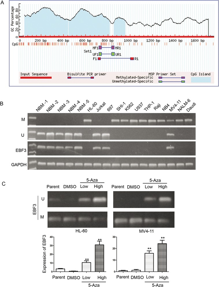
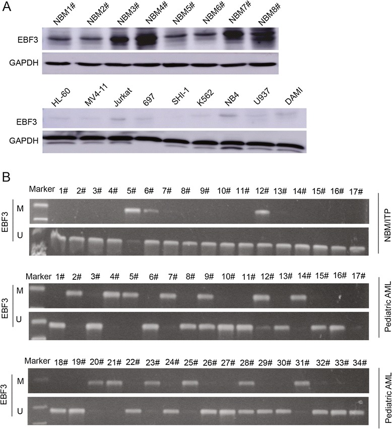
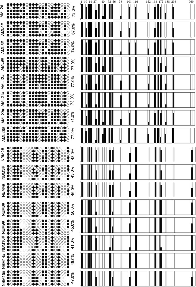
Methods: Twelve human acute leukemia cell lines, 105 pediatric AML samples and 30 normal bone marrow/idiopathic thrombocytopenic purpura (NBM/ITP) control samples were analyzed. Transcriptional level of EBF3 was evaluated by semi-quantitative and real-time PCR. EBF3 methylation status was determined by methylation specific PCR (MSP) and bisulfite genomic sequencing (BGS). The molecular mechanism of EBF3 was investigated by apoptosis assays and PCR array analysis.

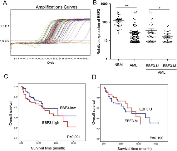
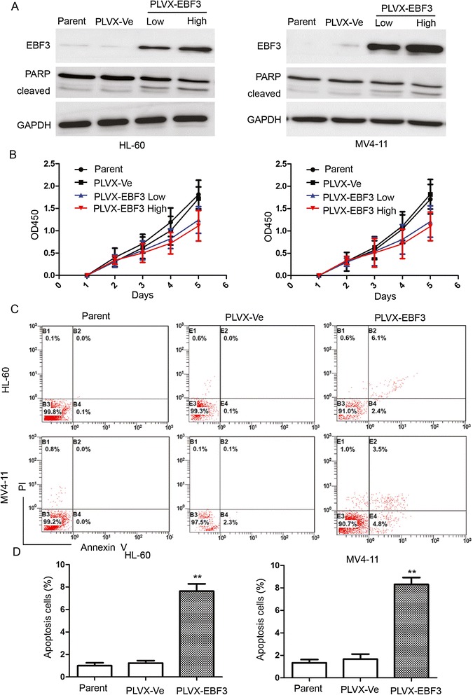
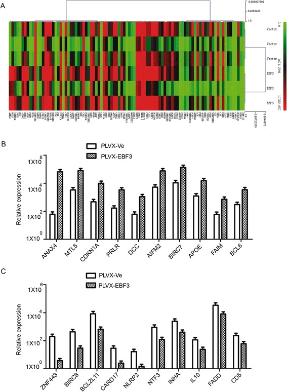
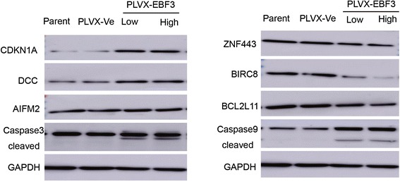
Results: EBF3 promoter was hypermethylated in 10/12 leukemia cell lines. Aberrant EBF3 methylation was observed in 42.9% (45/105) of the pediatric AML samples using MSP analysis, and the BGS results confirmed promoter methylation. EBF3 expression was decreased in the AML samples compared with control. Methylated samples revealed similar survival outcomes by Kaplan-Meier survival analysis. EBF3 overexpression significantly inhibited cell proliferation and increased apoptosis. Real-time PCR array analysis revealed 93 dysregulated genes possibly implicated in the apoptosis of EBF3-induced AML cells.

Conclusion: In this study, we firstly identified epigenetic inactivation of EBF3 in both AML cell lines and pediatric AML samples for the first time. Our findings also showed for the first time that transcriptional overexpression of EBF3 could inhibit proliferation and induce apoptosis in AML cells. We identified 93 dysregulated apoptosis-related genes in EBF3-overexpressing, including DCC, AIFM2 and DAPK1. Most of these genes have never been related with EBF3 over expression. These results may provide new insights into the molecular mechanism of EBF3-induced apoptosis; however, further research will be required to determine the underlying details. Our findings suggest that EBF3 may act as a putative tumor suppressor gene in pediatric AML.


Figures

Figure 1
Figure 1
Analysis of promoter methylation in pediatric AML by NimbleGen Human DNA Methylation arrays. Analysis of the methylation status of genes in five pediatric AML samples (M1, M2, M3, M4 and M5) and three NBM samples (N1, N2, and N3) using NimbleGen Human DNA Methylation arrays shows that the EBF3 promoter is significantly methylated in AML samples (5/5) and unmethylated in NBM samples (0/3). (A) Each red box represents the number of methylation peaks (PeakScore) overlapping the promoter region for the corresponding gene. The PeakScore is defined as the average -log10 (P value) from probes within the peak. (B) The scores reflect the probability of positive methylation enrichment.
Figure 2
Figure 2
The EBF3 promoter is methylated in AML cell lines. (A) Two CpG island regions can be identified in the promoter of EBF3. (B) MSP analysis of the methylation status of EBF3 in leukemia cell lines shows that the promoter is hypermethylated in 10/12 cell lines. M and U represents MSP results using primer sets for methylated and unmethylated EBF3 genes, respectively. (C) PCR analysis showed that the methylation status of EBF3 decreased in leukemia cells following 5-Aza treatment compared with control cells treated with DMSO. The transcript level of EBF3 is significantly upregulated in HL-60 and NB4 cells treated with 5-Aza compared with those treated with DMSO. *P < 0.05; **P < 0.01.
Figure 3
Figure 3
MSP analysis showing EBF3 promoter hypermethylation in AML samples. (A) Western blot analysis depicting the expression of EBF3 in eight NBM samples and nine leukemia cells. (B) MSP analysis the promoter methylation of EBF3 and aberrant EBF3 methylation was observed in 42.9% (45/105) of the pediatric AML samples compared with 13.3% (4/30) of the NBM control samples. M and U represent MSP results using primer sets for methylated and unmethylated EBF3 genes, respectively.
Figure 4
Figure 4
BGS analysis depicts EBF3 promoter hypermethylation in AML samples. Eight NBM samples and eight AML samples were selected for further analysis by BGS. The EBF3 promoter was methylated in the AML samples (67.0% - 77.0%); whereas the EBF3 promoter was methylated in only 41.0% - 50.0% in the NBM samples. ● methylated cytosines; ○ unmethylated cytosines.
Figure 5
Figure 5
The expression of EBF3 was downregulated in patients with pediatric AML. (A) Real-time PCR analysis of the transcript levels of EBF3 in 105 pediatric AML samples and 30 NBM control samples. (B) Quantification shows that EBF3 expression was found to be robustly decreased in the AML samples compared with the control samples (26.91 ± 50.86 vs. 121.14 ± 95.11, respectively; P <0.001). Those with methylated EBF3 showed significantly lower levels of EBF3 expression compared with unmethylated EBF3 (16.32 ± 12.93 vs. 34.86 ± 65.46, respectively; P = 0.043). (C) The prognostic significance of EBF3 expression was assessed in 105 Chinese pediatric AML patients with clinical follow-up records. Kaplan-Meier survival analysis revealed similar survival outcomes in tumors with high or low EBF3 expression among 105 pediatric AML patients (P = 0.091). (D) Samples with EBF3 promoter methylation revealed similar survival outcomes through Kaplan-Meier survival analysis (P = 0.190).
Figure 6
Figure 6
Overexpression of EBF3 inhibited proliferation and induced apoptosis in leukemia cells. (A) Western blot analysis of EBF3 expression in EBF3 transfected leukemia cells. Transfection with EBF3 lentivirus PLVX-EBF3 significantly upregulated expression of EBF3 in AML cells compared with mock-transfected cells. An expression level of cleaved PARP, a marker of apoptosis, was analyzed by Western blotting. (B) CCK-8 assays show that transfection with EBF3 lentivirus inhibits proliferation in HL-60 and MV4-11 cells compared with mock-transfected cells. (C) The number of cells displaying apoptotic features is higher in the HL-60 and MV4-11 cells transfected with PLVX-EBF3 compared with the mock-transfected cells. (D) Quantification shows that the proportion of apoptotic cells in the EBF3-overexpressing cells (PLVX-EBF3) was significantly greater than the vector control group (PLVX-Ve). **P < 0.01.
Figure 7
Figure 7
Real-time PCR array analysis shows dysregulated genes implicated in EBF3 over-expression. (A) Cluster of apoptosis genes related with EBF3 over-expression. We analyzed and clustered the expression of 370 key genes involved in apoptosis using the SABioscience Human Apoptosis PCR Array PAHS-3012 kit. (B) Relative expression of the genes most up-regulated in EBF3-overexpressing AML cells compared with mock-transfected cells. (C) Relative expression of the genes most down-regulated in EBF3-overexpressing cells compared with mock transfected cells.
Figure 8
Figure 8
Western-blot analysis verifying the dysregulated genes implicated in EBF3 -overexpressing cells. Western blot analysis of cells transfected with PLVX-EBF3 compared with PLVX-Ve control cells. The increase of cleaved caspase-3 and caspase-9 and up-regulation of CDKN1A, DCC, and AIFM2 and the down-regulation of ZNF443, BIRC8, and BCL2L11 in the EBF3-overexpressing group were verified by Western blot analysis.
Figure 9
Figure 9
IPA summary of the pathways regulated by EBF3 overexpression in HL-60 cells. Datasets representing 370 key genes involved in apoptosis with altered expression profiles that were obtained from real-time PCR arrays were imported into the IPA Tool and the following data is illustrated: (A) A list of the top five networks with their respective scores obtained from IPA. (B) A list of the top five molecular and cellular functions with their respective scores obtained from IPA. (C) A list of the top five canonical pathways with their respective scores obtained from IPA. (D) The network representation of the most highly rated network is shown. Genes that are shaded were determined to be statistically significant. A solid line represents a direct interaction between the two gene products and a dotted line means an indirect interaction.

References

    1. Appelbaum FR, Baer MR, Carabasi MH, Coutre SE, Erba HP, Estey E, et al. NCCN practice guidelines for acute myelogenous leukemia. Oncology (Williston Park) 2000;14:53–61. - PubMed
    1. Sanz GF, Sanz MA, Vallespi T, Canizo MC, Torrabadella M, Garcia S, et al. Two regression models and a scoring system for predicting survival and planning treatment in myelodysplastic syndromes: a multivariate analysis of prognostic factors in 370 patients. Blood. 1989;74:395–408. - PubMed
    1. Wan H, Zhu J, Chen F, Xiao F, Huang H, Han X, et al. SLC29A1 single nucleotide polymorphisms as independent prognostic predictors for survival of patients with acute myeloid leukemia: an in vitro study. J Exp Clin Cancer Res. 2014;33:90. doi: 10.1186/s13046-014-0090-9. - DOI - PMC - PubMed
    1. Im AP, Sehgal AR, Carroll MP, Smith BD, Tefferi A, Johnson DE, et al. DNMT3A and IDH mutations in acute myeloid leukemia and other myeloid malignancies: associations with prognosis and potential treatment strategies. Leukemia. 2014;28(9):1774–83. doi: 10.1038/leu.2014.124. - DOI - PMC - PubMed
    1. Abdel-Wahab O, Levine RL. Mutations in epigenetic modifiers in the pathogenesis and therapy of acute myeloid leukemia. Blood. 2013;121:3563–72. doi: 10.1182/blood-2013-01-451781. - DOI - PMC - PubMed

Publication types

MeSH terms