Review
. 2014 Apr 29;14(1):20.
doi: 10.1186/1470-7330-14-20.

The role of diffusion and perfusion weighted imaging in the differential diagnosis of cerebral tumors: a review and future perspectives

Review

The role of diffusion and perfusion weighted imaging in the differential diagnosis of cerebral tumors: a review and future perspectives

Patricia Svolos et al. Cancer Imaging. .

Abstract

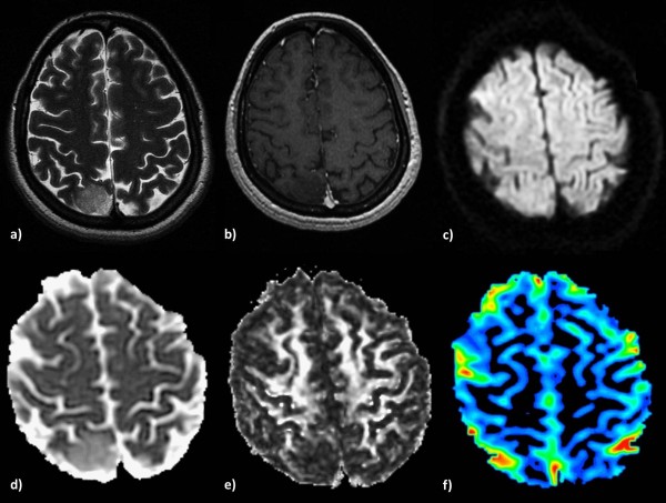
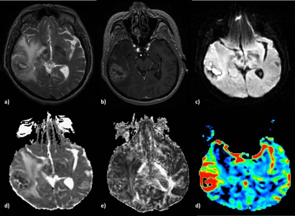
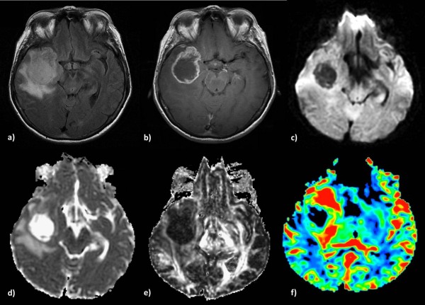
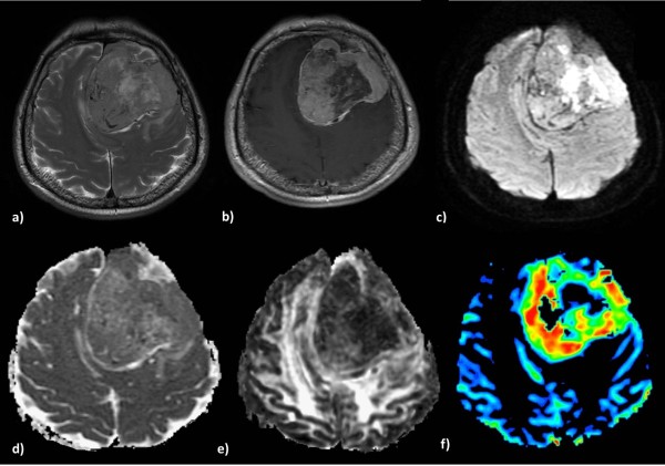
The role of conventional Magnetic Resonance Imaging (MRI) in the detection of cerebral tumors has been well established. However its excellent soft tissue visualization and variety of imaging sequences are in many cases non-specific for the assessment of brain tumor grading. Hence, advanced MRI techniques, like Diffusion-Weighted Imaging (DWI), Diffusion Tensor Imaging (DTI) and Dynamic-Susceptibility Contrast Imaging (DSCI), which are based on different contrast principles, have been used in the clinical routine to improve diagnostic accuracy. The variety of quantitative information derived from these techniques provides significant structural and functional information in a cellular level, highlighting aspects of the underlying brain pathophysiology. The present work, reviews physical principles and recent results obtained using DWI/DTI and DSCI, in tumor characterization and grading of the most common cerebral neoplasms, and discusses how the available MR quantitative data can be utilized through advanced methods of analysis, in order to optimize clinical decision making.


Figures

Figure 1
Figure 1
Low-grade glioma in a 55-year-old woman. a) High signal intensity on a T2-weighted image, b) no contrast enhancement on a 3D-SPGR image and c) an isointense signal on a diffusion-weighted image. The lesion shows increased ADC (d), lower FA (e) and no significant perfusion (f) on the corresponding parametric maps.
Figure 2
Figure 2
Anaplastic Astrocytoma in a 71-year-old man. a) T2-weighted image shows increased signal intensity with peritumoral edema, b) heterogeneous contrast enhancement on a post-contrast 3D-SPGR image and c) restricted diffusion in the solid portion of the tumor. The lesion is hypointense on the ADC map (d), presents low FA (e) and increased perfusion on the rCBV map (f).
Figure 3
Figure 3
Glioblastoma multiforme in a 65-year-old woman. Axial T2-weighted (a) and T1-weighted post contrast (b) images demonstrate a right temporal lesion with surrounding edema and ring-shaped enhancement. On the DW-image the lesion presents low signal intensity (c) resulting in higher intratumoral ADC (d), lower intratumoral FA (e), and high peritumoral rCBV (f), reflecting tumor infiltration in the surrounding parenchyma.
Figure 4
Figure 4
A typical Meningioma in a 60-year-old man. Axial T2-weighted (a) and postcontrast T1-weighted (b) images demonstrate a large heterogeneous enhanced left frontal mass with an intense mass effect. The lesion presents areas of restricted diffusion (c), isointensity on the ADC map (d), hypointensity on the FA map (e) and elevated blood volume (f).
Figure 5
Figure 5
Intracranial lung metastasis in a 68 year-old-man. a) T2-weighted image, b) ring-shaped enhancement on a T1-weighted post contrast edema, c) restricted diffusion in the periphery of the tumor. Increased intratumoral ADC (d), decreased FA (e) and elevated perfusion in the peripheral solid part of the lesion (f).
Figure 6
Figure 6
Primary Cerebral Lymphoma in a 59-year-old-woman. a) High signal intensity with peritumoral edema on a T2-weighted image, b) intense contrast-enhancement on a T1-weighted post contrast image and c) hyperintensity on a DW image. Decreased intratumoral ADC (d) and FA (e). The rCBV map shows moderate perfusion within the lesion (f).

References

    1. Hakyemez B, Erdogan C, Gokalp G, Dusak A, Parlak M. Solitary metastases and high-grade gliomas: radiological differentiation by morphometric analysis and perfusion-weighted MRI. Clin Radiol. 2010;65(1):15–20. doi: 10.1016/j.crad.2009.09.005. - DOI - PubMed
    1. Chang SC, Lai PH, Chen WL, Weng HH, Ho JT, Wang JS, Chang CY, Pan HB, Yang CF. Diffusion-weighted MRI features of brain abscess and cystic or necrotic brain tumors: comparison with conventional MRI. Clin Imaging. 2002;26(4):227–236. doi: 10.1016/S0899-7071(02)00436-9. - DOI - PubMed
    1. Liu X, Tian W, Kolar B, Yeaney GA, Qiu X, Johnson MD, Ekholm S. MR diffusion tensor and perfusion-weighted imaging in preoperative grading of supratentorial nonenhancing gliomas. Neuro Oncol. 2011;13(4):447–455. doi: 10.1093/neuonc/noq197. - DOI - PMC - PubMed
    1. De Belder FE, Oot AR, Van Hecke W, Venstermans C, Menovsky T, Van Marck V, Van Goethem J, Van den Hauwe L, Vandekerckhove M, Parizel PM. Diffusion tensor imaging provides an insight into the microstructure of meningiomas, high-grade gliomas, and peritumoral edema. J Comput Assist Tomogr. 2012;36(5):577–582. doi: 10.1097/RCT.0b013e318261e913. - DOI - PubMed
    1. Hakyemez B, Yildirim N, Erdogan C, Kocaeli H, Korfali E, Parlak M. Meningiomas with conventional MRI findings resembling intraaxial tumors: can perfusion-weighted MRI be helpful in differentiation? Neuroradiology. 2006;48(10):695–702. doi: 10.1007/s00234-006-0115-y. - DOI - PubMed