. 2015 Jan 21;35(3):1250-9.
doi: 10.1523/JNEUROSCI.2444-14.2015.

HDAC2 selectively regulates FOXO3a-mediated gene transcription during oxidative stress-induced neuronal cell death

Affiliations

HDAC2 selectively regulates FOXO3a-mediated gene transcription during oxidative stress-induced neuronal cell death

Shengyi Peng et al. J Neurosci. .

Abstract

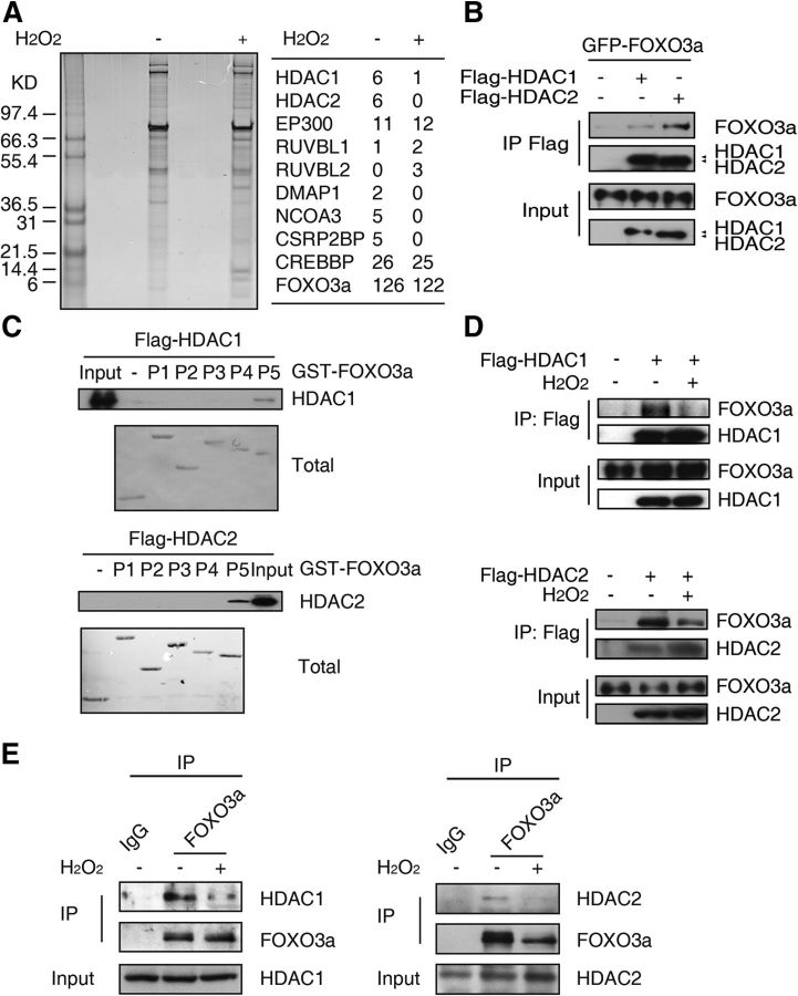
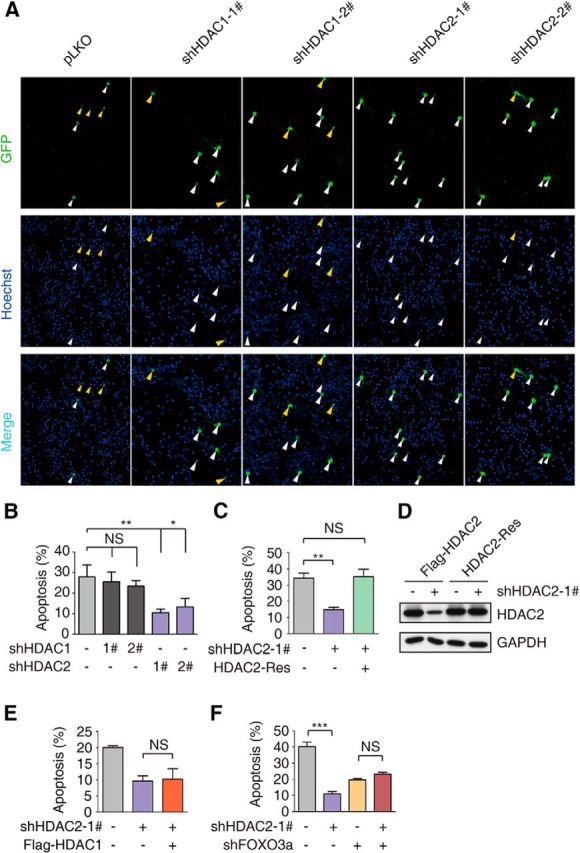
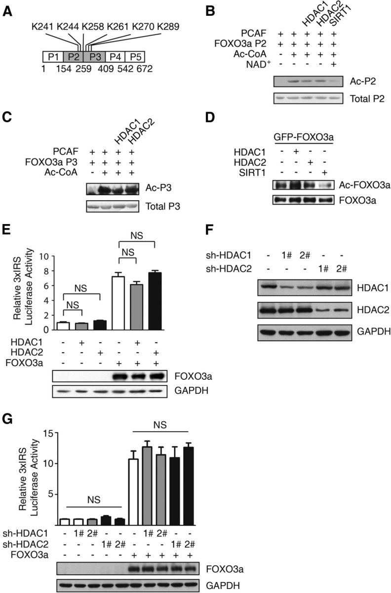
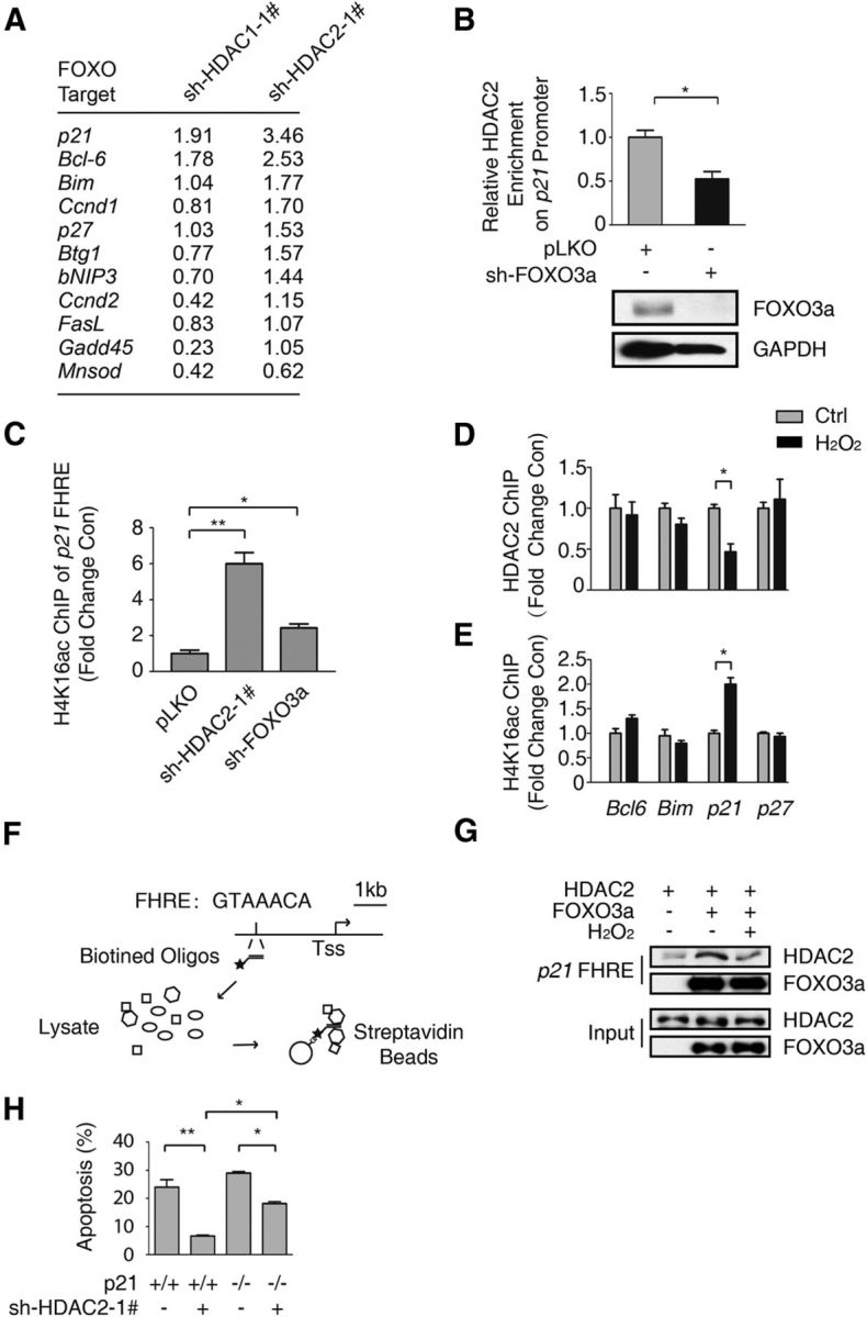
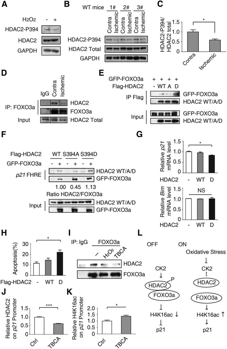
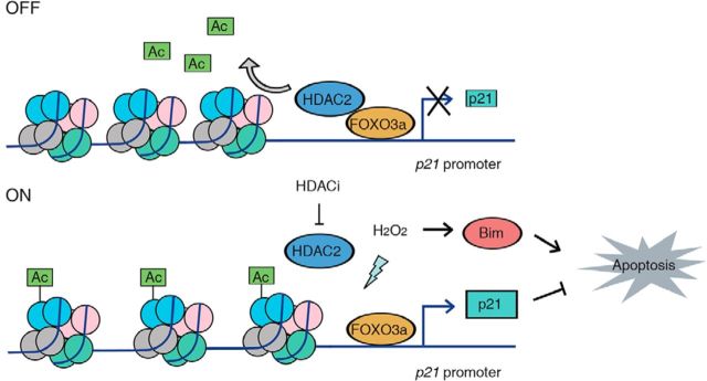
All neurodegenerative diseases are associated with oxidative stress-induced neuronal death. Forkhead box O3a (FOXO3a) is a key transcription factor involved in neuronal apoptosis. However, how FOXO3a forms complexes and functions in oxidative stress processing remains largely unknown. In the present study, we show that histone deacetylase 2 (HDAC2) forms a physical complex with FOXO3a, which plays an important role in FOXO3a-dependent gene transcription and oxidative stress-induced mouse cerebellar granule neuron (CGN) apoptosis. Interestingly, we also found that HDAC2 became selectively enriched in the promoter region of the p21 gene, but not those of other target genes, and inhibited FOXO3a-mediated p21 transcription. Furthermore, we found that oxidative stress reduced the interaction between FOXO3a and HDAC2, leading to an increased histone H4K16 acetylation level in the p21 promoter region and upregulated p21 expression in a manner independent of p53 or E2F1. Phosphorylation of HDAC2 at Ser 394 is important for the HDAC2-FOXO3a interaction, and we found that cerebral ischemia/reperfusion reduced phosphorylation of HDAC2 at Ser 394 and mitigated the HDAC2-FOXO3a interaction in mouse brain tissue. Our study reveals the novel regulation of FOXO3a-mediated selective gene transcription via epigenetic modification in the process of oxidative stress-induced cell death, which could be exploited therapeutically.

Keywords: FOXO3a; HDAC2; oxidative stress; p21; transcription.


Figures

Figure 1.
Figure 1.
FOXO3a interacts with HDAC1 and HDAC2. A, Suspended HeLa cells stably transfected with the pOZ-FOXO3a expression plasmid. A final concentration of 0 or 300 μm H2O2 was added to the medium for 12 h. The final HA-eluates from the nuclear fractions were resolved on a SDS-PAGE gel and silver-stained (left). Some of the identified FOXO3a complex components based on mass-spectral analysis are listed on the right. B, 293T cells were transiently transfected with the GFP-FOXO3a expression plasmid alone or together with the Flag-HDAC1 or Flag-HDAC2 expression plasmid, as indicated. Anti-Flag beads were used to immunoprecipitate the Flag-tagged proteins; anti-GFP and anti-Flag antibodies were used for immunoblotting. C, 293T cells were transiently transfected with the Flag-HDAC1 or Flag-HDAC2 expression plasmid. Cell lysates were incubated with the GST protein or a GST-tagged FOXO3a P1–P5 protein fragment. The anti-Flag antibody was used for immunoblotting. D, 293T cells were transfected with the Flag-HDAC1 or Flag-HDAC2 expression plasmid. Twenty-four hours after transfection, 300 μm H2O2 was added for 12 h, as indicated. The anti-Flag antibody was added to immunoprecipitate the HDAC proteins; the anti-Flag and anti-FOXO3a antibodies were used for immunoblotting. E, CGNs (7 DIV) were treated with 0 or 100 μm H2O2 for 1 h. Cell lysates were immunoprecipitated using an anti-FOXO3a antibody or IgG, and an immunoblot was performed using the indicated antibodies.
Figure 2.
Figure 2.
HDAC2 knockdown inhibits H2O2-induced neuronal death. A, CGNs were cotransfected with pEGFP-N1 and pLKO-HDAC1-1#/2#, pLKO-HDAC2-1#/2# or the empty vector pLKO at DIV 3. The neuronal apoptosis assay was performed 24 h after H2O2 (60–100 μm) treatment at DIV 6. Hoechst 33258 was used for nuclear staining, as indicated. The anti-GFP antibody was used for signal enhancement. Apoptotic cells are denoted by yellow arrows, and surviving cells are denoted by white arrows. B, Statistical analysis of A. HDAC2 knockdown protected neurons from oxidative stress-induced apoptosis. (ANOVA, n = 4 for the control group; n = 3 for the HDAC1 and HDAC2 knockdown groups; *p < 0.05, **p < 0.01). C, CGNs were transiently transfected with pEGFP-N1 together with the empty vector pLKO or pLKO-HDAC2–1# or the rescue vector HDAC2-Res as indicated. The neuronal apoptosis assay was performed as in A. HDAC2 knockdown protected neurons from apoptosis (ANOVA, n = 4 for the control and HDAC2 knockdown groups, **p < 0.01), but it did not protect the cells transfected with the rescue vector (ANOVA, n = 4 for the rescue-treated group, p > 0.1). A representative result of three independent experiments is shown. D, Lysates of 293T cells transfected with an expression vector encoding FLAG-HDAC2 or HDAC2-Res together with the pLKO-HDAC2–1# or control pLKO plasmid were immunoblotted with the FLAG and GAPDH antibodies. E, CGNs were transiently transfected with pEGFP-N1 together with the empty vector pLKO, pLKO-HDAC2–1#, or Flag-HDAC1 as indicated. The neuronal apoptosis assay was performed as in A. Overexpression of HDAC1 did not reverse the protective effect of HDAC2 knockdown (ANOVA, n = 3 for each group; p > 0.1). F, CGNs were transiently transfected with pEGFP-N1 together with the empty vector pLKO, pLKO-HDAC2–1#, or pLKO-FOXO3a. The neuronal apoptosis assay was performed as in A. In CGNs with FOXO3a knocked down, HDAC2 knockdown did not protect CGNs from H2O2-induced apoptosis (ANOVA, n = 3 for each group; p > 0.1). A representative result of three independent experiments is shown.
Figure 3.
Figure 3.
The FOXO3a acetylation and transcriptional activity levels are not directly affected by HDAC1 or HDAC2. A, The lysine sites of FOXO3a might be affected by HDACs. B, C, GST-FOXO3a P2 (B) and P3 fragments (C) were acetylated by PCAF in the presence or absence of Ac-CoA. The Flag-HDAC1, Flag-HDAC2, and Flag-SIRT1 proteins were purified from 293T cells transiently expressing the respective proteins using an anti-Flag antibody. Acetylated FOXO3a was incubated in purified HDAC1, HDAC2, or SIRT1 (supplemented with NAD+). The anti-acetyl antibody was used for immunoblotting. D, 293T cells were transfected with the GFP-FOXO3a expression plasmid alone or together with the Flag-HDAC1, Flag-HDAC2, or Flag-SIRT1 expression plasmid. The anti-GFP antibody was used to enrich the FOXO3a protein. The expression levels of the anti-acetylated FOXO3a and total FOXO3a proteins are shown. E, HT-22 cells were transiently transfected with 3xIRS, PR-TK together with the Flag-HDAC1, Flag-HDAC2, or Flag-FOXO3a expression plasmid as indicated. The relative 3xIRS luciferase activity was analyzed using a dual luciferase reporter assay system. The data are presented as the mean ± SEM. Firefly/Renilla luciferase activity was normalized to that of the control vector-transfected cells. No significant differences were detected between the control and HDAC1/2 groups with or without ectopic FOXO3a expression (ANOVA, n = 3; p > 0.05). The ectopic expressed FOXO3a protein level was blotted by anti-Flag antibody. F, Whole-cell extracts from HDAC1 or HDAC2 knockdown stable cells were resolved via SDS-PAGE; anti-HDAC1, anti-HDAC2, and anti-GAPDH antibodies were used for immunoblotting. G, HT-22 cells with HDAC1 or HDAC2 stably knocked down were transiently transfected with 3xIRS, PR-TK, and the Flag-FOXO3a expression plasmid as indicated. The relative 3xIRS luciferase activity was analyzed using the dual luciferase reporter assay system. No significant difference was detected between the knockdown and control groups. The data are presented as the mean ± SEM, and the firefly/Renilla luciferase activity level was normalized to that of the control cells (ANOVA, n = 3; p > 0.05). The ectopic expressed FOXO3a protein level was blotted by anti-Flag antibody.
Figure 4.
Figure 4.
HDAC2 is recruited to the p21 promoter by FOXO3a and regulates p21 expression. A, Total RNA was extracted from HT-22 cells with HDAC1 or HDAC2 stably knocked down. The relative mRNA level was detected via qRT-PCR. The fold-change in the RNA level compared with pLKO empty cells was determined. B, HT-22 cells with FOXO3a stably knocked down and control HT-22 cells were subjected to ChIP using an anti-HDAC2 antibody. qRT-PCR was performed to detect the abundance of HDAC2 at the p21 FHRE (top). The HDAC2 abundance was decreased due to FOXO3a knockdown (Student's t test, n = 3; *p < 0.05). Lysates from FOXO3a knockdown and pLKO-transfected control HT-22 cells were immunoblotted using anti-FOXO3a and anti-GAPDH antibodies (bottom). C, Lysates from HT-22 cells with HDAC2 or FOXO3a stably knocked down and pLKO control HT-22 cells were subjected to ChIP using an anti-H4K16ac antibody. The p21 promoter acetylation level was analyzed via qRT-PCR using primers specific to the p21 FHRE. HDAC2 knockdown (Student's t-test, n = 3, **p < 0.01) and FOXO3a knockdown (Student's t-test, n = 3, *p < 0.05) promoted H4K16ac enrichment in the p21 promoter. D, HT-22 cells were treated with 0 or 300 μm H2O2 for 1 h. Then, the samples were subjected to ChIP using the anti-HDAC2 antibody, which was followed by qRT-PCR analysis of the Bcl6, Bim, p21, and p27 promoter FHRE regions. HDAC2 was specifically enriched in the p21 promoter (Student's t test, n = 3; *p < 0.05). E, HT-22 cells were treated with 0 or 300 μm H2O2 for 1 h. Then, the samples were subjected to ChIP using an anti-H4K16ac antibody, which was followed by qRT-PCR analysis of the Bcl6, Bim, p21, and p27 promoter FHRE regions. The H4K16ac level was increased in the p21 promoter (Student's t test, n = 3, *p < 0.05). F, Flow chart of the DNA pull-down assay for the p21 FHRE. G, 293T cells were transfected with the Flag-HDAC2 or Flag-FOXO3a expression plasmid. The cells were treated with 300 μm H2O2 for 1 h before they were harvested, as indicated. The cell lysates were pulled down using the biotin-labeled p21 FHRE. Western blotting was performed using an anti-HDAC2 antibody. H, CGNs prepared from WT or p21 knock-out mice were transfected with pEGFP-N1 together with the pLKO-HDAC2–1# plasmid, as indicated. The neuronal apoptosis assay was performed as in Figure 2A. The neuronal protection of HDAC2 knockdown in p21−/− CGNs was less than in WT CGNs (ANOVA, n = 4 for each group; *p < 0.05).
Figure 5.
Figure 5.
HDAC2 knockdown can induce p21 upregulation in a p53-independent manner, and neuronal protection occurs independently of p53 and E2F1. A, HT-22 cells with p53 stably knocked down and control cells were treated with DMSO or 2 μm Ms-275 for 12 h. Total RNA was extracted, and the p21 mRNA level was detected. GAPDH served as a control. The p21 mRNA level was significantly higher when the cells in which p53 was knocked down were treated with Ms-275 (Student's t test, n = 3; ***p < 0.001). B, HT-22 cell lines were treated as in A, and cell lysates were resolved on an SDS-PAGE gel and analyzed (top). The statistical analysis of the protein level is shown in the bottom panel. In the cells in which p53 was knocked down as well as in the control cells, Ms-275 increased the p21 protein level (Student's t test, n = 3; **p < 0.01). C, CGNs were transfected with pEGFP-N1 together with pLKO-HDAC2–1#, pLKO-p53, and pLKO-FOXO3a, as indicated. A neuronal apoptosis assay was performed. Knocking down HDAC2 still protected the CGNs in the p53 knockdown groups (ANOVA, n = 4; ***p < 0.001). When FOXO3a was knocked down, HDAC2 RNAi could not protect the cells. (ANOVA, n = 3; p > 0.1). D, CGNs prepared from E2F1−/−, p53−/− mice were transfected with pEGFP-N1, together with pLKO-HDAC2–1#, and this step was followed by a neuronal apoptosis assay and analysis as in Figure 2A. HDAC2 knockdown protected CGNs from apoptosis (Student's t test, n = 3; *p < 0.05).
Figure 6.
Figure 6.
HDAC2 phosphorylation at S394 is important for the HDAC2-FOXO3a interaction. A, HT-22 cells were treated with 300 μm H2O2 for 1 h. The cell lysates were immunoblotted using an anti-HDAC2–p394, anti-HDAC2, or anti-GAPDH antibody. B, Brain lysates of the contralateral and ipsilateral hemispheres from MCAO model mice were resolved on a SDS-PAGE gel and immunoblotted using anti-HDAC2–p394, anti-HDAC2, and anti-GAPDH antibodies. Representative results of six independent mice are shown. C, Statistical analysis of B (Student's t test, n = 6, *p < 0.05). D, Brain lysates as in B were immunoprecipitated using an anti-FOXO3a antibody or normal mouse IgG. Anti-HDAC2 and anti-FOXO3a antibodies were used for immunoblotting. Representative results of three mice are shown. E, The Flag-HDAC2 WT, or S394A or S394D expression plasmids together with the GFP-FOXO3a plasmid were transiently transfected into 293T cells. Twenty-four hours after transfection, the cell lysates were immunoprecipitated using an anti-Flag antibody. Anti-GFP and anti-Flag antibodies were used for immunoblotting. F, 293T cells were transfected with the Flag-HDAC2 WT or S394A or S394D expression plasmid together with the GFP-FOXO3a plasmid. Then, the DNA pull-down assay was performed using biotin-labeled p21 FHRE DNA. The ratio of HDAC2/FOXO3a abundance was calculated. G, HT-22 cells were transfected with Flag-HDAC2 WT or S394D expression plasmid. Thirty-six hours after transfection, cells were harvested. The mRNA level of p21 and Bim were analyzed. HDAC2 S394D repressed p21 expression (Student's t test, n = 3; *p < 0.05), whereas Bim level remains no change (Student's t test, n = 3; p > 0.1). H, CGNs were transfected with pEGFP-N1 together with Flag-HDAC2 WT, S394D mutation or control vector as indicated. A neuronal apoptosis assay was performed. Ectopic HDAC2 S394D promotes oxidative stress-induced neuron apoptosis. (ANOVA, n = 3; *p < 0.05). I, HT-22 cells were treated with 300 μm H2O2 or 20 μm TBCA for 6 h. The cell lysates were immunoprecipitated using an anti-FOXO3a antibody or IgG, followed by an immunoblot using the indicated antibodies. J, K, HT-22 cells were treated with 20 μm TBCA for 6 h. Fixed cell lysates were immunoprecipitated using an anti-HDAC2 (J) or anti-H4K16ac antibody (K). qRT-PCR was performed. The relative abundance of HDAC2 (J; Student's t test, n = 3; ***p < 0.001) and H4K16ac (K; Student's t test, n = 3; *p < 0.05) was calculated. L, Model of the mechanism by which HDAC2 phosphorylation at S394 mediates the suppression of p21 expression via FOXO3a.
Figure 7.
Figure 7.
Working model by which oxidative stress regulates p21 expression via the HDAC2-FOXO3a complex. In “OFF” state, HDAC2 is recruited by FOXO3a and selectively enriched in p21 promoter region, which causes the hypoacetylation levels of histone and inhibits p21 transcription. In “ON” stage, the reduced interaction of HDAC2-FOXO3a by oxidative stress in p21 promoter region or HDAC inhibition increases histone acetylation and promotes p21 expression, which protects neuron from oxidative stress-induced neuronal cell death. (⊣ stands for inhibit and → stands for promote).

References

    1. Adenuga D, Rahman I. Protein kinase CK2-mediated phosphorylation of HDAC2 regulates co-repressor formation, deacetylase activity and acetylation of HDAC2 by cigarette smoke and aldehydes. Arch Biochem Biophys. 2010;498:62–73. doi: 10.1016/j.abb.2010.04.002. - DOI - PMC - PubMed
    1. Bi W, Xiao L, Jia Y, Wu J, Xie Q, Ren J, Ji G, Yuan Z. c-Jun N-terminal kinase enhances MST1-mediated pro-apoptotic signaling through phosphorylation at serine 82. J Biol Chem. 2010;285:6259–6264. doi: 10.1074/jbc.M109.038570. - DOI - PMC - PubMed
    1. Biswas SC, Liu DX, Greene LA. Bim is a direct target of a neuronal E2F-dependent apoptotic pathway. J Neurosci. 2005;25:8349–8358. doi: 10.1523/JNEUROSCI.1570-05.2005. - DOI - PMC - PubMed
    1. Brandl A, Wagner T, Uhlig KM, Knauer SK, Stauber RH, Melchior F, Schneider G, Heinzel T, Krämer OH. Dynamically regulated sumoylation of HDAC2 controls p53 deacetylation and restricts apoptosis following genotoxic stress. J Mol Cell Biol. 2012;4:284–293. doi: 10.1093/jmcb/mjs013. - DOI - PubMed
    1. Brochier C, Dennis G, Rivieccio MA, McLaughlin K, Coppola G, Ratan RR, Langley B. Specific acetylation of p53 by HDAC inhibition prevents DNA damage-induced apoptosis in neurons. J Neurosci. 2013;33:8621–8632. doi: 10.1523/JNEUROSCI.5214-12.2013. - DOI - PMC - PubMed

Publication types

MeSH terms