. 2015 Feb 15;128(4):728-40.
doi: 10.1242/jcs.160051. Epub 2015 Jan 20.

Subnuclear domain proteins in cancer cells support the functions of RUNX2 in the DNA damage response

Affiliations

Subnuclear domain proteins in cancer cells support the functions of RUNX2 in the DNA damage response

Seungchan Yang et al. J Cell Sci. .

Abstract

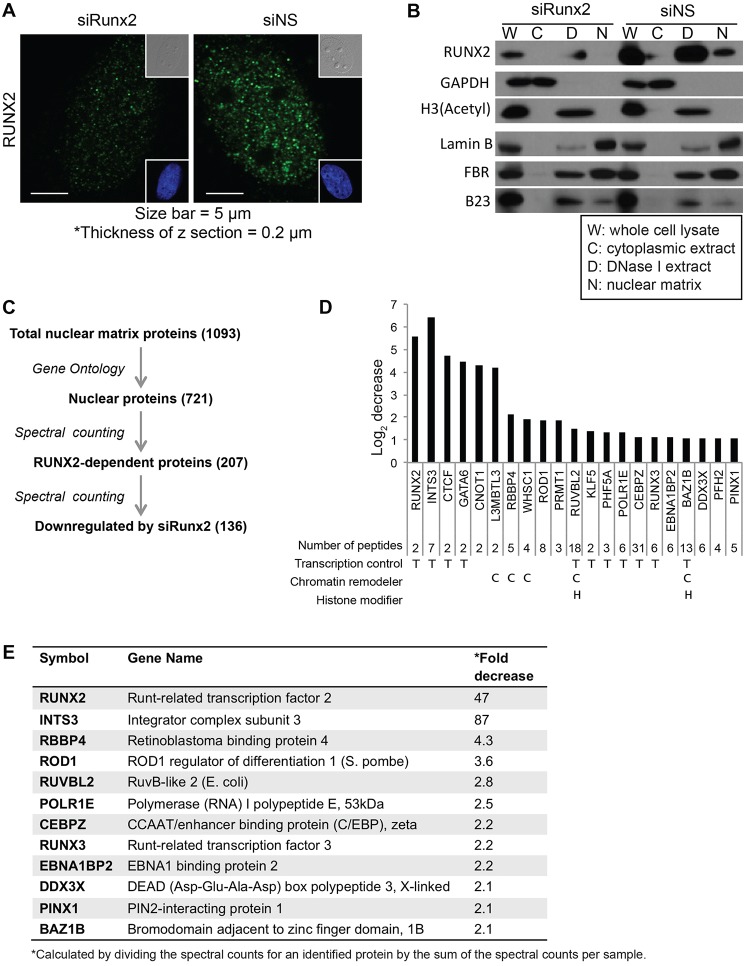
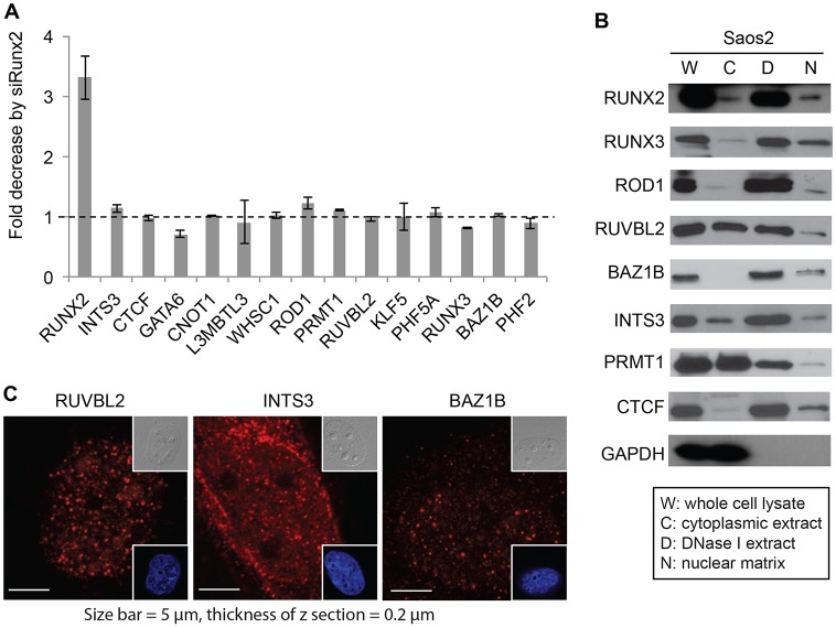
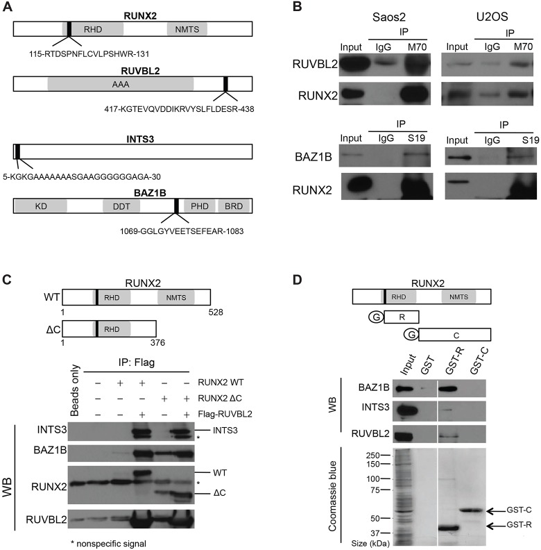
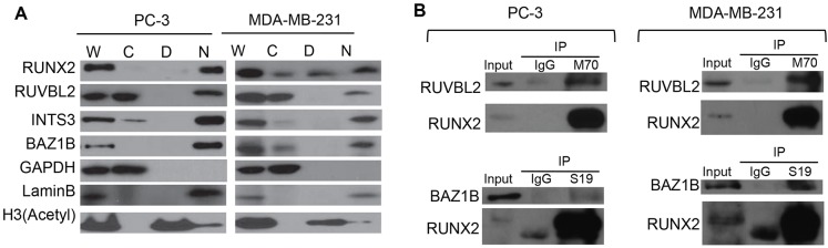
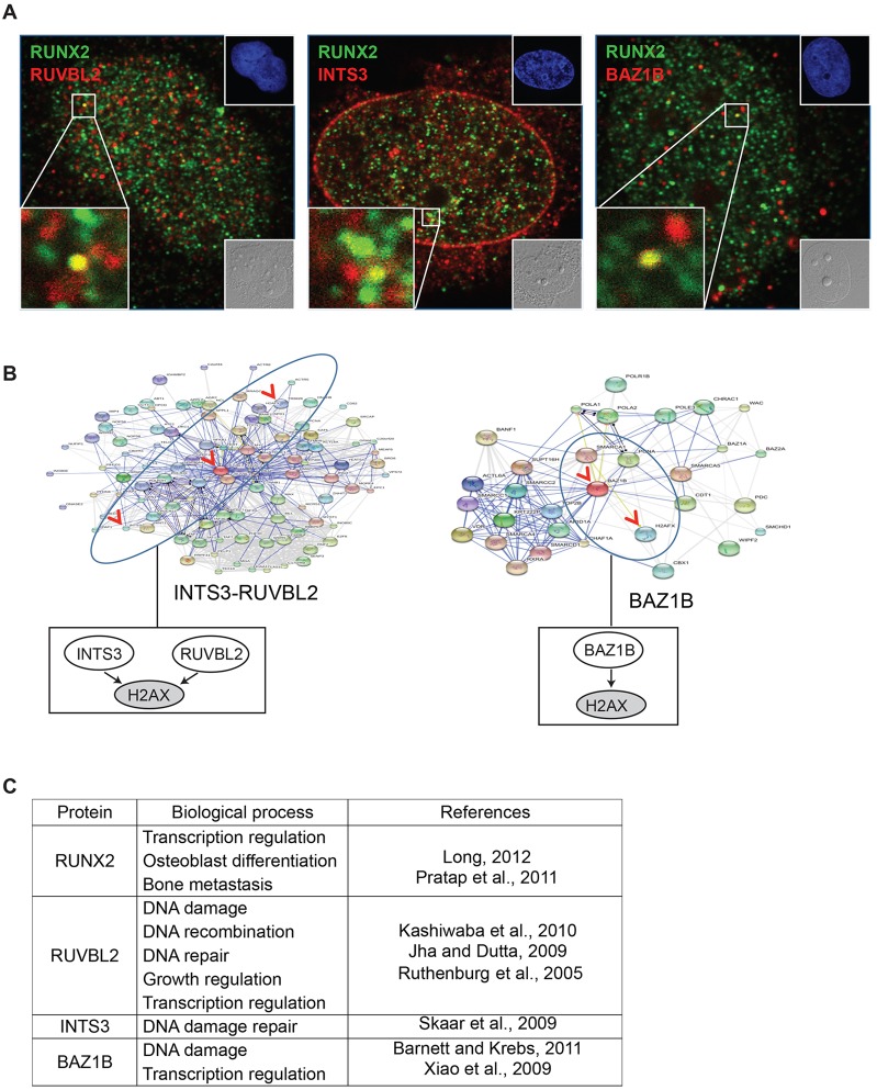
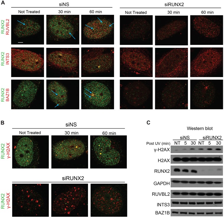
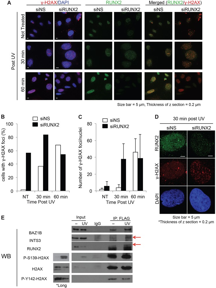
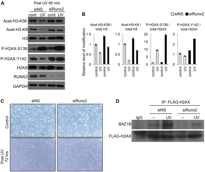
Cancer cells exhibit modifications in nuclear architecture and transcriptional control. Tumor growth and metastasis are supported by RUNX family transcriptional scaffolding proteins, which mediate the assembly of nuclear-matrix-associated gene-regulatory hubs. We used proteomic analysis to identify RUNX2-dependent protein-protein interactions associated with the nuclear matrix in bone, breast and prostate tumor cell types and found that RUNX2 interacts with three distinct proteins that respond to DNA damage - RUVBL2, INTS3 and BAZ1B. Subnuclear foci containing these proteins change in intensity or number following UV irradiation. Furthermore, RUNX2, INTS3 and BAZ1B form UV-responsive complexes with the serine-139-phosphorylated isoform of H2AX (γH2AX). UV irradiation increases the interaction of BAZ1B with γH2AX and decreases histone H3 lysine 9 acetylation levels, which mark accessible chromatin. RUNX2 depletion prevents the BAZ1B-γH2AX interaction and attenuates loss of H3K9 and H3K56 acetylation. Our data are consistent with a model in which RUNX2 forms functional complexes with BAZ1B, RUVBL2 and INTS3 to mount an integrated response to DNA damage. This proposed cytoprotective function for RUNX2 in cancer cells might clarify its expression in chemotherapy-resistant and/or metastatic tumors.

Keywords: BAZ1B; Breast; Cancer; DNA damage response; INTS3; Nuclear matrix; Osteosarcoma; Prostate; Proteomics; RUNX2.


Figures

Fig. 1.
Fig. 1.
Proteomic analysis of RUNX2-related nuclear matrix proteins in RUNX2-knockdown cells. (A) Immunofluorescence staining of Saos2 cells transfected with either nontargeting siRNA (siNS) or RUNX2-targeting siRNA (siRUNX2). Insets show differential interference contrast (DIC, upper right) and DAPI images (lower right). (B) Proteins in whole-cell lysates, cytoplasmic extracts, DNase I/salt extracts and nuclear matrix fractions, were resolved by 15% SDS-PAGE and analyzed by western blotting using primary antibodies specific for the indicated proteins. GAPDH, histone H3 and lamin B were used as markers for cytoplasmic extracts, DNase I/salt extracts and nuclear matrix fractions, respectively. FBR (fibrillarin) and B23 (nucleophosmin) are nuclear matrix components that are expected to be recovered in both DNase I/salt extracts and the nuclear matrix fraction. (C) Workflow for the proteomic screening of RUNX2-dependent nuclear matrix proteins. A total of 1093 proteins were identified by mass-spectrometry-assisted fingerprinting of the nuclear matrix fraction prepared from Saos2 cells transfected with nontargeting siRNA or RUNX2-targeting siRNA. Of these, 721 were identified as nuclear proteins by Gene Ontology (GO) analysis. Spectral counting obtained from mass spectrometry was used to compare the relative fold-change of protein levels; 136 proteins out of 207 RUNX2-dependent nuclear proteins were identified as nuclear proteins that were downregulated by RUNX2 knockdown (see supplementary material Table S1). A functional subset of proteins, including chromatin remodelers, epigenetic regulators, transcriptional controllers and most RUNX2-dependent nuclear proteins, was selected by further screening for proteins identified through ≥5 peptides (supplementary material Table S2). (D) The graph shows the log (base 2) fold-change in protein levels between nuclear matrix fractions of cells transfected with RUNX2-targeting siRNA or nontargeting siRNA determined by spectral counting obtained from mass spectrometry analysis. Results for a functional subset of proteins, including transcription regulators, chromatin remodelers and histone modifiers is shown. The number of unique peptides identified by mass spectrometry is indicated, as are the biological functions of proteins – T, transcription regulator; C, chromatin remodeler; H, histone modifier. (E) Functions and fold decreases due to RUNX2 knockdown of representative proteins from the mass spectrometry analysis of nuclear matrix proteins from siRNA-transfected Saos2 cells. The fold decrease in protein levels was calculated by dividing the spectral counts for an identified protein by the sum of the spectral counts per sample.
Fig. 2.
Fig. 2.
Quantitative analysis of RUNX2-dependent proteins. (A) The levels of mRNA for the indicated genes from Saos2 cells treated with nontargeting or RUNX2-targeting siRNA (siRUNX2) were analyzed by quantitative real time polymerase chain reaction (qRT-PCR). Two independent biological duplicates were performed and the data show the mean±s.d. (B) Western blot analysis of the subcellular localization of proteins identified by mass spectrometry confirms that all are present in the nuclear matrix fraction (‘N’) of Saos2 cells. (C) Immunofluorescent staining of RUVBL2, INTS3 and BAZ1B proteins in Saos2 cells. Insets show differential interference contrast (DIC, upper right) and DAPI images (lower right).
Fig. 3.
Fig. 3.
Interaction of RUNX2 with RUVBL2, INTS3 and BAZ1B. (A) The functional domains of RUNX2, RUVBL2, INTS3 and BAZ1B, and the location of peptide sequences identified by mass spectrometry. The peptide fragment identified by mass spectrometry is indicated as a closed bar. Functional domains in each peptide are indicated. RHD, Runt homolog domain; NMTS, nuclear matrix targeting sequences; AAA, ATPase associated with a variety of cellular activities; KD, kinase domain; DDT, DNA binding homeobox and different transcription factors; PHD, plant homeodomain; BRD, bromodomain. (B) Co-immunoprecipitation of RUNX2 with interacting proteins was analyzed by western blotting. To detect RUNX2–RUVBL2, RUNX2–INTS3 or RUNX2–BAZ1B endogenous interactions, 5 mg of whole-cell lysates from Saos2 or U2OS cells were immunoprecipitated (IP) with 5 µg of anti-RUNX2 antibodies or 5 µg of normal rabbit IgG as a negative control. Immunoprecipitation products were then analyzed by western blotting, using anti-RUVBL2, anti-INTS3 or anti-BAZ1B antibodies. Note that no clear immunoprecipitation products were seen using anti-INTS3 antibodies and the results are not shown. (C) Co-immunoprecipitation of FLAG–RUVBL2 protein with full-length RUNX2 [wildtype (WT), amino acids 1–528] or C-terminally deleted mutant (ΔC, amino acids 1–376). U2OS cells were transiently co-transfected with a FLAG–RUVBL2 expression construct and either full-length or C-terminally deleted RUNX2 construct. Whole-cell lysates were incubated with anti-FLAG M2 agarose beads (Sigma). Washed beads were subjected to SDS-PAGE and analyzed by western blotting (WB) using specific antibodies against the indicated proteins. Asterisks (*) mark bands caused by nonspecific interactions. (D) Bacterially expressed GST (‘G’), GST fused to the Runt homolog domain of RUNX2 (amino acids 107–241; GST-R) or GST fused to the C-terminus of RUNX2 (amino acids 240–528; GST-C) proteins were immobilized on glutathione beads and incubated with whole-cell lysates from Saos2 cells. After extensive washing, proteins bound to the beads were eluted in protein sample buffer and analyzed by western blotting with antibodies against the indicated proteins.
Fig. 4.
Fig. 4.
Interaction of RUNX2 with RUVBL2, INTS3 and BAZ1B in metastatic prostate PC3 or breast MDA-MB-231 cells. (A) Biochemical fractionation of PC3 and MDA-MB-231 cells. Proteins from whole-cell lysates (‘W’), cytoplasmic extracts, (‘C’), DNase I/salt extracts (‘D’) and nuclear matrix fraction (‘N’) from PC3 or MDA-MB-231 cells were resolved on 8% or 15% SDS-PAGE and analyzed by western blotting with the indicated primary antibodies. (B) Co-immunoprecipitation of RUNX2 with interacting proteins was analyzed by western blotting. To detect RUNX2–RUVBL2, RUNX2–INTS3 or RUNX2–BAZ1B endogenous interactions, 5 mg of whole-cell lysates from Saos2 or U2OS cells were immunoprecipitated (IP) with 5 µg of anti-RUNX2 antibodies or 5 µg of normal rabbit IgG as a negative control. Proteins were separated by SDS-PAGE and blotted, and immunoblots were probed with anti-RUVBL2, anti-INTS3 or anti-BAZ1B antibodies. Antibodies against GAPDH, histone H3 and lamin B were used as markers for cytoplasmic extracts, DNase I/salt extracts and the nuclear matrix fraction, respectively.
Fig. 5.
Fig. 5.
RUNX2 association with RUVBL2, INTS3 and BAZ1B in vivo. (A) Immunofluorescent staining of RUNX2 (Alexa Fluor 488, green) with RUVBL2, INTS3 or BAZ1B (Alexa Fluor 555, red) in Saos2 cells was analyzed by confocal microscopy (z-sections were 0.2 µm thickness; images were acquired using a 63x objective with oil immersion; 1.4 numerical aperture). Insets show differential interference contrast (DIC, lower right) and DAPI images (upper right). (B) The functional protein networks of RUVBL2, INTS3 and BAZ1B were analyzed using STRING (version 9.0). Boxes below the network diagrams show a simplified version of the model. (C) Reported biological functions of RUNX2, RUVBL2, INTS3 and BAZ1B, with references.
Fig. 6.
Fig. 6.
UV effect on RUNX2 and interacting proteins. Saos2 cells were transfected with nontargeting (siNS) or RUNX2-targeting siRNA (siRUNX2), and were UV irradiated (300 J/m2) or not treated with radiation (NT). (A,B) Confocal microscopy analysis of co-immunofluorescent staining. Non-irradiated (NT) cells and 30 or 60 min post-UV-irradiation cells were permeabilized, fixed and stained with RUNX2-specific antibody and RUVBL2, INTS3, BAZ1B (A) or γ-H2AX (B) antibodies. Blue arrows point to RUNX2-associated RUVBL2 or BAZ1B foci. The thickness of z-sections is 0.2 µm. Scale bar: 5 µm. (C) Western blot analysis of H2AX, γ-H2AX, RUNX2, RUVBL2, INTS3, BAZ1B and GAPDH in whole-cell lysates from non-irradiated and UV-irradiated Saos2 cells. UV-irradiated cells were harvested at 5 or 30 min after UV irradiation (300 J/m2).
Fig. 7.
Fig. 7.
RUNX2 association with γ-H2AX by UV irradiation. Saos2 cells transfected with nontargeting siRNA (siNS) or RUNX2-targeting siRNA (siRUNX2) were not treated with radiation (NT) or were UV-irradiated (300 J/m2). (A) After UV irradiation, cells were incubated for 30 or 60 min, then permeabilized, fixed and co-stained with RUNX2 (green) and γ-H2AX (red) antibodies and analyzed by confocal microscopy. (B) Cells showing γ-H2AX foci in the nucleus were counted. The graph shows the ratio of the number of cells with nuclear γ-H2AX foci to the total cells in the field; 3–14 cells were counted per field, and at least 9 fields were counted per treatment. (C) The number of γ-H2AX foci per nucleus was counted and plotted; ≥36 cells were analyzed for each data point. The data show the mean±s.d. (D) Representative confocal images from 30 min after UV irradiation are shown. (E) Saos2 cells were transfected with the FLAG–H2AX expression construct, UV irradiated or left untreated, then FLAG–H2AX protein was immunoprecipitated (IP) and analyzed by western blotting (WB). Because the intensity of ECL signal from Ser139 phosphorylation on H2AX from input lanes was low, we included longer exposures of the blot for Ser139 phosphorylation (P-S139-H2AX) and total H2AX input lanes. The upper band from the P-S139-H2AX and H2AX blots corresponds to overexpressed FLAG–H2AX and the lower band is from endogenous H2AX. *Long indicates data from a longer exposure of blots for ECL detection. Bands corresponding to INTS3 and RUNX2 are indicated with red arrows.
Fig. 8.
Fig. 8.
RUNX2-dependent histone modification in response to UV. (A) Saos2 cells transfected with nontargeting siRNA (siNS) or RUNX2-targeting siRNA (siRUNX2) were non- irradiated (cont) or were UV irradiated (300 J/m2). Then, cells were further incubated for 60 min, and K56 and K9 acetylation of histone H3 and S139 and Y142 phosphorylation of histone H2AX were analyzed by western blotting. (B) The graph shows quantification of the western blot data shown in A performed using Image J software. Each band from acetylation or phosphorylation of H3 or H2AX was measured and normalized to total H3 or H2AX levels. The image represents the analysis of at least three independent western blots. Data show the mean±s.d. (C) Saos2 cells transfected with siNS or siRUNX2 were non-irradiated (Control) or UV irradiated (UV, 300 J/m2), and images of cells were analyzed by Nikon phase-contrast microscopy (10×). (D) Immunoprecipitation (IP) analysis of UV irradiated (UV, 60 min) or non-irradiated (–) Saos2 cells co-transfected with FLAG–H2AX expression plasmid and the indicated siRNA using anti-FLAG antibody. Immunoprecipitation with normal IgG was used as a control.

References

    1. Albrethsen J., Knol J. C., Piersma S. R., Pham T. V., de Wit M., Mongera S., Carvalho B., Verheul H. M. W., Fijneman R. J. A., Meijer G. A. et al. (2010). Subnuclear proteomics in colorectal cancer: identification of proteins enriched in the nuclear matrix fraction and regulation in adenoma to carcinoma progression. Mol. Cell Proteomics 9, 988–1005 10.1074/mcp.M900546-MCP200 - DOI - PMC - PubMed
    1. Atanassov B., Gospodinov A., Stoimenov I., Mladenov E., Russev G., Tsaneva I., Anachkova B. (2005). Repair of DNA interstrand crosslinks may take place at the nuclear matrix. J. Cell. Biochem. 96, 126–136 10.1002/jcb.20518 - DOI - PubMed
    1. Barboro P., D'Arrigo C., Repaci E., Bagnasco L., Orecchia P., Carnemolla B., Patrone E., Balbi C. (2009). Proteomic analysis of the nuclear matrix in the early stages of rat liver carcinogenesis: identification of differentially expressed and MAR-binding proteins. Exp. Cell Res. 315, 226–239 10.1016/j.yexcr.2008.10.017 - DOI - PubMed
    1. Barnes L., Dumas M., Juan M., Noblesse E., Tesniere A., Schnebert S., Guillot B., Molès J. P. (2010). GammaH2AX, an accurate marker that analyzes UV genotoxic effects on human keratinocytes and on human skin. Photochem. Photobiol. 86, 933–941 10.1111/j.1751-1097.2010.00744.x - DOI - PubMed
    1. Barnett C., Krebs J. E. (2011). WSTF does it all: a multifunctional protein in transcription, repair, and replication. Biochem. Cell Biol. 89, 12–23 10.1139/O10-114 - DOI - PMC - PubMed

Publication types

MeSH terms