. 2015 Feb 17;84(7):668-79.
doi: 10.1212/WNL.0000000000001269. Epub 2015 Jan 21.

Novel mutations expand the clinical spectrum of DYNC1H1-associated spinal muscular atrophy

Affiliations

Novel mutations expand the clinical spectrum of DYNC1H1-associated spinal muscular atrophy

Mariacristina Scoto et al. Neurology. .

Abstract

Objective: To expand the clinical phenotype of autosomal dominant congenital spinal muscular atrophy with lower extremity predominance (SMA-LED) due to mutations in the dynein, cytoplasmic 1, heavy chain 1 (DYNC1H1) gene.

Methods: Patients with a phenotype suggestive of a motor, non-length-dependent neuronopathy predominantly affecting the lower limbs were identified at participating neuromuscular centers and referred for targeted sequencing of DYNC1H1.

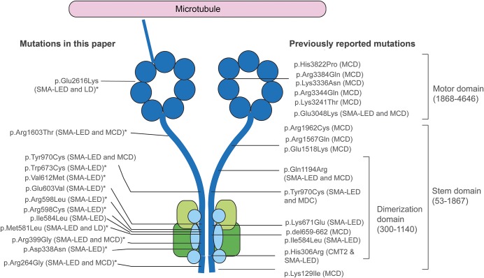
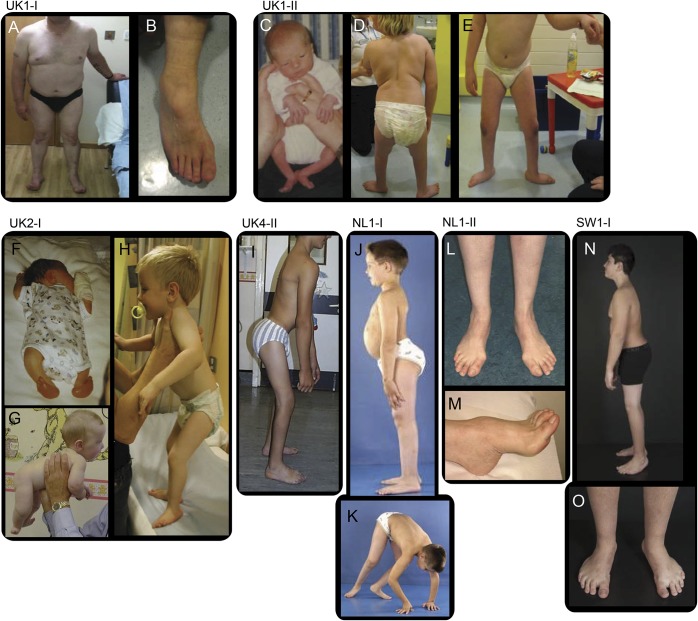
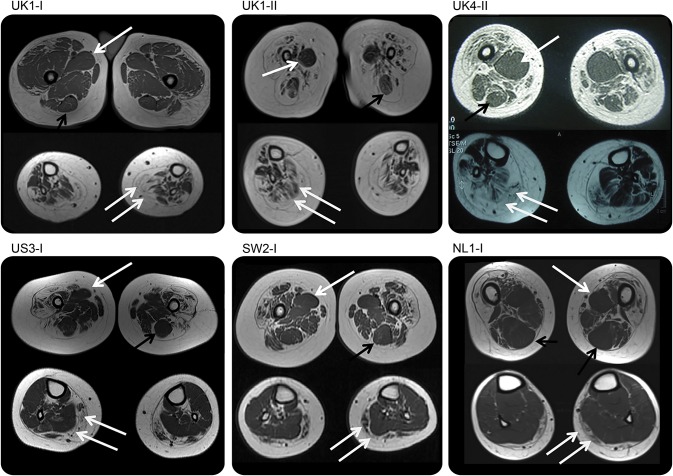
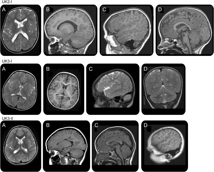
Results: We report a cohort of 30 cases of SMA-LED from 16 families, carrying mutations in the tail and motor domains of DYNC1H1, including 10 novel mutations. These patients are characterized by congenital or childhood-onset lower limb wasting and weakness frequently associated with cognitive impairment. The clinical severity is variable, ranging from generalized arthrogryposis and inability to ambulate to exclusive and mild lower limb weakness. In many individuals with cognitive impairment (9/30 had cognitive impairment) who underwent brain MRI, there was an underlying structural malformation resulting in polymicrogyric appearance. The lower limb muscle MRI shows a distinctive pattern suggestive of denervation characterized by sparing and relative hypertrophy of the adductor longus and semitendinosus muscles at the thigh level, and diffuse involvement with relative sparing of the anterior-medial muscles at the calf level. Proximal muscle histopathology did not always show classic neurogenic features.

Conclusion: Our report expands the clinical spectrum of DYNC1H1-related SMA-LED to include generalized arthrogryposis. In addition, we report that the neurogenic peripheral pathology and the CNS neuronal migration defects are often associated, reinforcing the importance of DYNC1H1 in both central and peripheral neuronal functions.


Figures

Figure 1
Figure 1. Position of mutations in DYNC1H1 relative to the structure of the dynein complex
DYNC1H1 is a large gene encoding the heavy chain 1 of the cytoplasmic dynein protein complex, a ubiquitously expressed multisubunit molecular motor involved in retrograde axonal transport, cell migration, nucleokinesis, Golgi localization, and autophagy. The dynein complex consists of 2 heavy chains (dark blue), 2 intermediate chains (dark green), 4 light intermediate chains (light green), and a number of light chains (light blue). The tail domain, located in the N-terminus, is required for heavy chain dimerization. The dynein heavy chain motor domain (C-terminus) possesses adenosine triphosphate hydrolase activity and is required for movement along microtubules. This figure shows the position of all mutations described in this report and in the published literature. The mutations identified in this study to cause both SMA-LED and MCD can be seen to span the entire length of the protein. The cluster of mutations in the dimerization domain may be explained by the selective screening for mutations in this domain. *Novel mutation. CMT2 = Charcot-Marie-Tooth disease type 2; LD = learning disability with cognitive/behavioral impairment; MCD = malformation of cortical development; SMA-LED = spinal muscular atrophy with lower extremity predominance.
Figure 2
Figure 2. Phenotypic characteristic of spinal muscular atrophy with lower extremity predominance caused by mutations in DYNC1H1
Distal wasting in lower limbs, Achilles tendon tightness, and foot deformity (A, B) in case UK1-I at the age of approximately 50 years; hypotonic at birth and proximal contractures at birth (C), distal muscle wasting, proximal weakness, stands with support at 9 years, disproportion between trunk and legs (D, E) in patient UK1-II; arthrogryposis affecting upper and lower limbs and talipes at birth (F), good neck control at 5 months (G), standing with support at 3 years of age, disproportion between trunk and legs, feet deformity (H) in case UK2-I; standing unaided, disproportion between trunk and legs, hips and knees contractures, foot deformity (I) in case UK4-II at the age of 15 years; hyperlordosis, disproportion between trunk and legs (J) and proximal weakness (K), in case NL1-I at the age of 8 years; distal wasting (L) and pes cavus (M) at the age of approximately 40 years in case NL1-II. (N, O) Case SW1-I at age 12 years, feet deformity (operated), short legs, proximal > distal weakness.
Figure 3
Figure 3. Muscle imaging of lower limbs
All cases demonstrate a distinctive feature of diffuse involvement of the quadriceps muscles and relative sparing of the adductor compartments with relative hypertrophy of the adductor longus (single white arrow) and of the semitendinosus muscle (black arrow) at the thigh level, while at the calf level there was diffuse involvement (double white arrow) with sparing of the anterior-medial muscles. Cases SW2.I and NL1.I show a milder pattern of involvement at the calf level but with relative sparing of the medial-anterior compartment.
Figure 4
Figure 4. Brain imaging
Brain MRI at the age of 3 years in case UK2.I shows a polymicrogyric pattern of frontal lobe cortex (A, B), sylvian fissure extending to the parietal lobe especially on the right side (C), and thin corpus callosum (D). Brain MRI at the age of 2 years in case UK3.I shows immature white matter, polymicrogyria-like pattern of sylvian and frontal cortex (A, B), posterior extension of sylvian fissure (C), and mild cerebellar hypoplasia (D). Brain MRI at the age of 4 years in case UK3.II shows a polymicrogyric pattern of the right frontal lobe (A, B), thin corpus callosum (C), and gyral overconvolution with posterior extension of the right sylvian fissure (D). All these subjects had underdeveloped white matter with thinning of the corpus callosum.

References

    1. Fleury P, Hageman G. A dominantly inherited lower motor neuron disorder presenting at birth with associated arthrogryposis. J Neurol Neurosurg Psychiatry 1985;48:1037–1048. - PMC - PubMed
    1. Frijns CJ, Van Deutekom J, Frants RR, Jennekens FG. Dominant congenital benign spinal muscular atrophy. Muscle Nerve 1994;17:192–197. - PubMed
    1. Van der Vleuten AJ, van Ravenswaaij-Arts CM, Frijns CJ, et al. Localisation of the gene for a dominant congenital spinal muscular atrophy predominantly affecting the lower limbs to chromosome 12q23-q24. Eur J Hum Genet 1998;6:376–382. - PubMed
    1. Mercuri E, Messina S, Kinali M, et al. Congenital form of spinal muscular atrophy predominantly affecting the lower limbs: a clinical and muscle MRI study. Neuromuscul Disord 2004;14:125–129. - PubMed
    1. Deng HX, Klein CJ, Yan J, et al. Scapuloperoneal spinal muscular atrophy and CMT2C are allelic disorders caused by alterations in TRPV4. Nat Genet 2010;42:165–169. - PMC - PubMed

Publication types