. 2015 Apr;89(7):3910-21.
doi: 10.1128/JVI.00089-15. Epub 2015 Jan 21.

Modulation of a pore in the capsid of JC polyomavirus reduces infectivity and prevents exposure of the minor capsid proteins

Affiliations

Modulation of a pore in the capsid of JC polyomavirus reduces infectivity and prevents exposure of the minor capsid proteins

Christian D S Nelson et al. J Virol. 2015 Apr.

Abstract

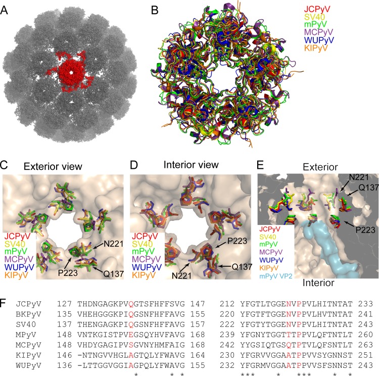
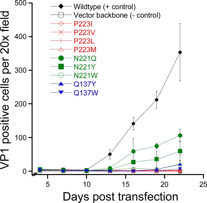
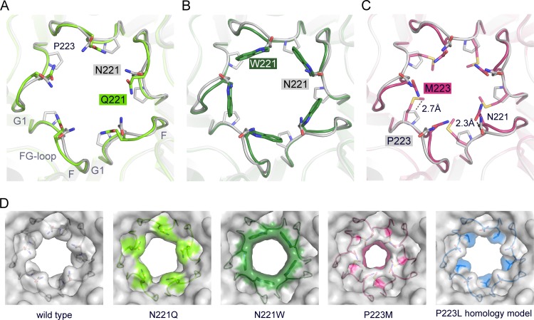
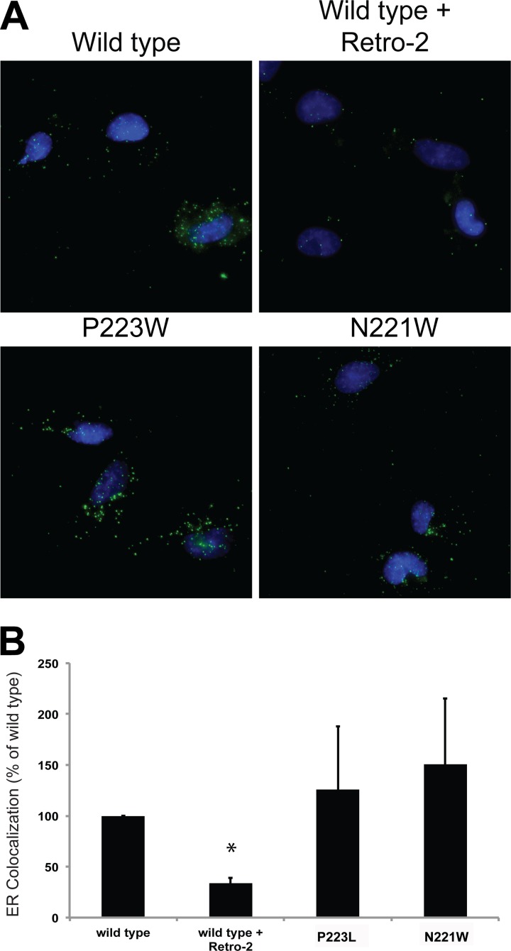
JC polyomavirus (JCPyV) infection of immunocompromised individuals results in the fatal demyelinating disease progressive multifocal leukoencephalopathy (PML). The viral capsid of JCPyV is composed primarily of the major capsid protein virus protein 1 (VP1), and pentameric arrangement of VP1 monomers results in the formation of a pore at the 5-fold axis of symmetry. While the presence of this pore is conserved among polyomaviruses, its functional role in infection or assembly is unknown. Here, we investigate the role of the 5-fold pore in assembly and infection of JCPyV by generating a panel of mutant viruses containing amino acid substitutions of the residues lining this pore. Multicycle growth assays demonstrated that the fitness of all mutants was reduced compared to that of the wild-type virus. Bacterial expression of VP1 pentamers containing substitutions to residues lining the 5-fold pore did not affect pentamer assembly or prevent association with the VP2 minor capsid protein. The X-ray crystal structures of selected pore mutants contained subtle changes to the 5-fold pore, and no other changes to VP1 were observed. Pore mutant pseudoviruses were not deficient in assembly, packaging of the minor capsid proteins, or binding to cells or in transport to the host cell endoplasmic reticulum. Instead, these mutant viruses were unable to expose VP2 upon arrival to the endoplasmic reticulum, a step that is critical for infection. This study demonstrated that the 5-fold pore is an important structural feature of JCPyV and that minor modifications to this structure have significant impacts on infectious entry.

Importance: JCPyV is an important human pathogen that causes a severe neurological disease in immunocompromised individuals. While the high-resolution X-ray structure of the major capsid protein of JCPyV has been solved, the importance of a major structural feature of the capsid, the 5-fold pore, remains poorly understood. This pore is conserved across polyomaviruses and suggests either that these viruses have limited structural plasticity in this region or that this pore is important in infection or assembly. Using a structure-guided mutational approach, we showed that modulation of this pore severely inhibits JCPyV infection. These mutants do not appear deficient in assembly or early steps in infectious entry and are instead reduced in their ability to expose a minor capsid protein in the host cell endoplasmic reticulum. Our work demonstrates that the 5-fold pore is an important structural feature for JCPyV.


Figures

FIG 1
FIG 1
The VP1 5-fold pore is conserved across polyomaviruses. (A) The X-ray crystal structure of SV40 (PDB: 1SVA), represented by a ball and stick rendering. A single VP1 pentamer, containing the 5-fold pore, is colored red. (B) Alignment of the VP1 structures from six divergent polyomaviruses: JCPyV (PDB: 3NXG), SV40 (PDB: 3BWQ), MPyV (PDB: 1VPN), Merkel cell polyomavirus (MCPyV) (PDB: 4FMG), unassembled KI polyomavirus (KIPyV) (PDB: 3S7V), and unassembled Washington University polyomavirus (WUPyV) (PDB: 3S7X). (C) Exterior view of the 5-fold pore, illustrating that residues Q137, N221, and P223 line the pore at its narrowest point. (D) Interior view of the 5-fold pore. (E) Cross-sectional view of the interior axial cavity, showing the relationship of the indicated residues to VP2 in the MPyV VP1-VP2 complex structure (PDB: 1CN3). (F) Alignment of VP1 amino acid residues from seven different polyomaviruses. Red letters indicate residues that were substituted in this study; asterisks indicate residues that are conserved in all seven polyomaviruses.
FIG 2
FIG 2
Amino acid substitutions to residues lining the 5-fold pore reduce infectivity. Infectious clones of wild-type JCPyV or pore mutants were transfected into SVGA cells, and viral spread was monitored over 22 days. Data represent the means of the results of three independent experiments, and the error bars indicate standard deviations.
FIG 3
FIG 3
VP1 pentamers with altered 5-fold pores are not deficient in assembly. (A) A truncated VP1 containing a hexahistidine tag was expressed in bacteria and purified in order to assess the ability of pore mutants to form pentamers. (B) Truncated VP1 containing a hexahistidine tag and full-length VP2 lacking affinity tags were expressed in bacteria and purified by IMAC and SEC. Western blotting was used to determine whether the indicated pore mutants were able to copurify VP2. (C) Differential scanning fluorometry of VP1 pore mutants. The indicated VP1 was mixed with Sypro Orange, and fluorescence was monitored over the indicated range of temperatures. Max, maximum.
FIG 4
FIG 4
Crystal structures of VP1 pentamers with N221Q, N221W, and P223M pore mutations. (A to C) Structural superposition of pore mutants N221Q (A), N221W (B), and P221M (C) with wild-type Mad-1 VP1. VP1 pentamers are shown in cartoon representations with side chains of key amino acid residues highlighted in stick representations and colored according to atom type (oxygens are shown in red, nitrogens in blue, and carbons in the colors assigned for the respective mutants). Wild-type VP1 is shown in gray. The accessible diameter at the narrowest constriction of the 5-fold pore was determined (28). The diameter was calculated to be 8.2 Å for wild-type VP1 and to be 7.1 Å and 8.6 Å for N221Q and N222W, respectively. The P223M mutation results in a constrained accessible pore diameter of 5.2 Å. (D) Surface representations of the 5-fold pore. Views are equivalent in all cases, and mutated amino acids are colored on the surface. A VP1 homology model for P223L was generated, and the most favorable rotamer conformation of Leu is shown in a surface representation using sticks.
FIG 5
FIG 5
PSVs containing pore mutations are not deficient in assembly or binding to cells. (A) Wild-type or mutant PSVs were produced, and assembly was assessed by TEM. Scale bars denote 50 nm. (B) Western blotting was performed to demonstrate that all of the mutants were able to package VP2 and VP3. (C) Pore mutant PSVs are deficient in viral entry. SVGA cells were inoculated with equivalent numbers of genome copies of the indicated PSV, and luciferase expression was determined 72 hpi. (D) Pore mutant PSVs are not deficient in binding to cells. SVGA cells were inoculated with equivalent numbers of genome copies of the indicated PSV, and binding was determined by flow cytometry.
FIG 6
FIG 6
Pore mutant PSVs are transported to the ER as efficiently as wild-type PSVs. (A) SVGA cells were inoculated with equivalent numbers of genome copies of each PSV, and ER colocalization was assessed at 8 hpi by proximity ligation assays. (B) Quantification of the data presented in panel A. Results represent the means of the results of three independent experiments, and error bars denote standard deviations. *, P < 0.05.
FIG 7
FIG 7
Pore mutants are deficient in exposing VP2 in the ER. (A) SVGA cells were inoculated with equivalent numbers of genome copies of each PSV, and VP2 exposure was assessed at 10 hpi by immunostaining for VP2. (B) Quantification of the data presented in panel A. Results represent the means of the results of three independent experiments, and error bars denote standard deviations. *, P < 0.05.

References

    1. Kean JM, Rao S, Wang M, Garcea RL. 2009. Seroepidemiology of human polyomaviruses. PLoS Pathog 5:e1000363. doi:10.1371/journal.ppat.1000363. - DOI - PMC - PubMed
    1. Ferenczy MW, Marshall LJ, Nelson CD, Atwood WJ, Nath A, Khalili K, Major EO. 2012. Molecular biology, epidemiology, and pathogenesis of progressive multifocal leukoencephalopathy, the JC virus-induced demyelinating disease of the human brain. Clin Microbiol Rev 25:471–506. doi:10.1128/CMR.05031-11. - DOI - PMC - PubMed
    1. Zurhein G, Chou SM. 1965. Particles resembling papova viruses in human cerebral demyelinating disease. Science 148:1477–1479. - PubMed
    1. Knipe DM, Howley PM, Griffin DE, Lamb RA, Martin MA, Roizman B, Straus SE (ed). 2007. Fields virology, 5th ed Lippincott Williams & Wilkins, Philadelphia, PA.
    1. Liddington RC, Yan Y, Moulai J, Sahli R, Benjamin TL, Harrison SC. 1991. Structure of simian virus 40 at 3.8-A resolution. Nature 354:278–284. doi:10.1038/354278a0. - DOI - PubMed

Publication types