. 2015 Apr;66(7):1851-63.
doi: 10.1093/jxb/eru522. Epub 2015 Jan 21.

Plant membrane assays with cytokinin receptors underpin the unique role of free cytokinin bases as biologically active ligands

Affiliations

Plant membrane assays with cytokinin receptors underpin the unique role of free cytokinin bases as biologically active ligands

Sergey N Lomin et al. J Exp Bot. 2015 Apr.

Abstract

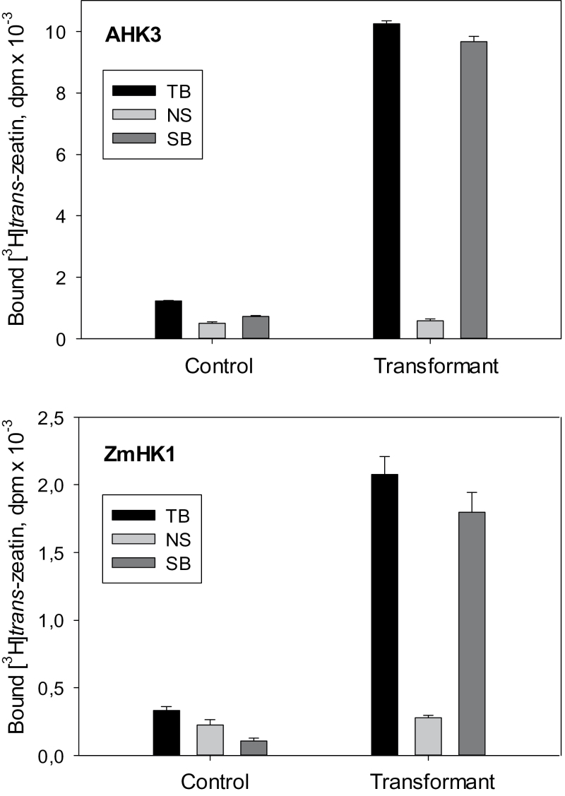
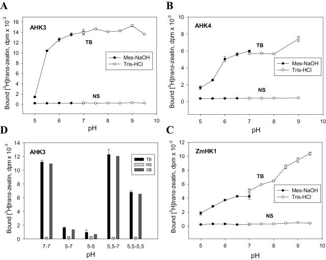
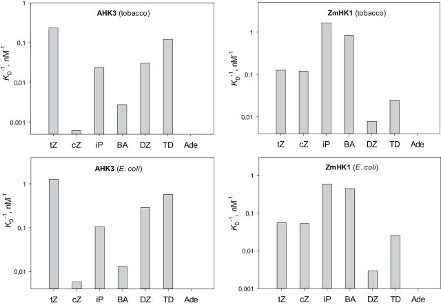
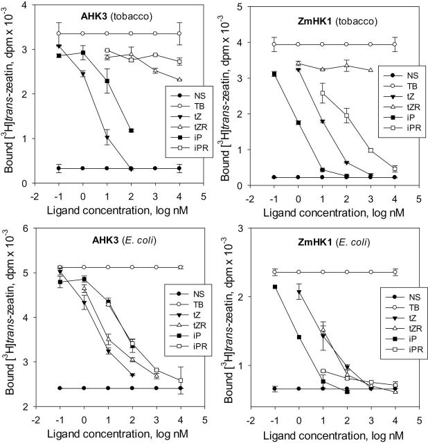
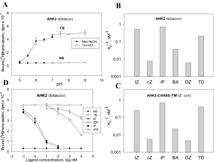
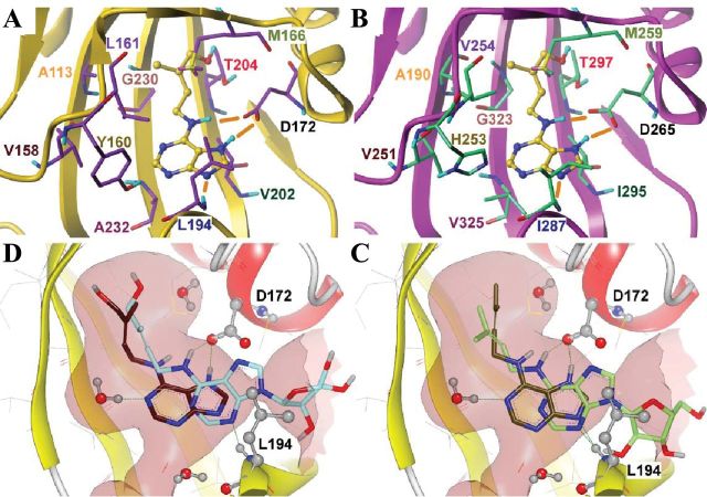
Cytokinin receptors play a key role in cytokinin-dependent processes regulating plant growth, development, and adaptation; therefore, the functional properties of these receptors are of great importance. Previously the properties of cytokinin receptors were investigated in heterologous assay systems using unicellular microorganisms, mainly bacteria, expressing receptor proteins. However, within microorganisms receptors reside in an alien environment that might distort the receptor properties. Therefore, a new assay system has been developed allowing studies of individual receptors within plant membranes (i.e. closer to their natural environment). The main ligand-binding characteristics of receptors from Arabidopsis [AHK2, AHK3, and AHK4] and maize (ZmHK1) were refined in this new system, and the properties of full-length Arabidopsis receptor AHK2 were characterized for the first time. Ligand specificity profiles of receptors towards cytokinin bases were comparable with the profiles retrieved in bacterial assay systems. In contrast, cytokinin-9-ribosides displayed a strongly reduced affinity for receptors in the plant assay system, indicating that ribosides as the common transport form of cytokinins have no or very weak cytokinin activity. This underpins the central role of free bases as the sole biologically active cytokinin compounds. According to molecular modelling and docking studies, N (9)-ribosylation alters the bonding pattern in cytokinin-receptor interaction and prevents β6-β7 loop movement important for tight hormone binding. A common feature of all receptors was a greatly reduced ligand binding at low (5.0-5.5) pH. The particularly high sensitivity of ZmHK1 to pH changes leads to the suggestion that some cytokinin receptors may play an additional role as pH sensors in the lumen of the endoplasmic reticulum.

Keywords: Arabidopsis thaliana; Zea mays.; cytokinin; cytokinin receptor; ligand specificity; pH sensor; plant assay system; receptor binding assay.


Figures

Fig. 1.
Fig. 1.
Increase in trans-zeatin-specific binding to plant membranes upon transient expression of cytokinin receptors in tobacco leaves. Microsomes from control and transiently transformed (‘Transformant’) plants are shown. TB, NS, and SB indicate total, non-specific, and specific binding, respectively.
Fig. 2.
Fig. 2.
pH dependence and reversibility of cytokinin–receptor interaction. Binding of trans-zeatin to AHK3 (A), AHK4 (B), and ZmHK1 (C) in the plant assay system at different pH values. Reversibility of ligand binding to AHK3 following a pH shift is shown in (D); initial and final pH values are indicated by numbers below the abscissa. TB, NS, and SB indicate total, non-specific, and specific binding, respectively.
Fig. 3.
Fig. 3.
Affinity constants of various cytokinin bases for AHK3 and ZmHK1 receptors in different assay systems. The affinity constants were measured in a plant (tobacco microsomes) or bacterial (E. coli spheroplasts) assay system as indicated. The ordinate (log scale) shows the apparent K A=K D –1 which positively correlates with the ligand affinity of the receptor. tZ, trans-zeatin; cZ, cis-zeatin; iP, isopentenyladenine; BA, N 6-benzyladenine; DZ, dihydrozeatin; TD, thidiazuron; Ade, adenine.
Fig. 4.
Fig. 4.
Interaction of cytokinin ribosides and bases with receptors AHK3 and ZmHK1. Receptor–ligand interaction was measured in a plant (tobacco microsomes) or bacterial (E. coli spheroplasts) assay system as indicated. tZ, trans-zeatin; tZR, trans-zeatin riboside; iP, isopentenyladenine; iPR, isopentenyladenosine; NS and TB indicate non-specific and total binding, respectively.
Fig. 5.
Fig. 5.
Ligand binding properties of full-length receptor AHK2 assayed in the plant assay system. Typical pH dependence of ligand binding is demonstrated in (A). Affinity constants for various cytokinin bases (B) are shown in comparison with constants of the same ligands for the AHK2 sensor module (AHK2–CHASE–TM) (C) studied in a bacterial (E. coli) assay system (Stolz et al., 2011). In contrast to free bases, cytokinin ribosides hardly bind to the receptor (D). For abbreviations, see legends to Figs 3 and 4.
Fig. 6.
Fig. 6.
Molecular modelling of cytokinin–receptor interaction. (A, B) Models for cytokinin (tZ) binding, predicted by molecular docking, to receptors ZmHK1 (A) and AHK3 (B). The ligand is rendered in a ball-and-stick representation with yellow carbon atoms; residues of the receptors forming the hormone-binding site surface are shown as sticks; the backbone is shown in cartoon representation. Residue numbers are coloured to indicate similar positions in the different receptors. Nitrogen atoms are coloured in blue, oxygen in red, hydrogen in cyan, and sulphur in light yellow. Hydrogen bonds are shown as orange sticks. Non-polar hydrogen atoms and water molecules are omitted for clarity. Both A and B have been produced using SybylX2.1. (C, D) Comparison of predicted binding modes of cytokinin bases and cytokinin ribosides, iP versus iPR (C) and tZ versus tZR (D), to ZmHK1. Hydrogen bonds are shown as green dashed lines. Important hydrogen-bonding residues and water molecules are shown in ball-and-stick representation. Conserved bond-forming residues (D172 and L194) in the binding cavity are indicated. The surface of the binding cavity is coloured rose. Both C and D have been produced using VIDA 4.2.1.

References

    1. Alvarez S, Marsh EL, Schroeder SG, Schachtman DP. 2008. Metabolomic and proteomic changes in the xylem sap of maize under drought. Plant, Cell and Environment 31, 325–340. - PubMed
    1. Berman HM, Westbrook J, Feng Z, Gilliland G, Bhat TN, Weissig H, Shindyalov IN, Bourne PE. 2000. The Protein Data Bank. Nucleic Acids Research 28, 235–242. - PMC - PubMed
    1. Bhargava A, Clabaugh I, To JP, Maxwell BB, Chiang Y-H, Schaller GE, Loraine A, Kieber JJ. 2013. Identification of cytokinin-responsive genes using microarray meta-analysis and RNA-seq in Arabidopsis. Plant Physiology 162, 272–294. - PMC - PubMed
    1. Bibikova TN, Jacob T, Dahse I, Gilroy S. 1998. Localized changes in apoplastic and cytoplasmic pH are associated with root hair development in Arabidopsis thaliana . Development 125, 2925–2934. - PubMed
    1. Caesar K, Thamm AM, Witthöft J, Elgass K, Huppenberger P, Grefen C, Horak J, Harter K. 2011. Evidence for the localization of the Arabidopsis cytokinin receptors AHK3 and AHK4 in the endoplasmic reticulum. Journal of Experimental Botany 62, 5571–5580. - PMC - PubMed

Publication types

MeSH terms