. 2015 Feb 15;194(4):1763-75.
doi: 10.4049/jimmunol.1401624. Epub 2015 Jan 21.

Neutrophil IL-1β processing induced by pneumolysin is mediated by the NLRP3/ASC inflammasome and caspase-1 activation and is dependent on K+ efflux

Affiliations

Neutrophil IL-1β processing induced by pneumolysin is mediated by the NLRP3/ASC inflammasome and caspase-1 activation and is dependent on K+ efflux

Mausita Karmakar et al. J Immunol. .

Abstract

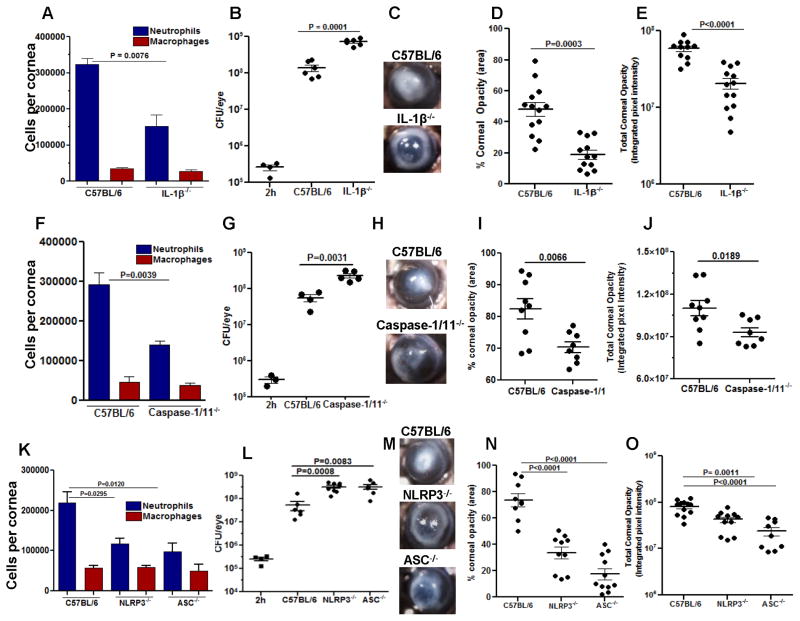
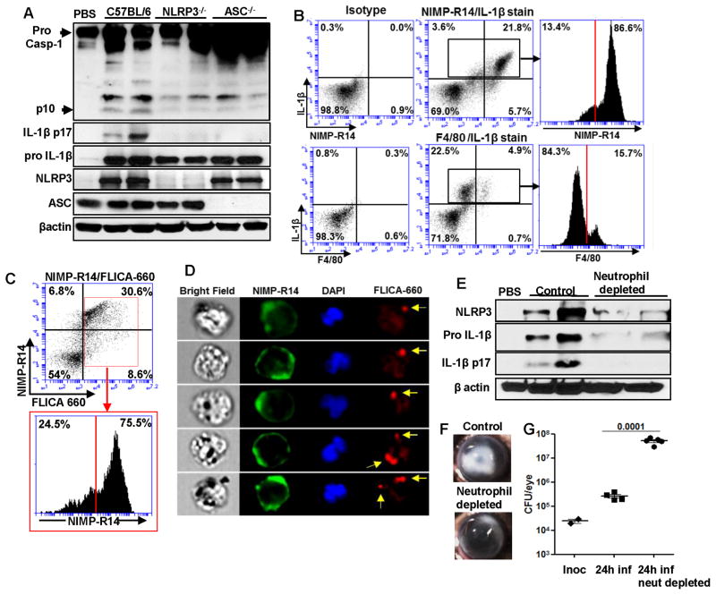
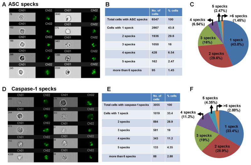
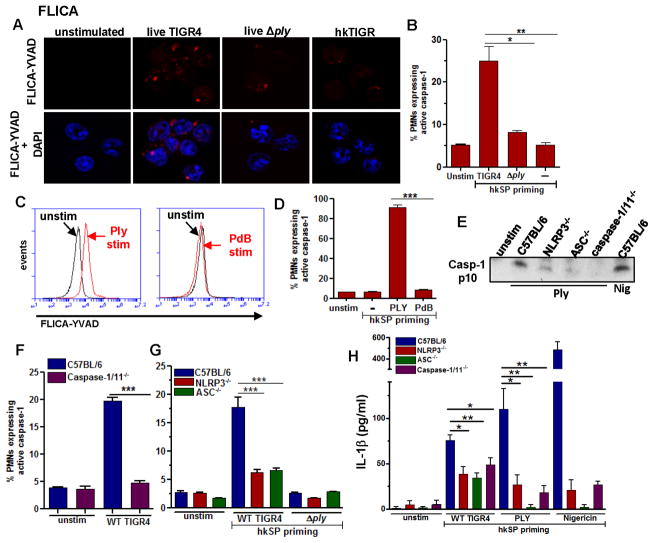
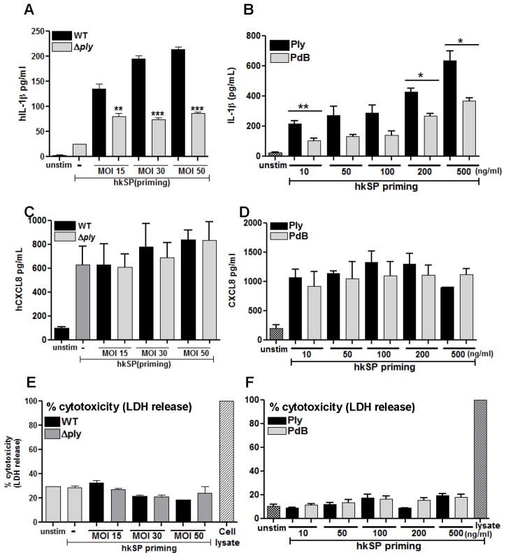
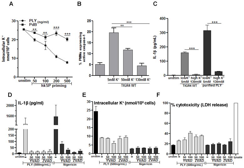
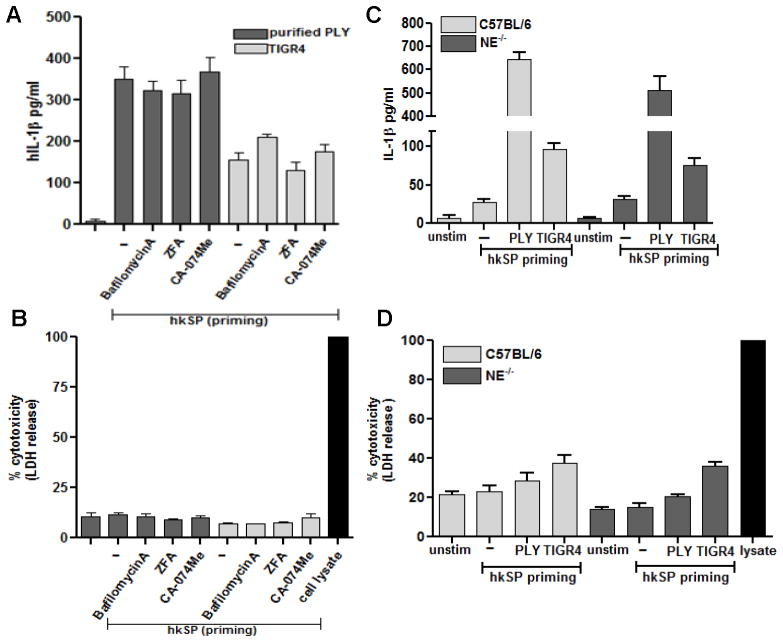
Although neutrophils are the most abundant cells in acute infection and inflammation, relatively little attention has been paid to their role in inflammasome formation and IL-1β processing. In the present study, we investigated the mechanism by which neutrophils process IL-1β in response to Streptococcus pneumoniae. Using a murine model of S. pneumoniae corneal infection, we demonstrated a requirement for IL-1β in bacterial clearance, and we showed that Nod-like receptor protein 3 (NLRP3), apoptosis-associated speck-like protein containing a caspase activation and recruitment domain (ASC), and caspase-1 are essential for IL-1β production and bacterial killing in the cornea. Neutrophils in infected corneas had multiple specks with enzymatically active caspase-1 (YVAD-FLICA 660), and bone marrow neutrophils stimulated with heat-killed S. pneumoniae (signal 1) and pneumolysin (signal 2) exhibited multiple specks when stained for NLRP3, ASC, or Caspase-1. High-molecular mass ASC complexes were also detected, consistent with oligomer formation. Pneumolysin induced K(+) efflux in neutrophils, and blocking K(+) efflux inhibited caspase-1 activation and IL-1β processing; however, neutrophils did not undergo pyroptosis, indicating that K(+) efflux and IL-1β processing is not a consequence of cell death. There was also no role for lysosomal destabilization or neutrophil elastase in pneumolysin-mediated IL-1β processing in neutrophils. Taken together, these findings demonstrate an essential role for neutrophil-derived IL-1β in S. pneumoniae infection, and they elucidate the role of the NLRP3 inflammasome in cleavage and secretion of IL-1β in neutrophils. Given the ubiquitous presence of neutrophils in acute bacterial and fungal infections, these findings will have implications for other microbial diseases.


Figures

Figure 1
Figure 1. The role of IL-1β NLRP3, ASC and Caspase-1 in S. pneumoniae keratitis
C57BL/6, IL-1β−/−, Caspase-1/11−/− NLRP3−/− and ASC−/− mice were infected in the corneal stroma with S. pneumoniae TIGR4. A, F, K: Total NIMP-R14+ neutrophils and F4/80+ macrophages in infected corneas were quantified by flow cytometry. B, G, L: Total CFU in eyes of infected mice after 2 h (inoculum) and after 24 h. C, H, M: Representative bright field images of corneas 24h post-infection; D, I, N: Percent, and E, J, O total corneal opacification calculated by Metamorph analysis as shown in Supplemental Figure S1. A, F, K: mean ±SD of 5 samples per group. For CFU and corneal opacification graphs, each data point represents a single cornea. Results are representative of three independent experiments with at least five mice per group.
Figure 2
Figure 2. NLRP3 and ASC are required for caspase-1 activation and IL-1β processing by neutrophils during S. pneumoniae corneal infection
C57BL/6, NLRP3−/− and ASC−/− mice were infected in the corneal stroma with S. pneumoniae TIGR4 A. 24h post infection, corneas were excised and homogenized in lysis buffer and western blot was performed for mature forms of Caspase 1 (p10) and IL-1β (p17) are indicated by arrow heads. B. Flow cytometric analysis of intracellular pro IL-1β in NIMP-R14+ and F4/80+ cells from infected C57BL/6 corneas after 24h. Gates were determined based on an isotype control. C–E. C57BL/6 mice were infected with S. pneumoniae and 24h later corneas were excised and digested in collagenase. Total corneal cells were incubated with NIMP-R14 and FLICA-660-YVAD to detect active caspase-1 producing neutrophils by flow cytometry (C). NIMP-R14 (green) and FLICA-660-YVAD (red) cells were detected by multi spectral imaging flow cytometry (MIFC) (D), original magnification is x60. EG. Neutrophils were depleted in C57BL/6 mice by i.p injection of NIMP-R14 antibody 24h prior to infection with S. pneumoniae TIGR4. 24h post infection, corneas were excised and homogenized in lysis buffer, and western blot was performed for NLRP3, pro- and mature IL-1β (E). Representative brightfield images show corneal opacification (F), (original magnification is x20). G. CFU in infected eyes was quantified 24h after infection. Data points represent individual eyes. These experiments were repeated twice with similar results.
Figure 3
Figure 3. NLRP3/ASC expression and oligomerization in neutrophils
A. NLRP3 protein expression by western blot of C57BL/6 bone marrow neutrophils after incubation with killed S. pneumoniae (hkSP) for 3h. B. NLRP3 protein expression in bone marrow neutrophils from C57BL/6 and TLR2−/− mice after stimulation with hkSP for 3h C, D. NLRP3 expression in C57BL/6 neutrophils incubated with either NFκB inhibitor JSH-23 (C) or the JNK/AP-1 inhibitor SP600125 (D) at the indicated concentrations (μM) for 30mins prior to stimulation with hkSP for 3h. Western blot was performed for NLRP3, ASC, p-JNK, total JNK and β-actin. E. NLRP3 expression in C57BL/6 bone marrow neutrophils after priming with hkSP for 3h followed by stimulation with WT TIGR4 (MOI 50), Ply or a non-hemolytic Ply (PdB) for 1.5h. Red is NLRP3 and blue is DAPI; scale bar is 10μm. F. ASC oligomers detected by western blot from C57BL/6 bone marrow neutrophils after priming with hkSP for 3h followed by stimulation with Ply (500ng/ml) for 2h or with Nigericin (10μM) for 45mins. Western blot was performed from the pellet and lysate (Lys) using anti-ASC antibody. Data are representative of two repeat experiments.
Figure 4
Figure 4. Multiple Caspase-1 and ASC specks in neutrophils
Bone marrow neutrophils were isolated from C57BL/6 mice and primed for 2hr with hkSP and stimulated with purified Ply for 2hr. Cells were then stained with antibody to ASC or with FAM-YVAD-FMK (for caspase-1). A. Representative bright field and fluorescent images (60x) of neutrophils showing ASC specks. B, C. 6547 neutrophils were analyzed by Amnis Imagestream X and spot counting was performed using IDEAS software, and Tables and pie charts were generated showing percentage and total number of neutrophils with one or multiple ASC specks. D. Representative bright field and fluorescent images (60x) of neutrophils showing caspase-1 speck (FAM-YVAD-FMK staining). E, F. Percentage and total number of neutrophils with one or multiple caspase-1specks (3055 cells were analyzed).
Figure 5
Figure 5. Active caspase-1 induction in murine neutrophils is dependent on NLRP3 and ASC, and requires active pneumolysin
C57BL/6 neutrophils were incubated 3h with heat killed S. pneumoniae (hkSP) (Signal 1), followed by 1.5 h stimulation with either live S. pneumoniae TIGR4 (WT), or the ply mutant (Signal 2), and active caspase-1 was detected using FLICA 660-YVAD. A. Representative neutrophils showing FLICA660-YVAD positive cells (red specks) (original magnification is x100). B. Percent FLICA660-YVAD positive neutrophils. C, D. Representative flow cytometry profiles (C) and percent (D) FLICA660-YVAD positive C57BL/6 neutrophils primed with hkSP and further incubated with Ply or the non-hemolytic Ply (PdB) for 1.5h. E. Mature p10 form of caspase-1 from supernatant of C57BL/6, NLRP3−/−, ASC−/− and Caspase-1/11−/− neutrophils after incubation with Ply (500ng/ml) for 2h or with nigericin as a positive control. F, G. Percent FLICA660-YVAD positive neutrophils from C57BL/6 and caspase-1/11−/− mice (F) and from NLRP3−/− and ASC−/− mice (G) following hkSP priming for 3h and stimulation with live WT or ply mutants for 1.5h. H. IL-1β secretion after hkSP priming and stimulation with WT TIGR4, purified Ply (500 ng/ml) for 2h or nigericin for 45mins. Data are representative of three repeat experiments.
Figure 6
Figure 6. Pneumolysin dependent caspase-1 activation and IL-1β secretion by human neutrophils
Human peripheral blood neutrophils were primed with hkSP for 3h followed by 2h stimulation with WT or Δply (A, C, E) or with Ply and PdB for 2h (B, D, F) and IL-1β (A, B) and CXCL-8 (C, D) in the supernatant was quantified by ELISA. Cell death was assayed by LDH release compared with lysed cells (E, F). Histograms are mean ±SD of atleast 5 samples per group and data shown are representative of three independent experiments. ** p< 0.001, *** p< 0.0001. Nig: Nigericin incubation was for 45mins.
Figure 7
Figure 7. Pneumolysin-induced caspase-1 activation and IL-1β secretion by neutrophils requires K+ efflux
A. Human peripheral blood neutrophils were hkSP primed with hkSP for 3h and stimulated with Ply or PdB for 2h. Total cell contents were extracted using 10% HNO3, and the cell associated K+ concentration was quantified by atomic absorbance spectroscopy. B. FLICA660-YVAD positive murine neutrophils quantified by flow cytometry after treatment with live TIGR4 in presence of increasing concentration of extracellular KCl.; C. hkSP primed neutrophils were stimulated 2h with either Ply (500ng/ml) or WT TIGR4 (50:1) in the presence of 5 mM or 130 mM KCl, and IL-1β secretion was measured. D–F. Neutrophils were primed and stimulated with Ply (500ng/ml), or were stimulated with Nigericin in the presence of the pan-caspase inhibitor ZVAD or the caspase-1 inhibitor YVAD at the indicated concentration (μM), and examined for IL-1β secretion after 2hr (D), intracellular K+ (E) and LDH (F). Histograms are mean ±SD of three samples per treatment condition, and are representative of two similar experiments with neutrophils from different donors. ** p< 0.001, *** p< 0.0001.
Figure 8
Figure 8. Pneumolysin induced IL-1β secretion by neutrophils does not require lysosomal disruption or serine protease activity
A, B. Primed human neutrophils were pretreated with bafilomycinA (200nM), CA-074-Me (100μM) or ZFA (50 μM) 30mins prior to stimulation with TIGR4 (50:1) or Ply (500ng/ml) for 2h and IL-1β (A) and LDH (B) release were quantified from the supernatant. C, D. Primed murine neutrophils from C57BL/6 and neutrophil elastase (NE)−/− were stimulated with TIGR4 (50:1) or Ply (500ng/ml) for 2h and IL-1β (C) and LDH (D) release were quantified. Histograms are mean ±SD of samples of 5 samples per treatment conditions, and are representative of three independent experiments with similar observations.

References

    1. Dinarello CA. Immunological and inflammatory functions of the interleukin-1 family. Annu Rev Immunol. 2009;27:519–550. - PubMed
    1. Dinarello CA, Simon A, van der Meer JW. Treating inflammation by blocking interleukin-1 in a broad spectrum of diseases. Nature reviews. Drug discovery. 2012;11:633–652. - PMC - PubMed
    1. Latz E, Xiao TS, Stutz A. Activation and regulation of the inflammasomes. Nat Rev Immunol. 2013;13:397–411. - PMC - PubMed
    1. Gross O, Thomas CJ, Guarda G, Tschopp J. The inflammasome: an integrated view. Immunological reviews. 2011;243:136–151. - PubMed
    1. Guma M, Ronacher L, Liu-Bryan R, Takai S, Karin M, Corr M. Caspase 1-independent activation of interleukin-1beta in neutrophil-predominant inflammation. Arthritis Rheum. 2009;60:3642–3650. - PMC - PubMed

Publication types

MeSH terms