

Progression to macula-off tractional retinal detachment after a contralateral intraoperative intravitreal bevacizumab injection for proliferative diabetic retinopathy
- PMID: 25609907
- PMCID: PMC4293918
- DOI: 10.2147/OPTH.S69466
Progression to macula-off tractional retinal detachment after a contralateral intraoperative intravitreal bevacizumab injection for proliferative diabetic retinopathy
Abstract
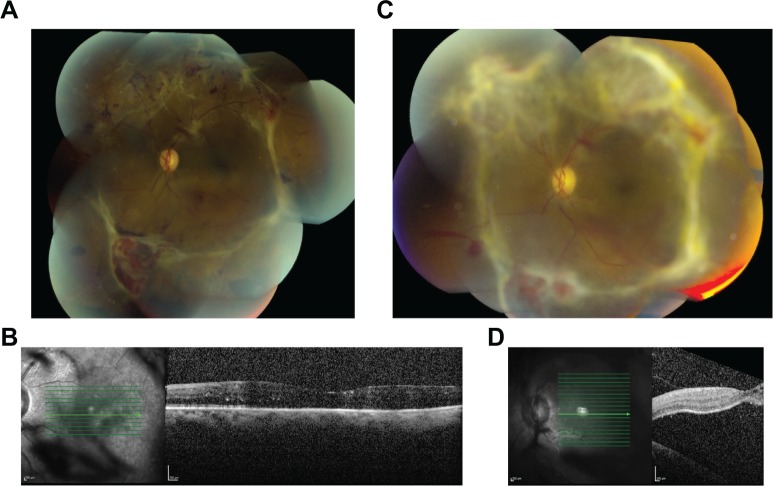
We report a patient with progression to a macula-off tractional retinal detachment in a fellow eye after a contralateral intraoperative intravitreal bevacizumab injection. A 32-year-old diabetic man noted decreased vision in his left eye 1 week following 25 gauge pars plana vitrectomy, gas tamponade, and intraoperative injection of bevacizumab in his right eye. Left eye visual acuity decreased from 20/80 to 20/200, and macula-off tractional retinal detachment was seen on clinical exam and imaging. Progression of tractional retinal detachment associated with proliferative diabetic retinopathy in a fellow eye after a contralateral intraoperative intravitreal bevacizumab injection may occur.
Keywords: anti-VEGF therapy; diabetes mellitus; fellow eye; tractional retinal detachment.
Figures
References
-
- Avery RL, Pearlman J, Pieramici DJ, et al. Intravitreal bevacizumab (Avastin) in the Treatment of proliferative diabetic retinopathy. Ophthalmology. 2006;113(10):1695–1705. - PubMed
-
- Arevalo JF, Fromow-Guerra J, Quiroz-Mercado H, et al. Pan-American Collaborative Retina Study Group Primary intravitreal bevacizumab (Avastin) for diabetic macular edema: results from the Pan-American Collaborative Retina Study Group at 6-month follow-up. Ophthalmology. 2007;114(4):743–750. - PubMed
-
- Ahn J, Woo SJ, Chung H, Park KH. The effect of adjunctive intravitreal bevacizumab for preventing postvitrectomy hemorrhage in proliferative diabetic retinopathy. Ophthalmology. 2011;118(11):2218–2226. - PubMed
-
- Arevalo JF, Maia M, Flynn HW, et al. Tractional retinal detachment following intravitreal bevacizumab (Avastin) in patients with severe proliferative diabetic retinopathy. Br J Ophthalmol. 2008;92(2):213–216. - PubMed
-
- Bakri SJ, Snyder MR, Reid JM, Pulido JS, Singh RJ. Pharmacokinetics of intravitreal bevacizumab (Avastin) Ophthalmology. 2007;114(5):855–859. - PubMed
Publication types
Grants and funding
LinkOut - more resources
Full Text Sources
Miscellaneous
