

Management of tracheobronchial injuries
- PMID: 25610327
- PMCID: PMC4299837
- DOI: 10.5152/eajm.2014.42
Management of tracheobronchial injuries
Abstract
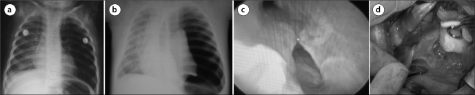
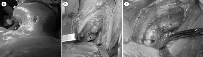
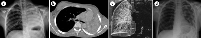
Tracheobronchial injury is one of cases which are relatively uncommon, but must be suspected to make the diagnosis and managed immediately. In such a case, primary initial goals are to stabilize the airway and localize the injury and then determine its extend. These can be possible mostly with flexible bronchoscopy conducted by a surgeon who can repair the injury. Most of the penetrating injuries occur in the cervical region. On the other hand, most of the blunt injuries occur in the distal trachea and right main bronchus and they can be best approached by right posterolateral thoracotomy. The selection of the manner and time of approaching depends on the existence and severity of additional injuries. Most of the injuries can be restored by deploying simple techniques such as individual sutures, while some of them requires complex reconstruction techniques. Apart from paying attention to the pulmonary toilet, follow-up is crucial for determination of anastomotic technique or stenosis. Conservative treatment may be considered an option with a high probability of success in patients meeting the criteria, especially in patients with iatrogenic tracheobronchial injury.
Trakeobronşiyal yaralanmalar göreceli olarak daha nadir görülen, ancak tanı konulabilmesi için şüphelenilmesi gereken ve sıklıkla anında müdahale gerektiren durumlardır. Bu durumda ilk yapılması gereken iki önemli amaç vardır; havayolunun stabilizasyonu ve yaralanmanın lokalizasyonu ve genişliğinin belirlenmesidir. Bunlar da sıklıkla yaralanmayı tedavi edebilecek bir cerrah tarafından yapılan fiberoptik bronkoskopi ile mümkün olur. Penetran yaralanmaların çoğu servikal bölgede olur. Künt yaralanmaların çoğu ise distal trakea ve sağ ana bronşda olur ve en iyi sağ posterolateral torakotomi ile yaklaşılır. Yaklaşım şeklinin seçimi ve zamanı ek yaralanmaların varlığı ve trakeobronşiyal yaralanmanın şiddetine bağlıdır. Yaralanmaların çoğu tek tek suturler kullanılarak yapılan basit tekniklerle onarılabilirken bazıları kompleks rekonstruksiyon teknikleri gerektirir. Pulmoner temizliğe dikkat edilmesi yanında anastomotik teknik veya stenozun tesbit edilmesi için takip önemlidir. Özellikle iatrojenik yaralanmalı hastalarda konservatif tedavi yaklaşımları da kabul edilebilir bir yaklaşım seçeneği olmaktadır.
Keywords: Trachea; bronchus; injury.
Figures
References
-
- Gussack GS, Jurkovich GJ, Luterman A. Laryngotracheal trauma: a protocol approach to a rare injury. Laryngoscope. 1986;96:660–5. - PubMed
-
- Lee RB. Traumatic injury of the cervicothoracic trachea and major bronchi. Chest Surg Clin N Am. 1997;7:285–304. - PubMed
-
- Karmy-Jones R, Wood ED. Traumatic Injury to the Trachea and Bronchus. Thorac Surg Clin. 2007;17:35–46. http://dx.doi.org/10.1016/j.thorsurg.2007.03.005. - DOI - PubMed
-
- Symbas PN, Justicz AG, Ricketts RR. Rupture of the airways from blunt trauma: treatment of complex injuries. Ann Thorac Surg. 1992;54:177–83. http://dx.doi.org/10.1016/0003-4975(92)91177-B. - DOI - PubMed
-
- Lupetin AR. Computed tomographic evaluation of laryngotracheal trauma. Curr Probl Diagn Radiol. 1997;26:185–206. http://dx.doi.org/10.1016/S0363-0188(97)90011-6. - DOI - PubMed
Publication types
LinkOut - more resources
Full Text Sources
Other Literature Sources
Medical