

Dermoscopic and clinical features of trunk melanomas
- PMID: 25610350
- PMCID: PMC4293392
- DOI: 10.5114/pdia.2014.47119
Dermoscopic and clinical features of trunk melanomas
Abstract
Introduction: Malignant melanomas account for 5% of all skin cancers and usually have a fatal clinical course. Additionally, the incidence of melanoma increases more rapidly than in any other cancer, and this has been attributed to the development of highly sensitive diagnostic techniques, mainly dermoscopy, which allows for early diagnosis. The phenotypic manifestations of gene/environment interactions, environmental factor and genetic factors may determine subtypes and anatomic localization of melanoma. Histopathologic subtypes, risk factors, and thickness of the skin are different in trunk melanomas.
Aim: To determine the frequency of dermatoscopic features in trunk melanomas. This study also investigates dermoscopic features according to the diameter of lesions.
Material and methods: Seventy-one trunk melanomas were included. Their dermoscopic and clinical images, histopathological and clinical data were assessed. The relations between the diameter, Breslow thickness and dermoscopic characteristics were evaluated.
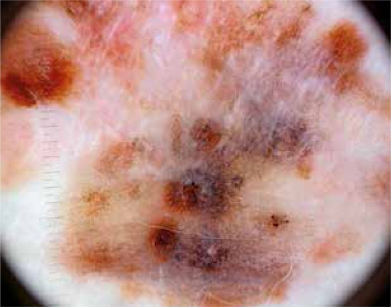
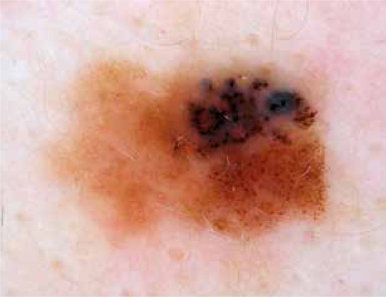
Results: The most common dermoscopic findings of trunk melanomas were the multicomponent pattern (55 patients, 77.5%), asymmetry (62 patients; 87.3%), blue-gray veil (59 patients, 83.1%), and color variety (56 patients, 78.8%). When dermoscopic findings were compared, a multicomponent pattern (p = 0.03), milky-red areas (p = 0.001), blue-gray veils (p = 0.023), and regression structures (p = 0.037) were more common in large melanomas than in small melanomas.
Conclusions: The most common dermoscopic findings of trunk melanomas were the multicomponent pattern, asymmetry and blue-gray veil, color variety. The multicomponent pattern, milky-red areas, blue-gray veils, regression structures were statistically significant dermoscopic features in a group of large-diameter melanomas, compared to small melanomas.
Keywords: dermoscopy; melanoma; trunk.
Figures
References
-
- Buyukpinarbasili N, Demirkesen C, Oguz O, et al. The prognostic factors in cutaneous malignant melanoma. Turk Derm. 2002;36:115–24.
-
- Aydemir EH. Treatment of malignant melanoma according to the stages of melanoma. Turk Derm. 2007;41:20–1.
-
- Ozdemir F. Diagnosis of nelanoma. Turk Derm. 2007;41:6–14.
-
- Stolz W, Riemann A, Cognetta AB, et al. ABCD rule of dermatoscopy: a new practical method for early recognition of malignant melanoma. Eur J Dermatol. 1994;4:521–7.
-
- Argenziano G, Fabbrocini G, Carli P, et al. Epiluminescence microscopy for the diagnosis of doubtful melanocytic skin lesions: comparison of the ABCD rule of dermatoscopy and a new 7-point checklist based on pattern analysis. Arch Dermatol. 1998;134:1563–70. - PubMed
LinkOut - more resources
Full Text Sources
Other Literature Sources