. 2014 Jul 3;3(7):e950163.
doi: 10.4161/15384101.2014.950163. eCollection 2014.

Human polymorphonuclear neutrophils specifically recognize and kill cancerous cells

Affiliations

Human polymorphonuclear neutrophils specifically recognize and kill cancerous cells

Jun Yan et al. Oncoimmunology. .

Abstract

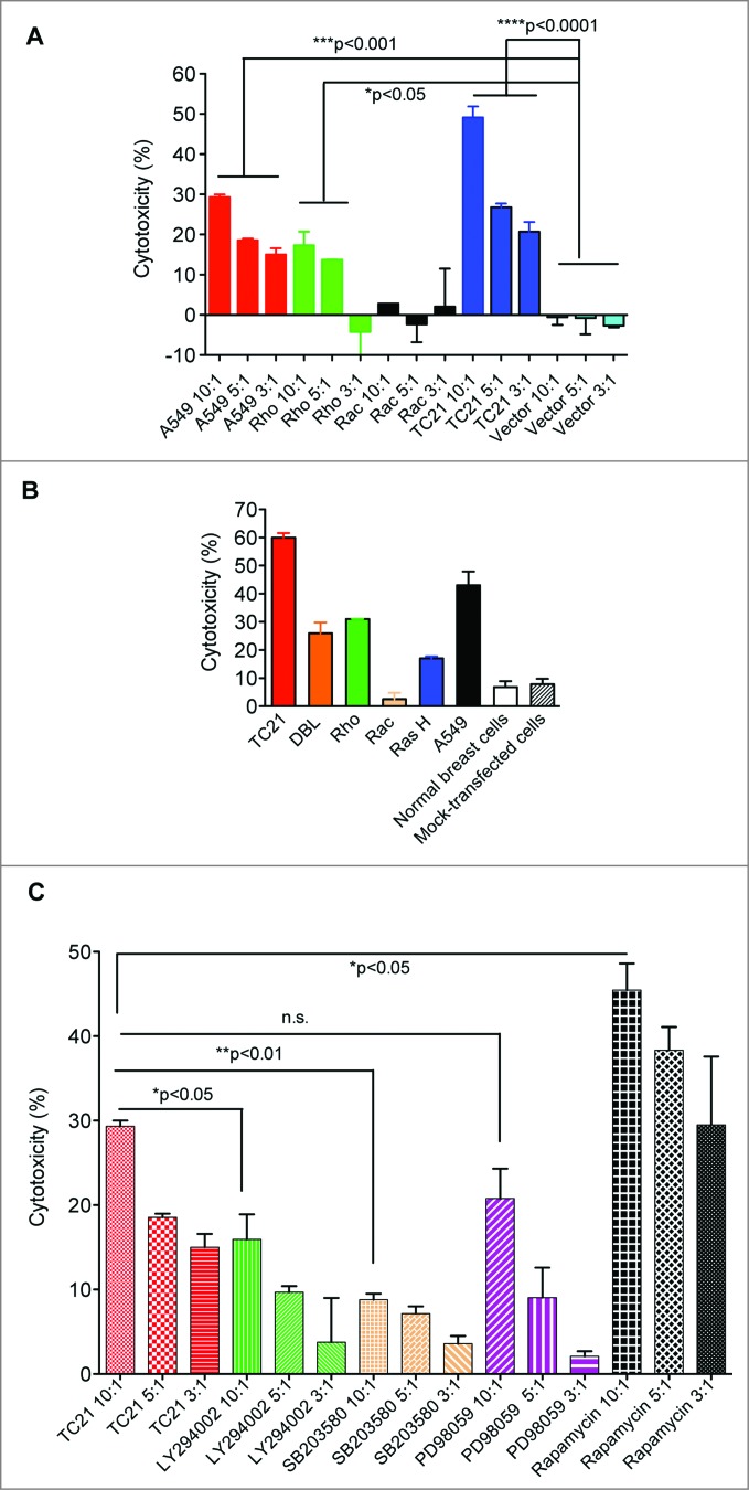
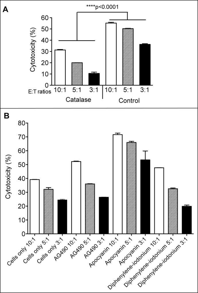
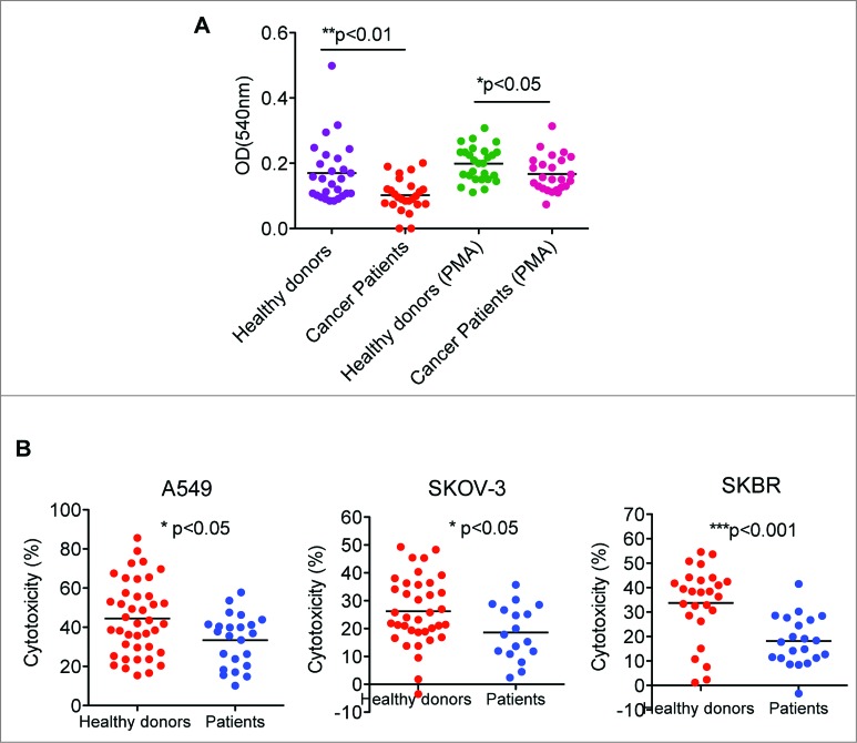
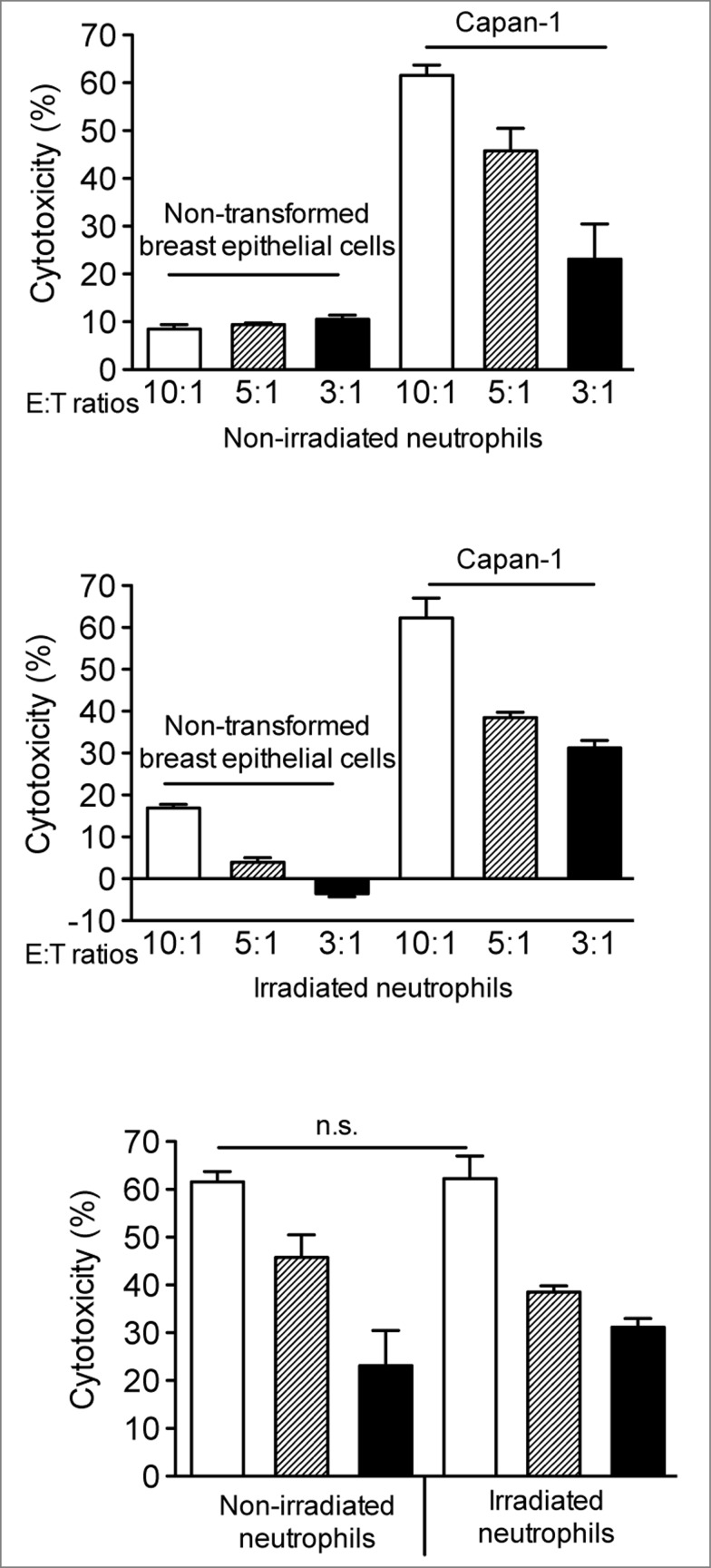
Polymorphonuclear neutrophils (PMNs), the main effectors of the innate immune system, have rarely been considered as an anticancer therapeutic tool. However, recent investigations using animal models and preliminary clinical studies have highlighted the potential antitumor efficacy of PMNs. In the current study, we find that PMNs from some healthy donors naturally have potent cancer-killing activity against 4 different human cancer cell lines. The killing activity appears to be cancer cell-specific since PMNs did not kill primary normal epithelial cells or an immortalized breast epithelial cell line. Transfecting the immortalized mammary cells with plasmids expressing activated forms of the rat sarcoma viral oncogene homolog (Ras) and teratocarcinoma oncogene 21 (TC21) oncogenes was sufficient to provoke aggressive attack by PMNs. However, transfection with activated Ras-related C3 botulinum toxin substrate (Rac1) was ineffective, suggesting specificity in PMN-targeting of neoplastic cells. Furthermore, PMNs from lung cancer patients were also found to exhibit relatively poor cancer-killing activity compared to the cytolytic activity of the average healthy donor. Taken together, our results suggest that PMN-based treatment regimens may represent a paradigm shift in cancer immunotherapy that may be easily introduced into the clinic to benefit a subset of patients with PMN-vulnerable tumors.

Keywords: BEN, benign ethnic neutropenia; DBL, proto-oncogene DBL; DPI, diphenyleneiodonium; E:T, effector:target; G-CSF, granulocyte-colony stimulating factor; GM-CSF, granulocyte macrophage-colony stimulating factor; GVHD, graft-versus-host disease; H-Ras, Harvey rat sarcoma viral oncogene homolog; MEK, mitogen-activated protein kinase kinase; NADPH, nicotinamide adenine dinucleotide phosphate; NBT, nitroblue tetrazolium; NSCLC, non-small cell lung carcinoma; PI3 kinase, phosphoinositide 3-kinase; PMN, polymorphonuclear neutrophils; ROS, reactive oxygen species; Rac1, Ras-related C3 botulinum toxin substrate 1; RhoA, Ras homolog family member A; TC-21, teratocarcinoma oncogene TC21; TGFβ, transforming growth factor; cytotoxicity; mAb, monoclonal antibody; mTOR, mammalian target of rapamycin; neutrophils; oncogene; tumor cells.


Figures

Figure 1.
Figure 1.
Neutrophils from healthy donors are potent cancer killing cells. AB. Neutrophil anticancer cytotoxicity was assayed by co-incubating 5 × 10³ cancerous or normal epithelial cells with human neutrophils from healthy donors at effector-to-target (E:T) cell ratios of 10:1, 5:1, and 3:1. Cells were incubated at 37°C in a humidified 5% CO2 incubator for 16 h. Cytotoxicity was calculated by measuring the relative decrease in current impedance in comparison to target cells only. (A). Cytotoxicity assays using neutrophils from 2 healthy donors were performed to measure killing activity against Hela, SKOV-3, Capan-1 and A549 cancer cells. (B). Cytotoxicity assays using healthy donor neutrophils to measure killing activity against human lung cancer cell line A549 versus primary epithelial cells or non-transformed, immortalized epithelial MCF-10A cells. All conditions were assayed in duplicate. Statistical analysis was performed by one way ANOVA; ***Pp < 0 .001, ****P < 0 .0001.
Figure 2.
Figure 2.
Neutrophils kill oncogene-transfected cells. A-C. The non-transformed, immortalized epithelial breast cell line MCF-10A was transfected with pZIP-Neo constructs encoding the indicated oncogenes. Stable cell lines were selected using G418 resistance and used in cytotoxicity assays using neutrophils from healthy donors. (A). Neutrophils from one of healthy donors were used to perform cytotoxicity assays against oncogene expressing MCF-10A cells. Vector only-transfected MCF-10A cells and A549 lung cancer cells were used as control target cells. Effector-to-target (E:T) cell ratios were at 10:1, 5:1, and 3:1. Statistical analysis was performed by one-way ANOVA;. *P < 0 .05, ***P < 0 .001, ****p<0 .0001 (B). Cytotoxicity assay using neutrophils from an independent healthy donor against MCF-10A cells stably transfected with plasmids expressing the indicated oncogenes. E:T ratio at 10:1. (C). MCF-10A cells transfected with the TC21 oncogene were used as target cells for neutrophil cytotoxicity assay in the presence or absence of different inhibitors including the PI3 kinase inhibitor LY294002 (1 μM), the p38 kinase inhibitor SB203580 (0.5 μM), the MEK inhibitor PD98059 (2 μM) and the mTOR inhibitor rapamycin (0.1 nM). Neutrophils were from healthy donor. E:T ratios were at 10:1, 5:1 and 3:1. Statistical analysis was performed by unpaired Student's t test; *P < 0 .05, **P < 0 .01, n.s., not significant. All conditions were assayed in duplicate.
Figure 3.
Figure 3.
H2O2 is essential for neutrophil-mediated anticancer cytotoxicity. A-B. Neutrophil anticancer cytotoxicity was assayed by co-incubating 5 × 10³ A549 cancer cells with human neutrophils from healthy donors at effector-to-target (E:T) cell ratios of 10:1, 5:1, and 3:1. Neutrophils were pretreated as indicated. Cells were incubated at 37°C in a humidified 5% CO2 incubator for 16 h. Cytotoxicity was calculated by measuring the relative decrease in current impedance in comparison to target cells only. (A). Neutrophils from healthy donors were treated with or without 5000 U/mL catalase for 30 min at 37°C. Cells were then washed and used for cytotoxicity assay with A549 lung cancer cells as target cells. (B). Neutrophil cytotoxicity assay in the presence or absence of the inhibitors 50 μM AG490, 100 μM apocyanin, or 10 μM diphenylene iodonium at indicated E:T ratios using A549 lung cancer cells as target cells. All conditions were assayed in duplicate. Statistical analysis was performed by one way ANOVA; ****P < 0 .0001.
Figure 4.
Figure 4.
Neutrophils derived from cancer patients have significantly decreased cancer cell killing potential. A-B. Neutrophils from healthy individuals (n = 27) or non-small cell lung cancer (NSCLC) patients (n = 25) were comparatively analyzed for cancer cell killing potential by analyzing superoxide anion potential via nitroblue tetrazolium (NBT) assay (A) and cytotoxicity assay (B). (A) Neutrophils were stimulated with or without 5 ng/mL phorbol myristate acetate (PMA) followed by the addition of NBT solution for 1 h. The reaction was stopped and plates read in a spectrophotometer at 540nm. (B) Cytotoxicity of neutrophils from healthy donors and NSCLC patients against A549, SKOV-3 and SKBR cancer cells at effector-to-target cell ratio of 10:1. Cells were incubated at 37°C in a humidified 5% CO2 incubator for 16 h. Cytotoxicity was calculated by measuring the relative decrease in current impedance in comparison to target cells only. All conditions were assayed in duplicate. Statistical analysis was performed by unpaired Student's t test; *p<0 .05, **p<0 .01, ***p<0 .001. The lines in the data indicate mean values.
Figure 5.
Figure 5.
Irradiation does not impact neutrophil cancer cell killing activity. Irradiated neutrophil anticancer cytotoxicity was assayed by co-incubating 5 × 10³ Capan-1 cancer cells with human neutrophils from healthy donors at effector-to-target (E:T) cell ratios of 10:1, 5:1, and 3:1. Neutrophils from healthy donors were irradiated (2500 rads) and non-irradiated neutrophils were used as controls. Cells were incubated at 37°C in a humidified 5% CO2 incubator for 16 h. Cytotoxicity was calculated by measuring the relative decrease in current impedance in comparison to target cells only. Statistical analysis was performed by unpaired Student's t test; n.s., not significant. All conditions were assayed in duplicate.

References

    1. Nathan C. Neutrophils and immunity:challenges and opportunities. Nat Rev Immunol 2006; 6:173-82; PMID:; http://dx.doi.org/10.1038/nri1785 - DOI - PubMed
    1. Weiss SJ. Tissue destruction by neutrophils. N Engl J Med 1989; 320:365-76; PMID:2536474 http://dx.doi.org/10.1056/NEJM198902093200606 - DOI - PubMed
    1. Mantovani A, Allavena P, Sica A, Balkwill F. Cancer-related inflammation. Nature 2008; 454:436-44; PMID:; http://dx.doi.org/10.1038/nature07205 - DOI - PubMed
    1. Coussens LM, Werb Z. Inflammation and cancer. Nature 2002; 420:860-7; PMID:; http://dx.doi.org/10.1038/nature01322 - DOI - PMC - PubMed
    1. Stoppacciaro A, Melani C, Parenza M, Mastracchio A, Bassi C, Baroni C, Parmiani G, Colombo MP. Regression of an established tumor genetically modified to release granulocyte colony-stimulating factor requires granulocyte-T cell cooperation and T cell-produced interferon gamma. J Exp Med 1993; 178:151-61; PMID:; http://dx.doi.org/10.1084/jem.178.1.151 - DOI - PMC - PubMed

Publication types