

Epicardial adipose tissue: far more than a fat depot
- PMID: 25610800
- PMCID: PMC4278038
- DOI: 10.3978/j.issn.2223-3652.2014.11.05
Epicardial adipose tissue: far more than a fat depot
Abstract
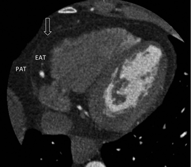
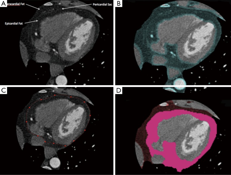
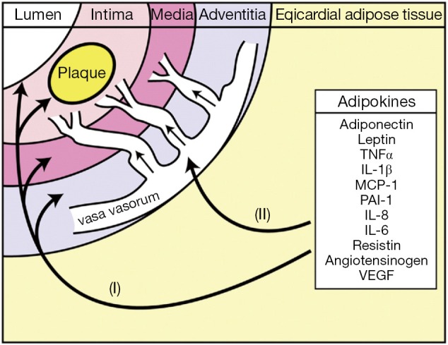
Epicardial adipose tissue (EAT) refers to the fat depot that exists on the surface of the myocardium and is contained entirely beneath the pericardium, thus surrounding and in direct contact with the major coronary arteries and their branches. EAT is a biologically active organ that may play a role in the association between obesity and coronary artery disease (CAD). Given recent advances in non-invasive imaging modalities such a multidetector computed tomography (MDCT), EAT can be accurately measured and quantified. In this review, we focus on the evidence suggesting a role for EAT as a quantifiable risk marker in CAD, as well as describe the role EAT may play in the development and vulnerability of coronary artery plaque.
Keywords: Epicardial adipose tissue (EAT); coronary artery disease (CAD); epicardial fat; multidetector computed tomography (MDCT).
Figures
References
-
- The 10 leading causes of death. World Health Organisation. Available online: http://www.who.int/mediacentre/factsheets/fs310/en/index.html. Accessed October 16, 2013.
-
- Calle EE, Thun MJ, Petrelli JM, et al. Body-mass index and mortality in a prospective cohort of U.S. adults. N Engl J Med 1999;341:1097-105. - PubMed
-
- Van Gaal LF, Mertens IL, De Block CE. Mechanisms linking obesity with cardiovascular disease. Nature 2006;444:875-80. - PubMed
-
- Fantuzzi G, Mazzone T.Adipose tissue and atherosclerosis: exploring the connection. Arterioscler Thromb Vasc Biol 2007;27:996-1003. - PubMed
-
- Okura T, Nakata Y, Yamabuki K, et al. Regional body composition changes exhibit opposing effects on coronary heart disease risk factors. Arterioscler Thromb Vasc Biol 2004;24:923-9. - PubMed
Publication types
LinkOut - more resources
Full Text Sources
Other Literature Sources
Miscellaneous