

Coxiella burnetii lipopolysaccharide blocks p38α-MAPK activation through the disruption of TLR-2 and TLR-4 association
- PMID: 25610812
- PMCID: PMC4285172
- DOI: 10.3389/fcimb.2014.00182
Coxiella burnetii lipopolysaccharide blocks p38α-MAPK activation through the disruption of TLR-2 and TLR-4 association
Abstract
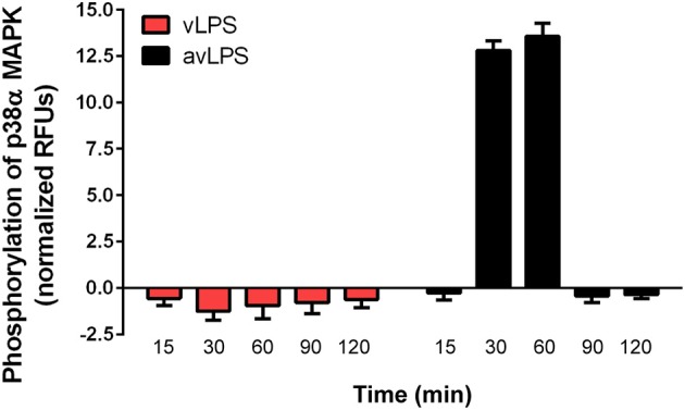
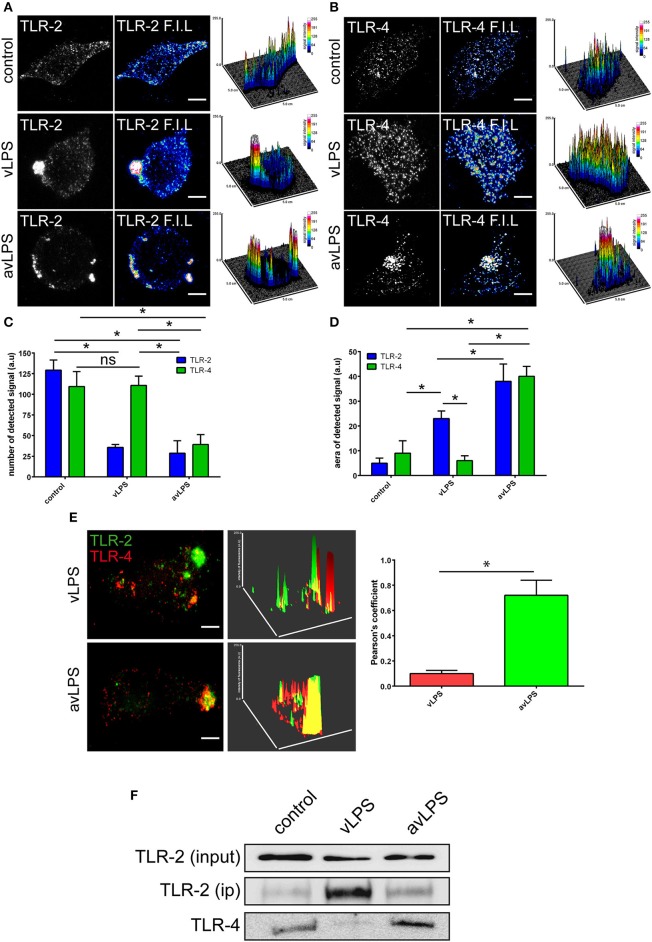
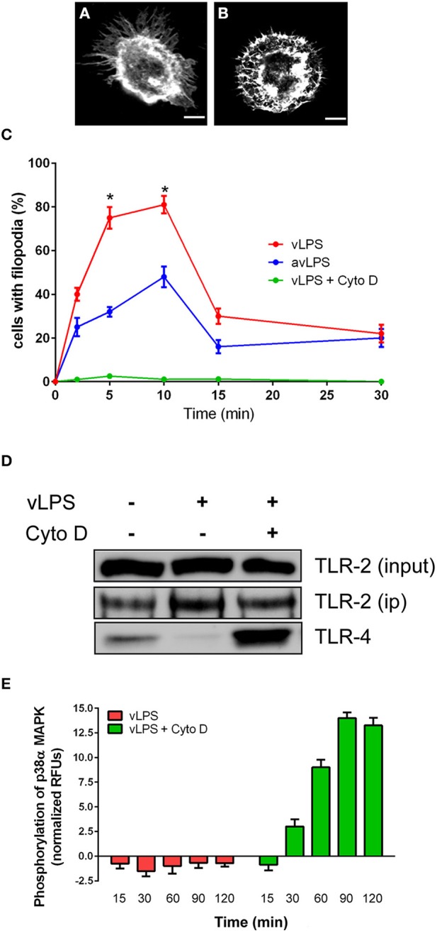
To survive in macrophages, Coxiella burnetii hijacks the activation pathway of macrophages. Recently, we have demonstrated that C. burnetii, via its lipopolysaccharide (LPS), avoids the activation of p38α-MAPK through an antagonistic engagement of Toll-like receptor (TLR)-4. We investigated the fine-tuned mechanism leading to the absence of activation of the p38α-MAPK despite TLR-4 engagement. In macrophages challenged with LPS from the avirulent variants of C. burnetii, TLR-4 and TLR-2 co-immunoprecipitated. This association was absent in cells challenged by the LPS of pathogenic C. burnetii. The disruption makes TLRs unable to signal during the recognition of the LPS of pathogenic C. burnetii. The disruption of TLR-2 and TLR-4 was induced by the re-organization of the macrophage cytoskeleton by C. burnetii LPS. Interestingly, blocking the actin cytoskeleton re-organization relieved the disruption of the association TLR-2/TLR-4 by pathogenic C. burnetii and rescued the p38α-MAPK activation by C. burnetii. We elucidated an unexpected mechanism allowing pathogenic C. burnetii to avoid macrophage activation by the disruption of the TLR-2 and TLR-4 association.
Keywords: Coxiella burnetii; TLR-2; TLR-4; cytoskeleton; macrophages.
Figures
Similar articles
-
Coxiella burnetii Lipopolysaccharide: What Do We Know?Int J Mol Sci. 2017 Nov 23;18(12):2509. doi: 10.3390/ijms18122509. Int J Mol Sci. 2017. PMID: 29168790 Free PMC article. Review.
-
Primary Role for Toll-Like Receptor-Driven Tumor Necrosis Factor Rather than Cytosolic Immune Detection in Restricting Coxiella burnetii Phase II Replication within Mouse Macrophages.Infect Immun. 2016 Mar 24;84(4):998-1015. doi: 10.1128/IAI.01536-15. Print 2016 Apr. Infect Immun. 2016. PMID: 26787725 Free PMC article.
-
Impaired stimulation of p38α-MAPK/Vps41-HOPS by LPS from pathogenic Coxiella burnetii prevents trafficking to microbicidal phagolysosomes.Cell Host Microbe. 2012 Dec 13;12(6):751-63. doi: 10.1016/j.chom.2012.10.015. Cell Host Microbe. 2012. PMID: 23245320
-
Lipopolysaccharide from Coxiella burnetii is involved in bacterial phagocytosis, filamentous actin reorganization, and inflammatory responses through Toll-like receptor 4.J Immunol. 2004 Mar 15;172(6):3695-703. doi: 10.4049/jimmunol.172.6.3695. J Immunol. 2004. PMID: 15004173
-
Intracellular life of Coxiella burnetii in macrophages.Ann N Y Acad Sci. 2009 May;1166:55-66. doi: 10.1111/j.1749-6632.2009.04515.x. Ann N Y Acad Sci. 2009. PMID: 19538264 Review.
Cited by
-
Mast Cell Cytonemes as a Defense Mechanism against Coxiella burnetii.mBio. 2019 Apr 16;10(2):e02669-18. doi: 10.1128/mBio.02669-18. mBio. 2019. PMID: 30992359 Free PMC article.
-
Coxiella burnetii Lipopolysaccharide: What Do We Know?Int J Mol Sci. 2017 Nov 23;18(12):2509. doi: 10.3390/ijms18122509. Int J Mol Sci. 2017. PMID: 29168790 Free PMC article. Review.
-
aYAP modRNA reduces cardiac inflammation and hypertrophy in a murine ischemia-reperfusion model.Life Sci Alliance. 2019 Dec 16;3(1):e201900424. doi: 10.26508/lsa.201900424. Print 2020 Jan. Life Sci Alliance. 2019. PMID: 31843959 Free PMC article.
-
Protective Effects of Lactobacillus plantarum Lac16 on Clostridium perfringens Infection-Associated Injury in IPEC-J2 Cells.Int J Mol Sci. 2021 Nov 17;22(22):12388. doi: 10.3390/ijms222212388. Int J Mol Sci. 2021. PMID: 34830269 Free PMC article.
-
The roles of the TLR/NF‑κB signaling pathway in the mutual interactions between the lung and the large intestine.Mol Med Rep. 2018 Aug;18(2):1387-1394. doi: 10.3892/mmr.2018.9111. Epub 2018 May 31. Mol Med Rep. 2018. PMID: 29901105 Free PMC article.
References
-
- Barry A. O., Boucherit N., Mottola G., Vadovic P., Trouplin V., Soubeyran P., et al. . (2012). Impaired stimulation of p38alpha-MAPK/Vps41-HOPS by LPS from pathogenic Coxiella burnetii prevents trafficking to microbicidal phagolysosomes. Cell Host Microbe 12, 751–763. 10.1016/j.chom.2012.10.015 - DOI - PubMed
Publication types
MeSH terms
Substances
LinkOut - more resources
Full Text Sources
Other Literature Sources