. 2015 Feb 11;137(5):2107-15.
doi: 10.1021/ja513080v. Epub 2015 Feb 2.

Structure and thermodynamics of N6-methyladenosine in RNA: a spring-loaded base modification

Affiliations

Structure and thermodynamics of N6-methyladenosine in RNA: a spring-loaded base modification

Caroline Roost et al. J Am Chem Soc. .

Erratum in

Abstract

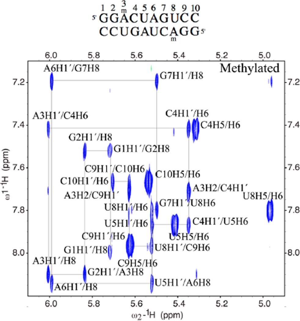
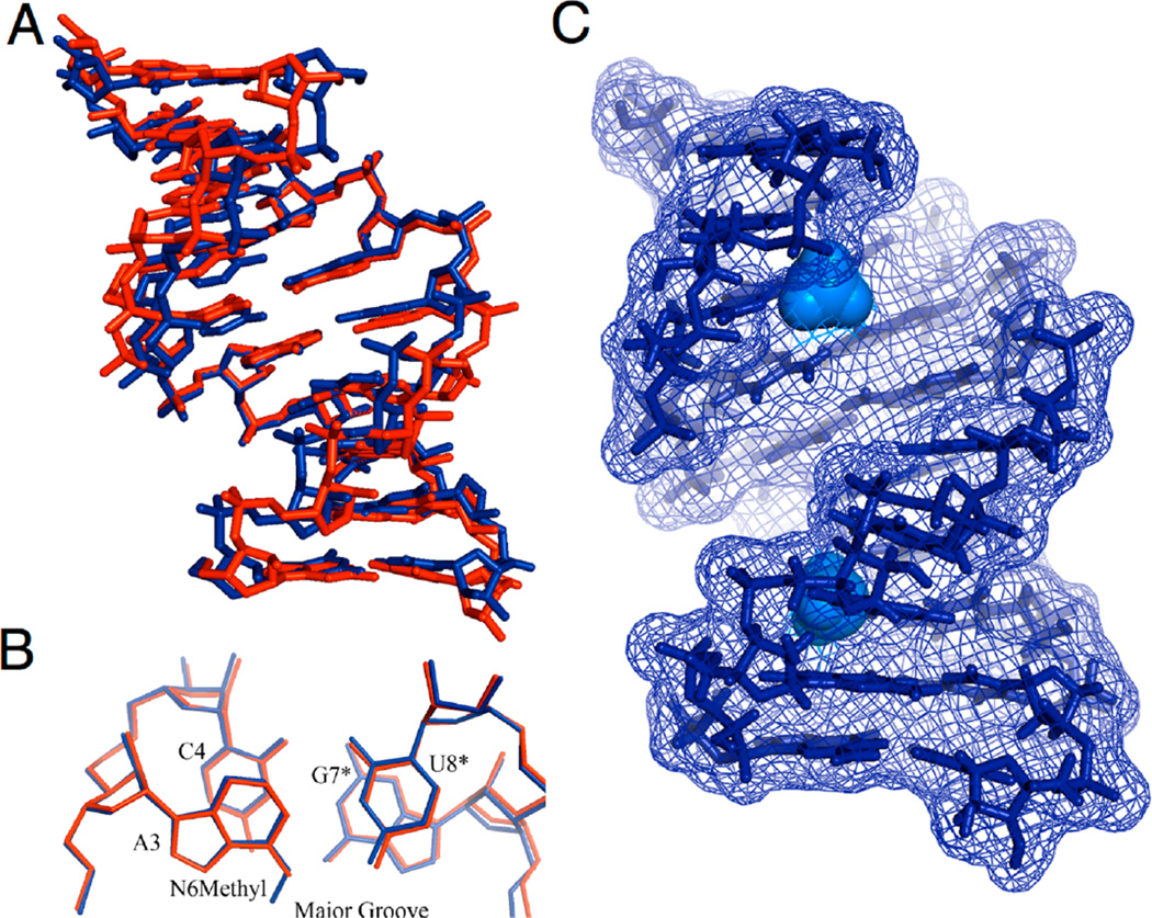
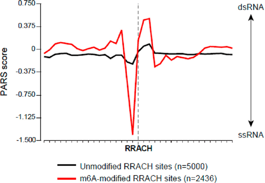
N(6)-Methyladenosine (m(6)A) modification is hypothesized to control processes such as RNA degradation, localization, and splicing. However, the molecular mechanisms by which this occurs are unclear. Here, we measured structures of an RNA duplex containing m(6)A in the GGACU consensus, along with an unmodified RNA control, by 2D NMR. The data show that m(6)A-U pairing in the double-stranded context is accompanied by the methylamino group rotating from its energetically preferred syn geometry on the Watson-Crick face to the higher-energy anti conformation, positioning the methyl group in the major groove. Thermodynamic measurements of m(6)A in duplexes reveal that it is destabilizing by 0.5-1.7 kcal/mol. In contrast, we show that m(6)A in unpaired positions base stacks considerably more strongly than the unmodified base, adding substantial stabilization in single-stranded locations. Transcriptome-wide nuclease mapping of methylated RNA secondary structure from human cells reveals a structural transition at methylated adenosines, with a tendency to single-stranded structure adjacent to the modified base.


Figures

Figure 1
Figure 1
Structures and conformations of m6A in RNA. (A) Syn methyl orientation is favored over anti when the base is unpaired as a result of a steric clash between the methyl group and N7. (B) Space-filling models of m6A in syn and anti conformations (N9 substituent is methyl). (C) Multiple pairing configurations are possible for m6A paired opposite U; these configurations vary on whether the adenine base and the methylamino group are syn or anti. The current work confirms the third (anti/anti) structure in an RNA duplex; the methyl group is spring-loaded into the high-energy anti conformation, trapped there by pairing with U and surrounding duplex structure.
Figure 2
Figure 2
Proton NMR spectra of RNA duplexes containing GGACU methylation motif, confirming fully paired structure. (A) 1D proton spectrum of methylated duplex; (B) spectrum of unmethylated duplex for comparison. Downfield regions of the 1D 1H NMR spectra are shown; they were acquired in 90% H2O/10% D2O at 15 °C with 11 solvent suppression and show evidence of 10 H-bonded base pairs (5 symmetrically unique) for methylated duplex MA (A) and unmodified AD (B).
Figure 3
Figure 3
H1′/aromatic portion of a 150 ms mixing time NOESY in D2O is shown for the methylated MA duplex, highlighting the sequential assignments of all bases. Sequence is 5′-GGACUAGUCC, where the bold base is methylated.
Figure 4
Figure 4
Structures of methylated and unmodified RNA duplexes, establishing that m6A is oriented anti in a paired duplex, with the methyl group anti as well. (A) The average structure of the entire 10 bp duplexes with the MA in blue and unmodified DA RNA in red. (B) Superimposed structures of the second, third, and fourth base pairs alone; the m6A3/U8*;A3/U8* and the neighboring base pair (C4/G7*) are shown for clarity. The second strands in the symmetric dimer are indicated with an asterisk (*). (C) Surface representation of the structure of the methylated RNA duplex, highlighting the adenine N6 methyl groups in the major groove (light blue).
Figure 5
Figure 5
Nuclease structural mapping reveals a structural transition from unpaired to paired structure surrounding m6A. Plot shows average PARS score for each nucleotide in the vicinity of methylated vs unmethylated adenines in the RRACH methylation consensus, as mapped for polyadenylated RNAs from GM12878 cells. Non-methylated sites were chosen as the 5000 sites with the lowest enrichment score after m6A IP. y-axis represents PARS score. Positive values indicate high probability of double-stranded conformation, as evidenced by RNase V1 cleavage, whereas negative values indicate high probability of single-stranded conformation, as evidenced by RNase S1 cleavage. x-axis represents nucleotide position.

References

    1. Schibler U, Kelley DE, Perry RP. J. Mol. Biol. 1977;115:695–714. - PubMed
    1. Dominissini D, Moshitch-Moshkovitz S, Schwartz S, Salmon-Divon M, Ungar L, Osenberg S, Cesarkas K, Jacob-Hirsch J, Amariglio N, Kupiec M, Sorek R, Rechavi G. Nature. 2012;485:201–206. - PubMed
    1. Meyer KD, Saletore Y, Zumbo P, Elemento O, Mason CE, Jaffrey SR. Cell. 2012;150:1–12. - PMC - PubMed
    1. Wang X, Lu Z, Gomez A, Hon GC, Yue Y, Han D, Fu Y, Parisien M, Dai Q, Jia G, Ren B, Pan T, He C. Nature. 2014;505:117–120. - PMC - PubMed
    1. Csepany T, Lin A, Baldick CJ, Jr, Beemon K. J. Biol. Chem. 1990;265:20117–20122. - PubMed

Publication types

LinkOut - more resources