. 2015 Sep;53(3):391-9.
doi: 10.1165/rcmb.2014-0150OC.

Pathologic Regulation of Collagen I by an Aberrant Protein Phosphatase 2A/Histone Deacetylase C4/MicroRNA-29 Signal Axis in Idiopathic Pulmonary Fibrosis Fibroblasts

Affiliations

Pathologic Regulation of Collagen I by an Aberrant Protein Phosphatase 2A/Histone Deacetylase C4/MicroRNA-29 Signal Axis in Idiopathic Pulmonary Fibrosis Fibroblasts

Wajahat Khalil et al. Am J Respir Cell Mol Biol. 2015 Sep.

Abstract

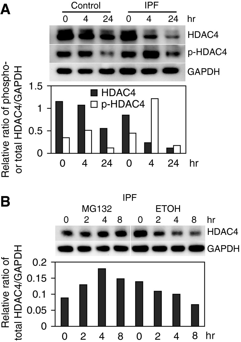
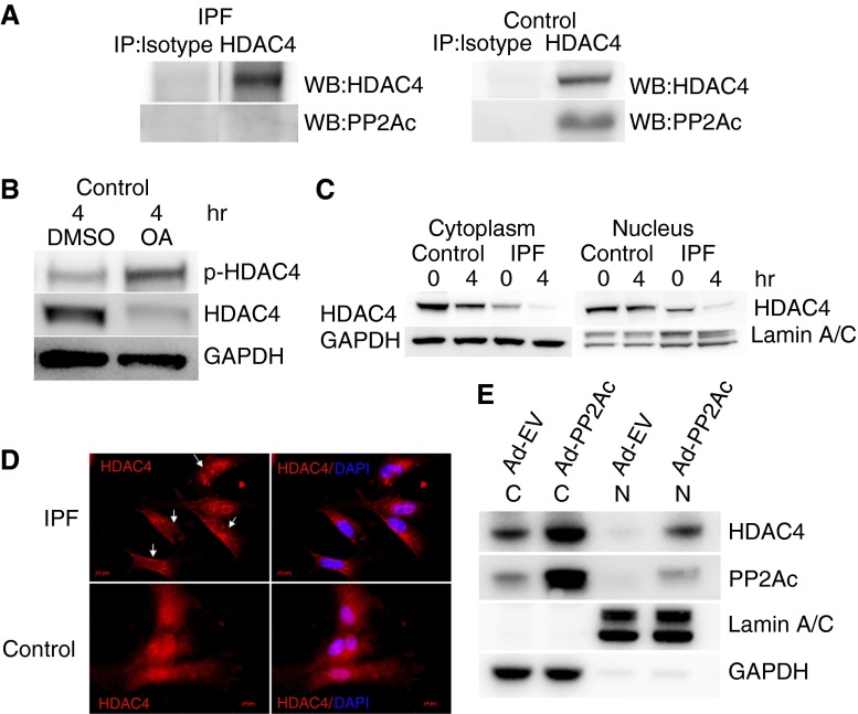
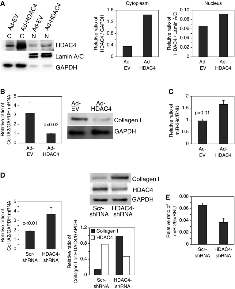
Idiopathic pulmonary fibrosis (IPF) is characterized by the relentless expansion of fibroblasts depositing type I collagen within the alveolar wall and obliterating the alveolar airspace. MicroRNA (miR)-29 is a potent regulator of collagen expression. In IPF, miR-29 levels are low, whereas type I collagen expression is high. However, the mechanism for suppression of miR-29 and increased type I collagen expression in IPF remains unclear. Here we show that when IPF fibroblasts are seeded on polymerized type I collagen, miR-29c levels are suppressed and type I collagen expression is high. In contrast, miR-29c is high and type I collagen expression is low in control fibroblasts. We demonstrate that the mechanism for suppression of miR-29 during IPF fibroblast interaction with polymerized collagen involves inappropriately low protein phosphatase (PP) 2A function, leading to histone deacetylase (HDA) C4 phosphorylation and decreased nuclear translocation of HDAC4. We demonstrate that overexpression of HDAC4 in IPF fibroblasts restored miR-29c levels and decreased type I collagen expression, whereas knocking down HDAC4 in control fibroblasts suppressed miR-29c levels and increased type I collagen expression. Our data indicate that IPF fibroblast interaction with polymerized type I collagen results in an aberrant PP2A/HDAC4 axis, which suppresses miR-29, causing a pathologic increase in type I collagen expression.

Keywords: HDAC4; IPF fibroblasts; PP2A; miR-29; type I collagen.


Figures

Figure 1.
Figure 1.
Type I collagen expression is high and microRNA (miR)-29c is low in idiopathic pulmonary fibrosis (IPF) fibroblasts. (A) Primary IPF (n = 6) and control human lung fibroblasts (n = 4) were seeded on polymerized type I collagen matrices for 4 hours. Left panel: Type I collagen protein expression was quantified by Western blot analysis. Glyceraldehyde 3-phosphate dehydrogenase (GAPDH) is shown as a loading control. Right panel: miR-29c levels were quantified by quantitative RT-PCR (qRT-PCR) and are shown as the ratio of miR-29 to U6-2 small nuclear RNA (RNU) expression. (B) IPF fibroblasts (n = 3) were plated on tissue culture plastic (TC) or polymerized type I collagen matrices (PC). Left panel: Type I collagen expression was quantified by Western analysis. GAPDH is shown as a loading control. Right panel: miR-29c levels were quantified by qRT-PCR and are shown as the ratio of miR-29 to RNU expression.
Figure 2.
Figure 2.
Histone deacetylase (HDAC) 4 protein levels are decreased in IPF fibroblasts. (A) IPF and control lung fibroblasts were seeded on polymerized type I collagen matrices as a function of time. Top panel: Phosphorylated HDAC4 (pHDAC4) and HDAC4 expression were examined by Western blot analysis. GAPDH is shown as a loading control. Bottom panel: pHDAC4 and HDAC4 levels were quantified by densitometry. (B) IPF fibroblasts were pretreated with the proteasome inhibitor MG132 (20 nM; 60 min) or vehicle control (ethanol [ETOH]) and seeded on polymerized collagen for 0, 2, 4, or 8 hours. HDAC4 protein levels were quantified by Western blot analysis. GAPDH is shown as a loading control. All experiments were repeated with three independent cell lines to confirm the findings.
Figure 3.
Figure 3.
Low protein phosphatase (PP) 2A in IPF fibroblasts results in HDAC4 hyperphosphorylation and decreases its nuclear localization. (A) IPF and control fibroblasts were plated on type I polymerized collagen for 4 hours. Immunoprecipitation of HDAC4 was performed, and samples were analyzed for association with PP2Ac. Immunoprecipitation with isotype antibody was used as a control. (B) Control lung fibroblasts were pretreated with the PP2A inhibitor okadaic acid (OA) (10 nM; 60 min) or DMSO as a control. The cells were then seeded on type I polymerized collagen matrices and phosphorylated, and HDAC4 protein expression was examined by Western blot analysis as a function of time. GAPDH is shown as a loading control. (C) IPF and control fibroblasts were seeded on polymerized collagen for 4 hours. The cells were lysed, and nuclear and cytoplasmic fractions were analyzed for HDAC4 expression by Western blot analysis. Lamin A/C is shown as a nuclear loading control; GAPDH is shown as a cytoplasmic loading control. (D) IPF and control fibroblasts were seeded on polymerized type I collagen for 4 hours. The cells were stained with HDAC4 antibody conjugated with Cy-3. 4′6-Diamidino-2-phenylindole (DAPI) indicates nuclear staining. Arrows point to four IPF fibroblasts with low nuclear HDAC4 expression. (E) PP2Ac was overexpressed in IPF fibroblasts using an adenoviral vector containing a wild-type PP2Ac construct (Ad-PP2Ac). Cells infected with empty vector served as control (Ad-EV). The cells were seeded on polymerized collagen for 4 hours and then lysed. Nuclear (N) and cytoplasmic (C) fractions were analyzed for PP2Ac and HDAC4 expression by Western blot analysis. Lamin A/C is shown as a nuclear loading control; GAPDH is shown as a cytoplasmic loading control.
Figure 4.
Figure 4.
HDAC4 regulates type I collagen and miR-29 expression. (AC) HDAC4 was overexpressed in IPF fibroblasts using an adenoviral vector containing a wild-type HDAC4 construct (Ad-HDAC4). Controls consisted of cells expressing empty vector (Ad-EV). (A) Cells were seeded on polymerized type I collagen matrices for 4 hours. Cells were lysed and nuclear (N) and cytoplasmic (C) fractions were analyzed for HDAC4 protein levels by Western analysis (left panel). Nuclear and cytoplasmic HDAC4 levels were quantified by densitometric analysis (middle and right panels). (B) COL1A2 messenger RNA (mRNA) (left panel) and collagen I protein (right panel) expression were examined by qRT-PCR and Western blot analysis, respectively. GAPDH is shown as a loading control. (C) miR-29c levels were quantified by qRT-PCR. (D and E) HDAC4 was knocked down in control fibroblasts using a lentiviral vector containing HDAC4 short hairpin RNA (shRNA). Cells infected with lentiviral vector containing scrambled shRNA (Scr-shRNA) were used as a control. The cells were plated on polymerized type I collagen for 4 hours. (D) COL1A2 mRNA (left panel) and collagen I and HDAC4 protein levels (top right panel) were examined by qRT-PCR and Western blot analysis, respectively. GAPDH is shown as a loading control. Collagen I and HDAC4 protein levels were quantified by densitometric analysis (bottom right panel). (E) miR-29c levels were quantified by qRT-PCR.
Figure 5.
Figure 5.
PP2A/HDAC4 axis regulates type I collagen expression via miR-29. (A and B) HDAC4 was knocked down by HDAC4 shRNA in IPF fibroblasts in which PP2Ac was overexpressed using an adenoviral vector (Ad-PP2Ac/HDAC4-shRNA). Controls consisted of IPF fibroblasts in which PP2A was overexpressed and treated with scrambled shRNA (Ad-PP2Ac/Scr-shRNA), cells infected with empty vector (control for PP2A) and treated with HDAC4-shRNA (Ad-EV/HDAC4-shRNA), and cells infected with empty vector and treated with scrambled shRNA (Ad-EV/Scr-shRNA). qRT-PCR was done to assess mRNA levels of Col1A1 (A) and miR-29c (B). (C and D) Control fibroblasts in which HDAC4 had been knocked down were infected with a lentiviral vector containing a miR-29c construct to overexpress miR-29 (HDAC4-shRNA/miR-29 overexpression [OE]). Controls consisted of control fibroblasts in which HDAC4 had been knocked down and then treated with empty vector (HDAC4-shRNA/EV), control fibroblasts treated with scrambled shRNA in which miR-29c had been overexpressed (Scr-shRNA/miR-29 OE), and control fibroblasts treated with scrambled shRNA and empty vector (Scr-shRNA/EV). The cells were seeded on polymerized type I collagen matrices for 4 hours. (C) Left panel: To confirm HDAC4 knockdown, HDAC4 expression was quantified by Western blot analysis. Shown is the ratio of HDAC4 to GAPDH. Right panel: To confirm miR-29 overexpression, miR-29c expression was quantified by qRT-PCR. Shown is the ratio of miR-29 to RNU. EV, empty vector. (D) Type I collagen expression was assessed by Western blot analysis. GAPDH is shown as a loading control (top panel). Collagen I protein expression was quantified by densitometric analysis (bottom panel).
Figure 6.
Figure 6.
IPF fibroblastic foci contain a paucity of HDAC4 immunoreactive cells. IPF (A and B) and control (C and D) human lung tissue specimens (n = 3 each) were analyzed for HDAC4 immunoreactivity by immunohistochemistry. (A) Representative image of immunohistochemistry of IPF lung tissue demonstrating a paucity of HDAC4-expressing cells in the IPF fibrotic reticulum. (B) Negative control: IPF lung tissue stained with secondary antibody only. (C) Numerous HDAC4-expressing cells were detected in alveolar structures of anatomically normal control lung tissue. Arrows point to HDAC4 immunoreactive cells. Inset: High-power image of a cell displaying nuclear HDAC4 immunoreactivity. (D) Negative control: control lung tissue stained with secondary antibody only. Scale bar, 50 μm.

References

    1. Coultas DB, Zumwalt RE, Black WC, Sobonya RE. The epidemiology of interstitial lung diseases. Am J Respir Crit Care Med. 1994;150:967–972. - PubMed
    1. Mannino DM, Etzel RA, Parrish RG. Pulmonary fibrosis deaths in the United States, 1979–1991: an analysis of multiple-cause mortality data. Am J Respir Crit Care Med. 1996;153:1548–1552. - PubMed
    1. Noble PW. Idiopathic pulmonary fibrosis: natural history and prognosis. Clin Chest Med. 2006;27(1) Suppl 1:S11–S16. - PubMed
    1. Raghu G, Weycker D, Edelsberg J, Bradford WZ, Oster G. Incidence and prevalence of idiopathic pulmonary fibrosis. Am J Respir Crit Care Med. 2006;174:810–816. - PubMed
    1. King TE, Costabel U, Cordier JF, DoPico GA, DuBois RM, Lynch D, Lynch JP, III, Myers J, Panos R, Raghu G, et al. American Thoracic Society. Idiopathic pulmonary fibrosis: diagnosis and treatment: International consensus statement. American Thoracic Society (ATS), and the European Respiratory Society (ERS) Am J Respir Crit Care Med. 2000;161:646–664. - PubMed

Publication types