

The development and initial evaluation of a realistic simulated SPECT dataset with simultaneous respiratory and cardiac motion for gated myocardial perfusion SPECT
- PMID: 25612263
- PMCID: PMC4356536
- DOI: 10.1088/0031-9155/60/4/1399
The development and initial evaluation of a realistic simulated SPECT dataset with simultaneous respiratory and cardiac motion for gated myocardial perfusion SPECT
Abstract
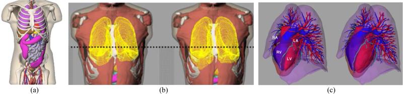
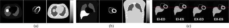
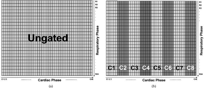
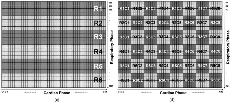
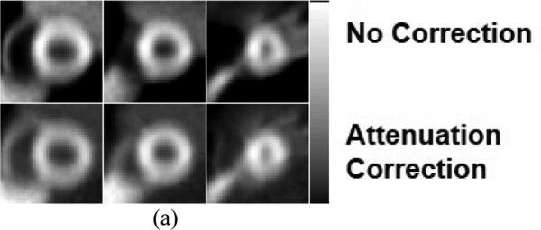
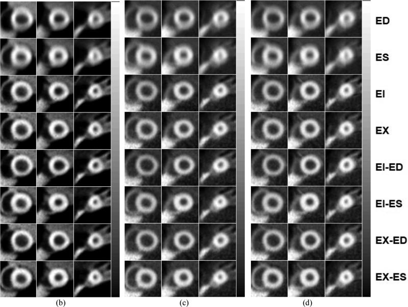
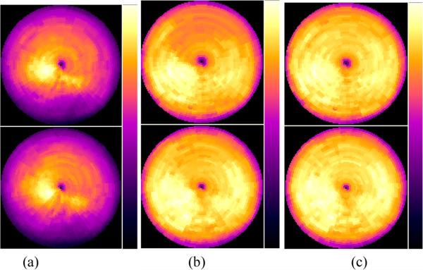
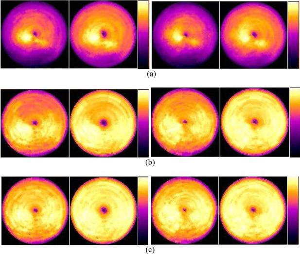
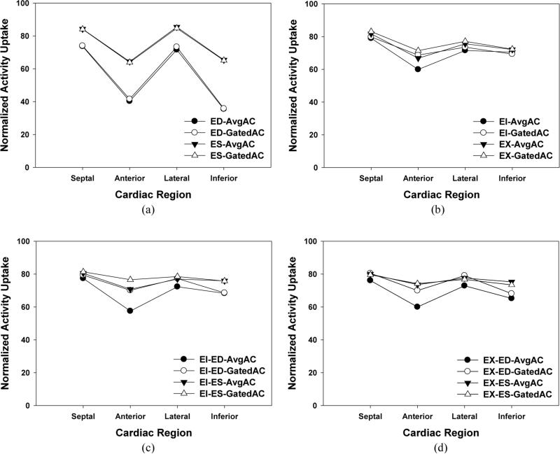
We developed a realistic simulation dataset for simultaneous respiratory and cardiac (R&C) gated SPECT/CT using the 4D NURBS-based Cardiac-Torso (NCAT) Phantom and Monte Carlo simulation methods, and evaluated it for a sample application study. The 4D NCAT phantom included realistic respiratory motion and beating heart motion based on respiratory gated CT and cardiac tagged MRI data of normal human subjects. To model the respiratory motion, a set of 24 separate 3D NCAT phantoms excluding the heart was generated over a respiratory cycle. The beating heart motion was modeled separately with 48 frames per cardiac cycle for each of the 24 respiratory phases. The resultant set of 24 × 48 3D NCAT phantoms provides a realistic model of a normal human subject at different phases of combined R&C motions. An almost noise-free SPECT projection dataset for each of the 1152 3D NCAT phantoms was generated using Monte Carlo simulation techniques and the radioactivity uptake distribution of (99m)Tc sestamibi in different organs. By grouping and summing the separate projection datasets, separate or simultaneous R&C gated acquired data with different gating schemes could be simulated. In the initial evaluation, we combined the projection datasets into ungated, 6 respiratory-gates only, 8 cardiac-gates only, and combined 6 respiratory-gates & 8 cardiac-gates projection datasets. Each dataset was reconstructed using 3D OS-EM without and with attenuation correction using the averaged and respiratory-gated attenuation maps, and the resulting reconstructed images were compared. These results were used to demonstrate the effects of R&C motions and the reduction of image artifact due to R&C motions by gating and attenuation corrections. We concluded that the realistic 4D NCAT phantom and Monte Carlo simulated SPECT projection datasets with R&C motions are powerful tools in the study of the effects of R&C motions, as well as in the development of R&C gating schemes and motion correction methods for improved SPECT/CT imaging.
Figures
Similar articles
-
Task-based evaluation of a 4D MAP-RBI-EM image reconstruction method for gated myocardial perfusion SPECT using a human observer study.Phys Med Biol. 2015 Sep 7;60(17):6789-809. doi: 10.1088/0031-9155/60/17/6789. Epub 2015 Aug 24. Phys Med Biol. 2015. PMID: 26301337 Free PMC article.
-
Development of 4D mathematical observer models for the task-based evaluation of gated myocardial perfusion SPECT.Phys Med Biol. 2015 Apr 7;60(7):2751-63. doi: 10.1088/0031-9155/60/7/2751. Epub 2015 Mar 13. Phys Med Biol. 2015. PMID: 25768980
-
A Simulation Study of the Effect of Phase-Shift on Dual Gated Myocardial Perfusion ECT.IEEE Nucl Sci Symp Conf Rec (1997). 2011 Oct;2011:2728-2732. doi: 10.1109/NSSMIC.2011.6152957. IEEE Nucl Sci Symp Conf Rec (1997). 2011. PMID: 26536654 Free PMC article.
-
Attenuation compensation for cardiac single-photon emission computed tomographic imaging: Part 2. Attenuation compensation algorithms.J Nucl Cardiol. 1996 Jan-Feb;3(1):55-64. doi: 10.1016/s1071-3581(96)90024-0. J Nucl Cardiol. 1996. PMID: 8799228 Review.
-
Application of Monte Carlo Algorithms to Cardiac Imaging Reconstruction.Curr Pharm Des. 2021;27(16):1960-1972. doi: 10.2174/1381612826999201228215225. Curr Pharm Des. 2021. PMID: 33371829 Review.
Cited by
-
4-D Reconstruction With Respiratory Correction for Gated Myocardial Perfusion SPECT.IEEE Trans Med Imaging. 2017 Aug;36(8):1626-1635. doi: 10.1109/TMI.2017.2690819. Epub 2017 Apr 4. IEEE Trans Med Imaging. 2017. PMID: 28391190 Free PMC article.
-
Gated SPECT myocardial perfusion imaging quality assurance in current and future practice.J Nucl Cardiol. 2017 Apr;24(2):543-545. doi: 10.1007/s12350-016-0752-4. Epub 2016 Dec 21. J Nucl Cardiol. 2017. PMID: 28004313 No abstract available.
-
Impact of data-driven cardiac respiratory motion correction on the extent and severity of myocardial perfusion defects with free-breathing CZT SPECT.J Nucl Cardiol. 2018 Aug;25(4):1299-1309. doi: 10.1007/s12350-017-0806-2. Epub 2017 Feb 3. J Nucl Cardiol. 2018. PMID: 28160264
-
[Research on non-contact respiratory rate measurement method based on video information].Sheng Wu Yi Xue Gong Cheng Xue Za Zhi. 2021 Dec 25;38(6):1173-1180. doi: 10.7507/1001-5515.202011029. Sheng Wu Yi Xue Gong Cheng Xue Za Zhi. 2021. PMID: 34970901 Free PMC article. Chinese.
-
Image reconstruction in higher dimensions: myocardial perfusion imaging of tracer dynamics with cardiac motion due to deformation and respiration.Phys Med Biol. 2015 Nov 7;60(21):8275-301. doi: 10.1088/0031-9155/60/21/8275. Epub 2015 Oct 9. Phys Med Biol. 2015. PMID: 26450115 Free PMC article.
References
-
- Achtert AD, King MA, Dahlberg ST, Pretorius PH, LaCroix KJ, Tsui BM. An investigation of the estimation of ejection fractions and cardiac volumes by a quantitative gated SPECT software package in simulated gated SPECT images. J. Nucl. Cardiol. 1998;5:144–52. - PubMed
-
- Blume M, Martinez-Moller A, Keil A, Navab N, Rafecas M. Joint reconstruction of image and motion in gated positron emission tomography. IEEE Trans. Med. Imaging. 2010;29:1892–906. - PubMed
-
- Bundschuh RA, Martinez-Moller A, Essler M, Nekolla SG, Ziegler SI, Schwaiger M. Local motion correction for lung tumours in PET/CT--first results. Eur. J. Nucl. Med. Mol. 2008;35:1981–8. - PubMed
-
- Büther F, Dawood M, Stegger L, Wübbeling F, Schäfers M, Schober O, P SK. List mode-driven cardiac and respiratory gating in PET. J. Nucl. Med. 2009;50:674–81. - PubMed
-
- Chen S, Tsui BMW. Accuracy Analysis of Image Registration Based Respiratory Motion Compensation in Respiratory-Gated FDG Oncologcial PET Reconstruction. Proc. IEEE Medical Imaging Conf. (Dresden, Germany, Oct. 2008) 2008:M06–417.
Publication types
MeSH terms
Grants and funding
LinkOut - more resources
Full Text Sources
Other Literature Sources