Review
. 2015 May 15;334(1):35-44.
doi: 10.1016/j.yexcr.2015.01.004. Epub 2015 Jan 19.

Regulation of mitochondrial transport in neurons

Affiliations
Review

Regulation of mitochondrial transport in neurons

Mei-Yao Lin et al. Exp Cell Res. .

Abstract

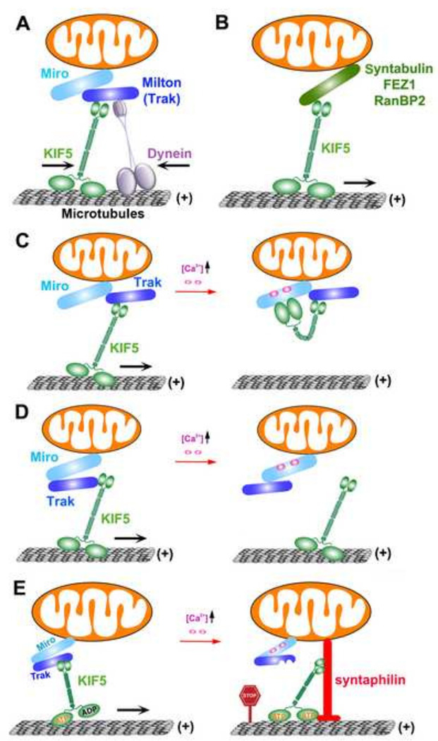
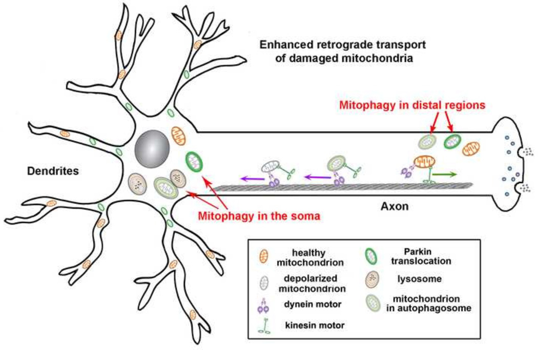
Mitochondria are cellular power plants that supply ATP to power various biological activities essential for neuronal growth, survival, and function. Due to unique morphological features, neurons face exceptional challenges to maintain ATP and Ca(2+) homeostasis. Neurons require specialized mechanisms distributing mitochondria to distal areas where energy and Ca(2+) buffering are in high demand, such as synapses and axonal branches. These distal compartments also undergo development- and activity-dependent remodeling, thereby altering mitochondrial trafficking and distribution. Mitochondria move bi-directionally, pause briefly, and move again, frequently changing direction. In mature neurons, only one-third of axonal mitochondria are motile. Stationary mitochondria serve as local energy sources and buffer intracellular Ca(2+). The balance between motile and stationary mitochondria responds quickly to changes in axonal and synaptic physiology. Furthermore, neurons are postmitotic cells surviving for the lifetime of the organism; thus, mitochondria need to be removed when they become aged or dysfunction. Mitochondria also alter their motility under stress conditions or when their integrity is impaired. Therefore, regulation of mitochondrial transport is essential to meet altered metabolic requirements and to remove aged and damaged mitochondria or replenish healthy ones to distal terminals. Defects in mitochondrial transport and altered distribution are implicated in the pathogenesis of several major neurological disorders. Thus, research into the mechanisms regulating mitochondrial motility is an important emerging frontier in neurobiology. This short review provides an updated overview on motor-adaptor machineries that drive and regulate mitochondrial transport and docking receptors that anchor axonal mitochondria in response to the changes in synaptic activity, metabolic requirement, and altered mitochondrial integrity. The review focuses on microtubule (MT)-based mitochondrial trafficking and anchoring. Additional insight from different perspectives can be found in other in-depth reviews.

Keywords: Dynein motors; Kinesin motors; Mitochondrial docking; Mitochondrial transport; Mitophagy; Motile mitochondria; Stationary mitochondria; Synaptic activity; Syntaphilin.


Figures

Figure 1
Figure 1. Regulation of mitochondrial transport by motor-adaptor complexes and docking receptor
(A). The Miro-Milton (Miro-TRAK) complexes link KIF5 motors for driving mitochondrial transport (Glater et al., 2006; (Koutsopoulos et al., 2010; Macaskill et al., 2009a). Miro and TRAK may also serve as a receptor for dynein for mediating retrograde transport (Franker and Hoogenraad, 2013; Russo et al., 2009; Nguyen et al., 2014; Russo et al., 2009). Thus, relative motility of the opposite motors can be coordinated by mitochondrial adaptor/receptor complexes. (B) Syntabulin serves as an alternative KIF5 motor adaptor for driving mitochondrial anterograde transport (Cai et al., 2005). (C, D) Miro-Ca2+ sensing models for activity-dependent regulation of mitochondrial motility. The C-terminal cargo-binding domain of KIF5 motors binds to the Miro-TRAK adaptor complex. Ca2+ binding to Miro’s EF-hands induces the motor domain to disconnect with MTs and thus prevents motor–MT engagement (C) (Wang and Schwarz, 2009). Alternatively, Ca2+ binding releases KIF5 motors from mitochondria (D) (Macaskill et al., 2009b). Thus, Ca2+ influx upon synaptic activity arrests motile mitochondria at activated synapses. (E) Syntaphilin-mediated “engine-switch and brake” model. A Miro-Ca2+ sensing pathway triggers the binding switch of KIF5 motors from the Miro-TRAK adaptor complex to docking receptor syntaphilin, which immobilizes axonal mitochondria via inhibiting motor ATPase activity. Thus, syntaphilin turns off the “Engine” (KIF5 motor) by sensing a “Stop Sign” (elevated Ca2+) and putting a brake on mitochondria (Chen and Sheng, 2013). Figure is modified from Sheng (2014) originally published in Journal of Cell Biology, 204, 1087–1098 (doi/10.1083/jcb.201312123)
Figure 2
Figure 2. Mitochondrial integrity impacts their transport
Chronically depolarized mitochondria, by dissipating mitochondrial membrane potential (Δψm) with uncoupling reagents alters motility with reduced anterograde and enhanced retrograde transport, thus resulting in accumulation of Parkin-targeted mitochondria in the soma and proximal regions (Cai et al., 2012). This spatial process allows neurons to efficiently remove dysfunctional mitochondria from distal axons to the soma where mature acidic lysosomes are relatively enriched. Damaged mitochondria at axonal terminals can also recruit Parkin for mitophagy once they are anchored by syntaphilin (Cai et al., 2012) or immobilized by turnover of the motor adaptor Miro on the mitochondrial surface (Ashrafi et al, 2014). Autophagosomes engulfing damaged mitochondria at axonal terminals transport predominantly to the soma for maturation and degradation within acidic lysosomes (Maday et al., 2012). Figure is modified from Sheng (2014) originally published in Journal of Cell Biology, 204, 1087–1098 (doi/10.1083/jcb.201312123).

Similar articles

Cited by

References

    1. Ashrafi G, Schlehe JS, LaVoie MJ, Schwarz TL. Mitophagy of damaged mitochondria occurs locally in distal neuronal axons and requires PINK1 and Parkin. J. Cell Biol. 2014;98:15294. - PMC - PubMed
    1. Blasius TL, Cai D, Jih GT, Toret CP, Verhey KJ. Two binding partners cooperate to activate the molecular motor Kinesin-1. J. Cell Biol. 2007;176:11–17. - PMC - PubMed
    1. Brickley K, Stephenson FA. Trafficking kinesin protein (TRAK)-mediated transport of mitochondria in axons of hippocampal neurons. J. Biol. Chem. 2011;286:18079–18092. - PMC - PubMed
    1. Cai Q, Gerwin C, Sheng Z-H. Syntabulin-mediated anterograde transport of mitochondria along neuronal processes. J. Cell Biol. 2005;170:959–969. - PMC - PubMed
    1. Cai Q, Zakaria HM, Simone A, Sheng Z-H. Spatial parkin translocation and degradation of damaged mitochondria via mitophagy in live cortical neurons. Curr. Biol. 2012;22:545–552. - PMC - PubMed

Publication types

Substances