

Corneal stroma microfibrils
- PMID: 25613072
- PMCID: PMC4379971
- DOI: 10.1016/j.exer.2015.01.014
Corneal stroma microfibrils
Abstract
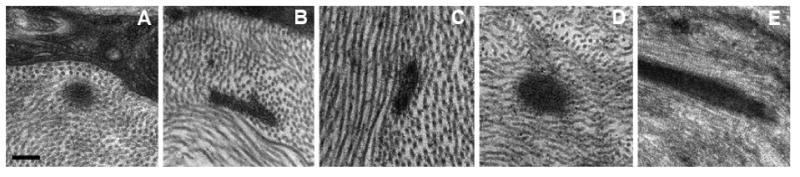
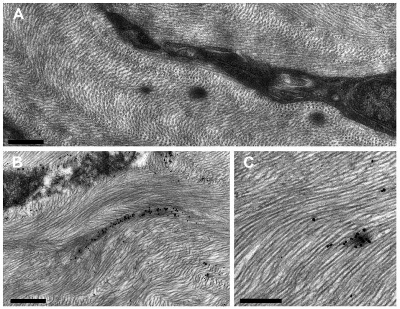
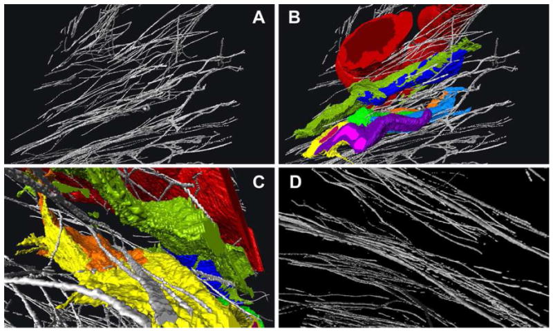
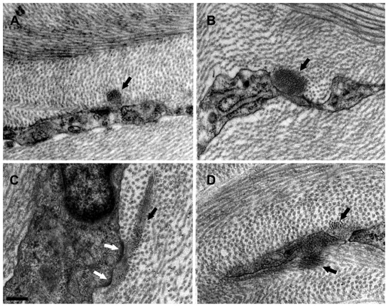
Elastic tissue was first described well over a hundred years ago and has since been identified in nearly every part of the body. In this review, we examine elastic tissue in the corneal stroma with some mention of other ocular structures which have been more thoroughly described in the past. True elastic fibers consist of an elastin core surrounded by fibrillin microfibrils. However, the presence of elastin fibers is not a requirement and some elastic tissue is comprised of non-elastin-containing bundles of microfibrils. Fibers containing a higher relative amount of elastin are associated with greater elasticity and those without elastin, with structural support. Recently it has been shown that the microfibrils, not only serve mechanical roles, but are also involved in cell signaling through force transduction and the release of TGF-β. A well characterized example of elastin-free microfibril bundles (EFMBs) is found in the ciliary zonules which suspend the crystalline lens in the eye. Through contraction of the ciliary muscle they exert enough force to reshape the lens and thereby change its focal point. It is believed that the molecules comprising these fibers do not turn-over and yet retain their tensile strength for the life of the animal. The mechanical properties of the cornea (strength, elasticity, resiliency) would suggest that EFMBs are present there as well. However, many authors have reported that, although present during embryonic and early postnatal development, EFMBs are generally not present in adults. Serial-block-face imaging with a scanning electron microscope enabled 3D reconstruction of elements in murine corneas. Among these elements were found fibers that formed an extensive network throughout the cornea. In single sections these fibers appeared as electron dense patches. Transmission electron microscopy provided additional detail of these patches and showed them to be composed of fibrils (∼10 nm diameter). Immunogold evidence clearly identified these fibrils as fibrillin EFMBs and EFMBs were also observed with TEM (without immunogold) in adult mammals of several species. Evidence of the presence of EFMBs in adult corneas will hopefully pique an interest in further studies that will ultimately improve our understanding of the cornea's biomechanical properties and its capacity to repair.
Keywords: Cornea; Elastic tissue; Fibrillin; Microfibrils; Oxytalan.
Copyright © 2015 Elsevier Ltd. All rights reserved.
Figures
References
-
- Abelsdorff G. Tartuferi. On the elastic tissue of the cornea and a particular method of metallic impregnation. Horstmann C, editor. Systematic Report on the Progress of Ophthalmology in the Third Quarter of the Year 1903. Archives of Ophthalmology. 1904;33:561.
-
- Akhtar S, Alkatan H, Kirat O, Almubrad T. Ultrastructural and three-dimensional study of post-LASIK ectasia cornea. Microscopy research and technique 2013 - PubMed
-
- Alexander RA, Clayton DC, Howes RC, Garner A. Effect of oxidation upon demonstration of corneal oxytalan fibres: a light and electron microscopical study. Medical laboratory sciences. 1981;38:91–101. - PubMed
-
- Alexander RA, Garner A. Elastic and precursor fibres in the normal human eye. Experimental eye research. 1983;36:305–315. - PubMed
Publication types
MeSH terms
Substances
Grants and funding
LinkOut - more resources
Full Text Sources
Other Literature Sources
Miscellaneous
