Review
. 2015:4:208-14.
doi: 10.1016/j.redox.2015.01.001. Epub 2015 Jan 13.

Oxidative stress and autophagy: crucial modulators of kidney injury

Affiliations
Review

Oxidative stress and autophagy: crucial modulators of kidney injury

Angara Sureshbabu et al. Redox Biol. 2015.

Abstract

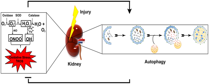
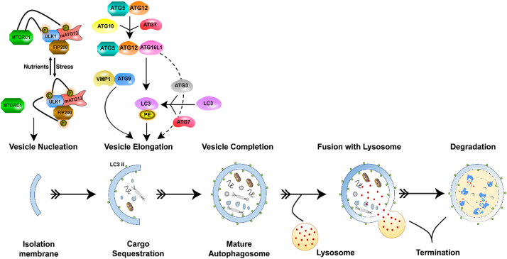
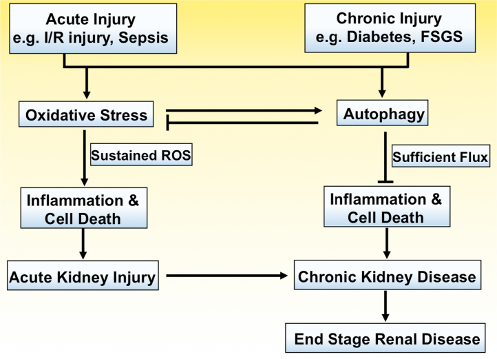
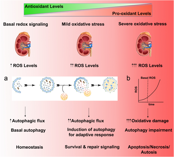
Both acute kidney injury (AKI) and chronic kidney disease (CKD) that lead to diminished kidney function are interdependent risk factors for increased mortality. If untreated over time, end stage renal disease (ESRD) is an inevitable outcome. Acute and chronic kidney diseases occur partly due to imbalance between the molecular mechanisms that govern oxidative stress, inflammation, autophagy and cell death. Oxidative stress refers to the cumulative effects of highly reactive oxidizing molecules that cause cellular damage. Autophagy removes damaged organelles, protein aggregates and pathogens by recruiting these substrates into double membrane vesicles called autophagosomes which subsequently fuse with lysosomes. Mounting evidence suggests that both oxidative stress and autophagy are significantly involved in kidney health and disease. However, very little is known about the signaling processes that link them. This review is focused on understanding the role of oxidative stress and autophagy in kidney diseases. In this review, we also discuss the potential relationships between oxidative stress and autophagy that may enable the development of better therapeutic intervention to halt the progression of kidney disease and promote its repair and resolution.

Keywords: Autophagy; Inflammation cell death; Kidney disease; Kidney injury; Oxidative stress.


Figures

None
Graphical abstract
Fig. 1
Fig. 1
Schematic depiction illustrating the molecular machinery of autophagy with the major autophagy-related proteins in the autophagy signaling pathway. Molecular components that include the ULK1/2-mAtg13-FIP200 complex are necessary for the induction of the autophagy pathway. Under a nutrient-rich environment, mTORC1 phosphorylates ULK1/2 and mAtg13 and leads to association and subsequent inhibition. During stress or starvation, mTORC1 dephosphorylates ULK1/2 and leads to the phosphorylation of FIP200, resulting in downstream activation of the autophagy pathway. E1-like ATG7 and E2-like ATG3 enzymes mediate the lipidation of ubiquitin-like enzyme LC3 with phosphatidylethanolamine (PE) to form LC3-PE. ATG5-ATG12-ATG16L1 complex and VMP1-ATG9 and LC3-PE systems function in cargo sequestration.
Fig. 2
Fig. 2
Interplay of redox signaling and autophagy in acute and chronic kidney injury. In general, both acute and chronic kidney injuries increase oxidative stress and autophagic flux. The proposed mechanism by which oxidative stress contributes to inflammation and cell death involves sustained levels of ROS generation. This excessive oxidative stress, depending upon intensity and/or duration, induces cellular dysfunction and the pathogenesis of AKI and CKD, and ultimately kidney failure and ESRD. Autophagy protects renal tissue by clearing damaged organelles such as mitochondria and thereby indirectly inhibits excessive ROS production, inflammation and cell death.
Fig. 3
Fig. 3
Diagrammatic representation illustrating the induction of oxidative stress and autophagy leading to normal cell growth or cell death at various levels of stress in the kidney. Basal levels of redox signaling and autophagy are necessary for the homeostasis. Mild oxidative stress triggers cell survival and repair mechanisms such as the autophagy pathway. a) Schematic overview of the autophagy pathway b) increased ROS levels above basal values for a prolonged period. In the case of severe oxidative stress, ROS/RNS levels are excessive for a prolonged period leading to oxidative damage and ultimately cell demise. For example, lysosomal permeabilization occurs due to excessive ROS levels can contribute to autophagic impairment and autosis.

References

    1. Bellomo R., Kellum J.A., Ronco C. Acute kidney injury. Lancet. 2012;380(9843):756–766. 22617274 - PubMed
    1. National Kidney and Urologic Diseases Information Clearinghouse (NKUDIC): Kidney Disease Statistics for the United States, 2012 http://kidney.niddk.nih.gov/kudiseases/pubs/kustats/KU_Diseases_Stats_508.pdf
    1. Chawla L.S., Eggers P.W., Star R.A., Kimmel P.L. Acute kidney injury and chronic kidney disease as interconnected syndromes. New England Journal of Medicine. 2014;371(1):58–66. 24988558 - PMC - PubMed
    1. De Duve C. The lysosome. Scientific American. 1963;208:64–72. 14025755 - PubMed
    1. Choi A.M., Ryter S.W., Levine B. Autophagy in human health and disease. New England Journal of Medicine. 2013;368(7):651–662. 23406030 - PubMed

Publication types

MeSH terms