. 2015 Apr;8(2):284-93.
doi: 10.1161/CIRCGENETICS.113.000587. Epub 2015 Jan 22.

GATA-Binding Factor 6 Contributes to Atrioventricular Node Development and Function

Affiliations

GATA-Binding Factor 6 Contributes to Atrioventricular Node Development and Function

Fang Liu et al. Circ Cardiovasc Genet. 2015 Apr.

Abstract

Background: Several transcription factors regulate cardiac conduction system (CCS) development and function but the role of each in specifying distinct CCS components remains unclear. GATA-binding factor 6 (GATA6) is a zinc-finger transcription factor that is critical for patterning the cardiovascular system. However, the role of GATA6 in the embryonic heart and CCS has never been shown.

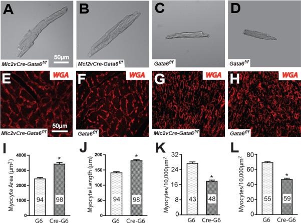
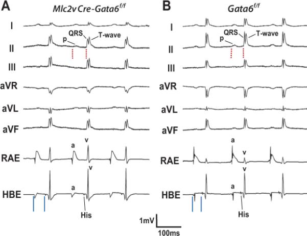
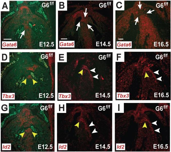
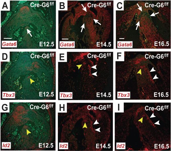
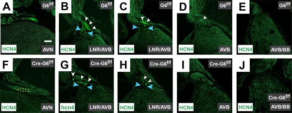
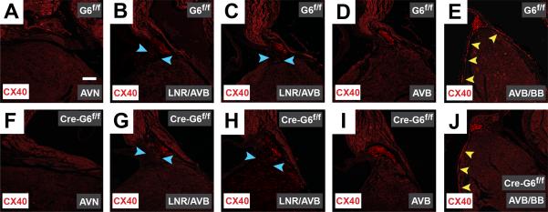
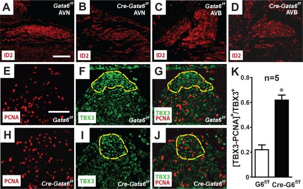
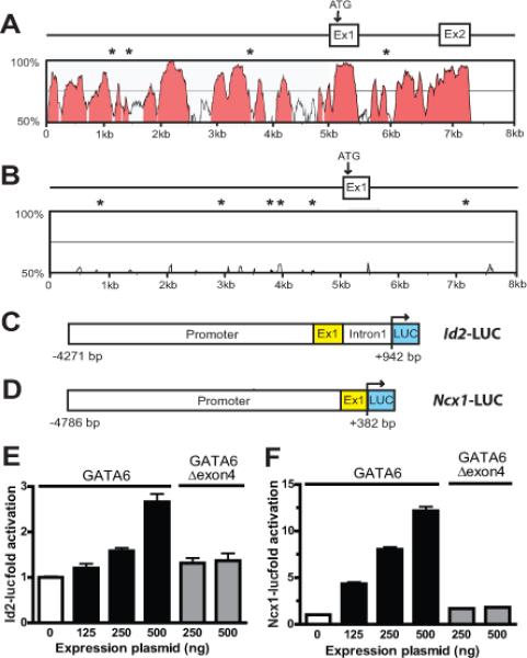
Methods and results: We report that Gata6 is expressed abundantly in the proximal CCS during midgestation in mice. Myocardial-specific deletion of the carboxyl zinc-finger of Gata6 induces loss of hyperpolarizing cyclic nucleotide-gated channel, subtype 4 staining in the compact atrioventricular node with some retention of hyperpolarizing cyclic nucleotide-gated channel, subtype 4 staining in the atrioventricular bundle, but has no significant effect on the connexin-40-positive bundle branches. Furthermore, myocardial-specific deletion of the carboxyl zinc-finger of Gata6 alters atrioventricular conduction in postnatal life as assessed by surface and invasive electrophysiological evaluation, as well as decreasing the number of ventricular myocytes and inducing compensatory myocyte hypertrophy. Myocardial-specific deletion of the carboxyl zinc-finger of Gata6 is also associated with downregulation of the transcriptional repressor ID2 and the cardiac sodium-calcium exchanger NCX1 in the proximal CCS, where GATA6 transactivates both of these factors. Finally, carboxyl zinc-finger deletion of Gata6 reduces cell-cycle exit of TBX3+ myocytes in the developing atrioventricular bundle during the period of atrioventricular node specification, which results in fewer TBX3+ cells in the proximal CCS of mature mutant mice.

Conclusions: GATA6 contributes to the development and postnatal function of the murine atrioventricular node by promoting cell-cycle exit of specified cardiomyocytes toward a conduction system lineage.

Keywords: GATA6 transcription factor; atrioventricular node; cardiac electrophysiology.


Figures

Figure 1
Figure 1
Gata6 carboxyl zinc-finger deletion increases ventricular myocyte size. (A-D) Bright field images of isolated ventricular myocytes from two mature Mlc2vCre-Gata6f/f mice (A,B) and Gata6f/f mice (C,D). Ventricular myocytes without the Gata6 carboxyl zinc-finger are significantly larger. Scale bar in panel A applies to panels B-D. (E-H) Wheat germ agglutinin (WGA) staining of ventricular sections of mature Gata6f/f and mutant mice (E,F) and E16 Gata6f/f and mutant mice (G,H). Ventricular myocyte cross-sections are larger in the mutant ventricles at both ages. Scale bar in panel E applies to panels F-H. The plots show that mature ventricular myocyte area (I) and length (J) are increased in the Gata6 mutant hearts. Myocytes were isolated from 3 different mice of each genotype. The number of myocytes in each group is shown within the bars. Panels K and L show the number of ventricular myocyte cross-sections in each 10,000μm2 region. The number of regions analyzed from three different hearts in each group is shown within the bars. G6=Gata6f/f; Cre-G6f/f=Mlc2vCre-Gata6f/f. *p<0.01.
Figure 2
Figure 2
Conduction defects in mature mice without the Gata6 carboxyl zinc-finger. (A, B) Surface ECG leads I, II, III. aVR, aVL & aVF along with intracardiac recordings of the right atrial electrogram (RAE) and His-bundle electrogram (HBE) from an adult Mlc2vCre-Gata6f/f (A) and Gata6f/f (B) mouse. Note the PR-interval is longer (red dashed lines) in the mutant mouse, with no gross defects in overall QRS-complex morphology or other intervals on the surface ECG leads. The Mlc2vCre-Gata6f/f mouse has a PR-interval of 57 ms vs. 46 ms for the Gata6f/f mouse. Recordings from the HBE show atriohisian (AH) interval prolongation (solid blue lines) without functional Gata6. The Mlc2v-CreGata6f/f mouse has an AH-interval of 42 ms vs. 31 ms for the Gata6f/f mouse. P=P-wave; QRS=QRS-complex; T=T-wave; a=atrial electrogram; v=ventricular electrogram.
Figure 3
Figure 3
Gata6 expression in the developing murine AV conduction system. In situ hybridization with probes against Gata6-exon4, Tbx3 and Id2 mRNA in frontal sections from Gata6f/f mouse embryos are shown. At E12.5 with functional Gata6 (A), Gata6 expression overlaps with Tbx3 (D) and Id2 (G). At E14.5 Gata6 transcript expression (B) is weaker in the regions that overlap with Tbx3 (E) and Id2 (H). By E16.5 Gata6 expression (C) is lower in the regions where it overlaps with Tbx3 (F) and Id2 (I), but Gata6 is more robustly expressed in the Tbx3-negative ventricular myocardium. The white arrows point to regions of developing CCS that are either marked or devoid of Gata6+ staining. The yellow arrowheads point to regions of positive signal within the developing AV node and AV bundle. The white arrowheads point to regions of positive signal within the developing bundle branches. Scale bar=100 μm. Scale bar in (A) applies to all E12.5 images; scale bar in (B) applies to all E14.5 images; scale bar in (C) applies to all E16.5 images.
Figure 4
Figure 4
Effects of Gata6 carboxyl zinc-finger deletion in the developing mouse AV conduction system. In situ hybridization with probes against Gata6-exon4, Tbx3 and Id2 mRNA in frontal sections from Mlc2v-CreGata6f/f mouse embryos are shown. In the absence of Gata6 with a carboxyl zinc-finger, Gata6 expression at E12.5 (A) overlaps with that of Tbx3 (D) and Id2 (G), but the region stained by each of these transcripts is smaller compared to similar anatomic sections from hearts with intact Gata6. A reduced area stained by Gata6 (B), Tbx3 (E) and Id2 (H) persists at E14.5, as well at E16.5 (C, F & I). The white arrows point to regions of the developing CCS that are either marked or devoid of Gata6+ staining. The yellow arrowheads point to regions of positive signal within the developing AV node and AV bundle. The white arrowheads point to regions of positive signal within the developing bundle branches. Scale bar=100 μm. Scale bar in (A) applies to all E12.5 images; scale bar in (B) applies to all E14.5 images; scale bar in (C) applies to all E16.5 images.
Figure 5
Figure 5
Gata6 carboxyl zinc-finger deletion reduces HCN4 expression in the AV node. Immunohistochemical staining of frontal sections from mature murine hearts against HCN4 (green) in control hearts (A-E), and Gata6 mutant hearts (F-J), shows HCN4 expression is significantly diminished within the AV node in the absence of Gata6 with a carboxyl zinc-finger (A & F). The dotted yellow line in panel F represents the region of TBX3+ staining in an adjacent section as shown in Supplemental Figure 2. In both the presence and absence of Gata6 with a carboxyl zinc-finger, HCN4-positive cells are expressed in the lower nodal region and portions of the AV bundle (B-D & G-I). The expected lack of HCN4 staining in the bundle branches is seen in the control and mutant hearts (E & J). Blue arrowheads point to the lower nodal region; white arrowheads point to HCN4+ staining in the AV bundle. Scale bar=250 μm and applies to all panels. AVN=atrioventricular node; AVB=atrioventricular bundle; BB=bundle branches; LNR=lower nodal region.
Figure 6
Figure 6
Gata6 carboxyl zinc-finger deletion does not affect CX40 expression in the mouse CCS. Immunohistochemical staining of frontal sections from mature murine hearts against connexin40 (CX40, red) in Gata6f/f hearts (A-E) and those without the Gata6 carboxyl zinc-finger (F-J) reveals the expected lack of CX40 staining in the AV node, despite the presence of Gata6 (A & F). In the presence and absence of Gata6 with a carboxyl zinc-finger, the expected expression of CX40 within the AV bundle is observed (B-D & G-I). Deletion of the Gata6 carboxyl zinc-finger does not appear to effect the expression of CX40-positive cells within the bundle branches (E & J). The blue arrowheads point to the lower nodal region; the yellow arrowheads point to regions of CX40+ staining in the bundle branches. Scale bar=250 μm and applies to all panels. AVN=atrioventricular node; AVB=atrioventricular bundle; BB=bundle branches; LNR=lower nodal region.
Figure 7
Figure 7
Gata6 carboxyl zinc-finger deletion reduces ID2 expression and cardiomyocyte cell-cycle exit in the developing AV bundle. Immunohistochemical staining in adult murine hearts against ID2 in Gata6f/f hearts (A & C) shows ID2 expression is higher in the proximal CCS compared to the working myocardium. In the absence of Gata6 with a carboxyl zinc-finger (B & D), ID2 expression is reduced in the AV node and AV bundle. Panels E-K show Gata6 with a carboxyl zinc-finger is required for cell-cycle exit of cardiomyocytes in the developing AV bundle. Immunohistochemical staining of E12.5 wild-type hearts (E-G) against PCNA (red) and TBX3 (green), as well as similarly staged Gata6 mutant hearts (H-J). The dashed yellow outline indicates the TBX3-expressing AV bundle. Note there are fewer PCNA/TBX3-positive cells in the Gata6f/f heart compared to the mutant heart. (K) PCNA-positive nuclei in the crest of the septum that overlap with TBX3-expressing cells averaged from five hearts of each genotype. *P<0.01. Scale bar=100 μm. Scale bar in (A) applies to (A-D); scale bar in (E) applies to (E-J). AVN=atrioventricular node; AVB=atrioventricular bundle.
Figure 8
Figure 8
GATA6 transactivates Id2 and Ncx1. (A) VISTA comparison of the murine and human Id2 proximal promoter and exon 1, and (B) of the murine and human Ncx1 proximal promoter and exon 1. In panels A and B the x and y axes indicate sequence length (kb) and percent homology (≥ 75%, pink), respectively. GATA6 binding sites conserved in the mouse and human sequence are indicated by asterisks. (C) Schematic of the Id2-LUC reporter containing the 5.2-kb proximal promoter upstream of firefly luciferase (LUC), and (D) the Ncx1-LUC reporter containing the 5.1-kb proximal promoter that is also upstream of firefly luciferase. (E) Activation of the Id2-LUC reporter by GATA6. HL-1 cells were transiently transfected with Id2-LUC and 125–500 ng of expression plasmid encoding wild-type GATA6 or GATA6 lacking exon 4 (Gata6Δexon4). The reporter is activated by wild-type GATA6 but not by GATA6Δexon4. (F). Activation of the Ncx1-LUC reporter by GATA6. HL-1 cells were similarly transiently transfected with Ncx1-LUC and 125–500 ng of expression plasmid encoding wild-type GATA6 or GATA6Δexon4. As with Id2-LUC, the Ncx1-LUC reporter was activated by the expression of wild-type GATA6 but not by GATA6Δexon4. The transient transection studies in panels E and F show the mean change in report activity from at least 3 different experiments at each concentration of expression plasmid.

Comment in

  • Guiding Cardiac Conduction With GATA.
    Nemer M, Gharibeh L. Nemer M, et al. Circ Cardiovasc Genet. 2015 Apr;8(2):247-9. doi: 10.1161/CIRCGENETICS.115.001039. Circ Cardiovasc Genet. 2015. PMID: 25901035 No abstract available.

References

    1. Pashmforoush M, Lu JT, Chen H, Amand TS, Kondo R, Pradervand S, et al. Nkx2-5 pathways and congenital heart disease: loss of ventricular myocyte lineage specification leads to progressive cardiomyopathy and complete heart block. Cell. 2004;117:378–386. - PubMed
    1. Moskowitz IP, Kim JB, Moore ML, Wolf CM, Peterson MA, Shendure J, et al. A molecular pathway including Id2, Tbx5, and Nkx2-5 required for cardiac conduction system development. Cell. 2007;129:1365–1376. - PubMed
    1. Ismat FA, Zhang M, Kook H, Huang B, Zhou R, Ferrari VA, et al. Homeobox protein Hop functions in the adult cardiac conduction system. Circ Res. 2005;96:898–903. - PMC - PubMed
    1. Bakker ML, Boukens BJ, Mommersteeg MT, Brons JF, Wakker V, Moorman AF, et al. Transcription factor Tbx3 is required for the specification of the atrioventricular conduction system. Circ Res. 2008;102:1340–1349. - PubMed
    1. Aanhaanen WT, Brons JF, Domínguez JN, Rana MS, Norden J, Airik R, et al. The Tbx2+ primary myocardium of the atrioventricular canal forms the atrioventricular node and the base of the left ventricle. Circ Res. 2009;104:1264–1274. - PubMed

Publication types

MeSH terms

Substances