. 2015 Sep;141(9):1563-74.
doi: 10.1007/s00432-015-1917-2. Epub 2015 Jan 23.

Role of tumor cell surface lysosome-associated membrane protein-1 (LAMP1) and its associated carbohydrates in lung metastasis

Affiliations

Role of tumor cell surface lysosome-associated membrane protein-1 (LAMP1) and its associated carbohydrates in lung metastasis

Akhil Kumar Agarwal et al. J Cancer Res Clin Oncol. 2015 Sep.

Abstract

Purpose: Expression of lysosome-associated membrane protein-1 (LAMP1) on the surface correlates with metastatic potential of B16 melanoma cells. Downregulation of their expression in high metastatic (B16F10) cells reduced their surface expression and metastatic potential. Present investigations explore if overexpression of LAMP1 on the surface of low metastatic (B16F1) cells augment their metastatic ability, and if so, how?

Methods: B16F1 cells were transduced with lentiviral vector carrying mutant-LAMP1 (Y386A) (mutLAMP1). Surface expression of LAMP1 and carbohydrates was analyzed by flow cytometry, immunofluorescence and/or immunoprecipitation and Western blotting. Cell spreading and motility were assessed on components of extracellular matrix (ECM) (fibronectin) and basement membrane (BM) (matrigel), and galectin-3-coated coverslips/plates. Metastatic potential was assessed using experimental metastasis assay.

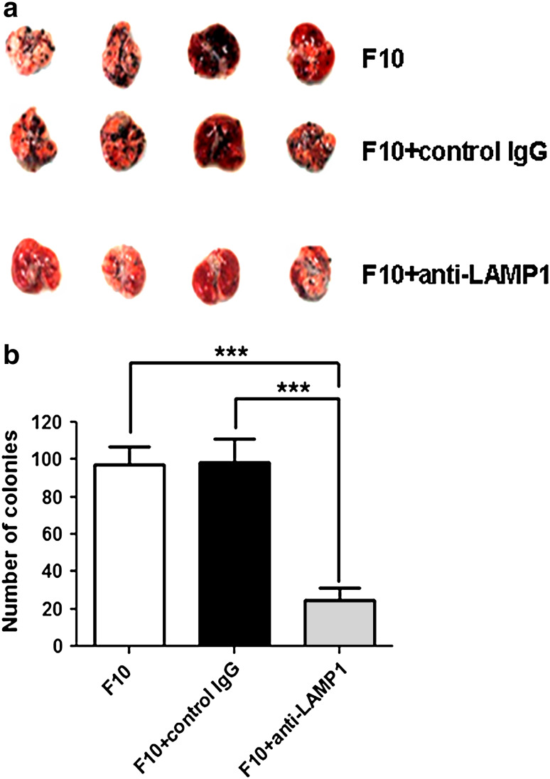
Results: Pre-incubation with anti-LAMP1 antibodies significantly reduced lung metastasis of B16F10 cells. Overexpression of mutLAMP1 significantly increased its surface expression on B16F1 cells, resulting in increased cellular spreading and motility on fibronectin and matrigel. LAMP1 is the major carrier of poly-N-acetyllactosamine (polyLacNAc) on B16F10 cells. However, significantly higher expression of mutLAMP1 had no effect on galectin-3 binding on cell surface or on spreading or motility of cells on galectin-3-coated coverslips/plates. These cells also failed to show any gain in metastatic ability. This could be because LAMP1 from these cells carried significantly lower levels of polyLacNAc in comparison with B16F10 cells.

Conclusions: PolyLacNAc on B16F10 cells and galectin-3 on lungs are the major participants in melanoma metastasis. Although surface LAMP1 promotes interactions with organ ECM and BM, carbohydrates on LAMP1 play a decisive role in dictating lung metastasis.


Conflict of interest statement

We declare that we have no conflict of interest.

Figures

Fig. 1
Fig. 1
Cell surface LAMP1 plays an important role in lung-specific metastasis. a Melanoma colonies on lungs of C57BL/6 mice injected with untreated B16F10 cells (F10), and those treated with 10 μg/ml of either control rat IgG (F10 + control IgG) or blocking antibodies to LAMP1 (F10 + anti-LAMP1). Four mice were taken in each group. b Graphical representation of mean number of lung colonies. One-way analysis of variance followed by Bonferroni’s multiple comparison test was performed to compare significance (denoted by *** p value <0.001)
Fig. 2
Fig. 2
Analysis of LAMP1 expression on the surface of melanoma cells. Comparison of surface expression of LAMP1 by flow cytometry, in a B16F1 cells (gray shade F1), B16F1 cells infected with viruses having empty vector control (pink dotted lines VC) and B16F10 cells (green solid lines F10) and b comparison of the same between vector control (pink dotted lines VC) and B16F1 clones expressing mutLAMP1 (gray shade C1) and (orange solid lines C11). Cells treated with only anti-rat FITC served as control (__). The flow cytometry overlays have been split into two for better understanding of the data. c Graphical representation of the mean fluorescence intensities of surface LAMP1 of the cells shown in a and b. d Immunofluorescence images of the cells stained with anti-LAMP1 antibody and FITC-labelled secondary antibody (green). Nuclei were stained with DAPI (blue). Scale bar 5 μm
Fig. 3
Fig. 3
Effect of increased surface expression of LAMP1 on spreading of melanoma cells on fibronectin and matrigel. Spreading of B16F1 cells infected with viruses having empty vector (VC) or those having mutLAMP1 (C1 and C11) and F10 cells on a fibronectin (FN) and b matrigel (Mat)-coated coverslips as assessed by staining with Phalloidin-FITC (green). DAPI was used to stain the nuclei (blue). Scale bar 5 μm. c Each bar represents ratio of cytoplasmic to nuclear (C/N) area for around 100 cells from two different experiments for spreading on fibronectin (FN) and d for spreading on matrigel (Mat). One-way analysis of variance followed by Bonferroni’s multiple comparison test was performed to compare significance (denoted by *** p value <0.001)
Fig. 4
Fig. 4
Effect of increased surface expression of LAMP1 on motility of melanoma cells on fibronectin and matrigel. Motility of B16F1 cells infected with viruses having empty vector (VC) or those having mutLAMP1 (C1 and C11) and F10 cells on a fibronectin and b matrigel-coated plates as represented by time lapse video microscopy images at 0 and 16 h of wound closure. c, d Represent mean percent wound closure at 4-h interval on fibronectin and matrigel, respectively. Area of wound closure was measured by Image J software and each image from two different experiments was analyzed at three different positions. Two-way analysis of variance followed by Bonferroni’s multiple comparison test was performed to compare significance from VC (denoted by **** p value <0.0001, *** p value <0.001)
Fig. 5
Fig. 5
Effect of increased surface expression of LAMP1 on spreading of melanoma cells on galectin-3. Spreading of B16F1 cells infected with viruses having empty vector (VC) or those having mutLAMP1 (C1 and C11) and F10 cells on a uncoated (Un) and b galectin-3 (Gal3)-coated coverslips as assessed by staining with Phalloidin-FITC (green). DAPI was used to stain the nuclei (blue). Scale bar 5 μm. c, d Each bar represents ratio of cytoplasmic to nuclear (C/N) area for around 100 cells from two different experiments for spreading on uncoated c and galectin-3-coated coverslips d. One-way analysis of variance followed by Bonferroni’s multiple comparison test was performed to compare significance
Fig. 6
Fig. 6
Effect of increased surface expression of LAMP1 on motility of melanoma cells on galectin-3. Motility of B16F1 cells infected with viruses having empty vector (VC) or those having mutLAMP1 (C1 and C11) and F10 cells on a BSA and b galectin-3-coated plates as represented by time lapse video microscopy images at 0 and 16 h of wound closure. c, d Represent mean percent wound closure at 4-h interval on BSA and galectin-3, respectively. Area of wound closure was measured by Image J software and each image from two different experiments was analyzed at three different positions. Two-way analysis of variance followed by Bonferroni’s multiple comparison test was performed to compare significance from VC (denoted by *** p value <0.001)
Fig. 7
Fig. 7
Increased surface expression of LAMP1 on B16F1 cells has no effect on lung metastasis. a Melanoma colonies on lungs of C57BL/6 mice injected with F1, VC, C1, C11 and F10 cells. Five mice were taken in each group. b Comparison of galectin-3 binding by flow cytometry using biotinylated galectin-3 in uninfected B16F1 cells (red dotted lines F1) or those infected with viruses having empty vector as control (pink dotted lines VC) with B16F1 clones expressing mutLAMP1 (blue dotted lines C1) and (orange dotted lines C11) and with B16F10 cells (green solid line F10). Cells treated with only extra-avidin FITC (__) served as control. c Graphical representation of the mean fluorescence intensities of galectin-3 binding of all the cells. d Comparison of β1,6 branched N-oligosaccharides (LPHA) and polyLacNAc (LEA) on normalized amounts of immunoprecipitated LAMP1 from B16F10 (F10) cells and B16F1 cells having either empty vector (VC) or mutLAMP1 (C1), by Western blotting

References

    1. Agarwal AK, Kalraiya RD (2014) Glycosylation regulates the expression of Lysosome Associated Membrane Protein-1 (LAMP1) on the cell surface. J Biosci Technol 5:556–563
    1. Agarwal AK, Gude RP, Kalraiya RD (2014) Regulation of melanoma metastasis to lungs by cell surface Lysosome Associated Membrane Protein-1 (LAMP1) via galectin-3. Biochem Biophys Res Commun 449:332–337 - PubMed
    1. Alter G, Malenfant JM, Altfeld M (2004) CD107a as a functional marker for the identification of natural killer cell activity. J Immunol Methods 294:15–22 - PubMed
    1. Bayer EA, Wilchek M (1990) Protein biotinylation. Methods Enzymol 184:138–160 - PubMed
    1. Betts MR, Brenchley JM, Price DA, De Rosa SC, Douek DC, Roederer M, Koup RA (2003) Sensitive and viable identification of antigen-specific CD8+ T cells by a flow cytometric assay for degranulation. J Immunol Methods 281:65–78 - PubMed

Publication types