Comparative Study
. 2015 Mar;35(3):645-54.
doi: 10.1161/ATVBAHA.114.305054. Epub 2015 Jan 22.

Fibrin, γ'-fibrinogen, and transclot pressure gradient control hemostatic clot growth during human blood flow over a collagen/tissue factor wound

Affiliations
Comparative Study

Fibrin, γ'-fibrinogen, and transclot pressure gradient control hemostatic clot growth during human blood flow over a collagen/tissue factor wound

Ryan W Muthard et al. Arterioscler Thromb Vasc Biol. 2015 Mar.

Abstract

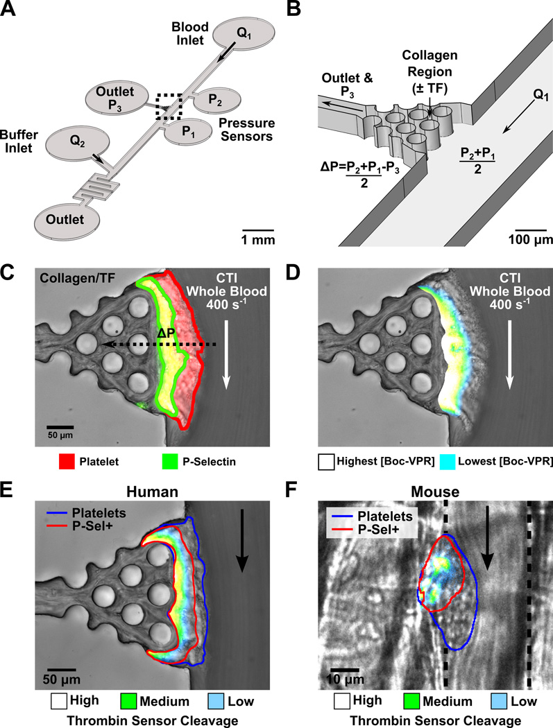
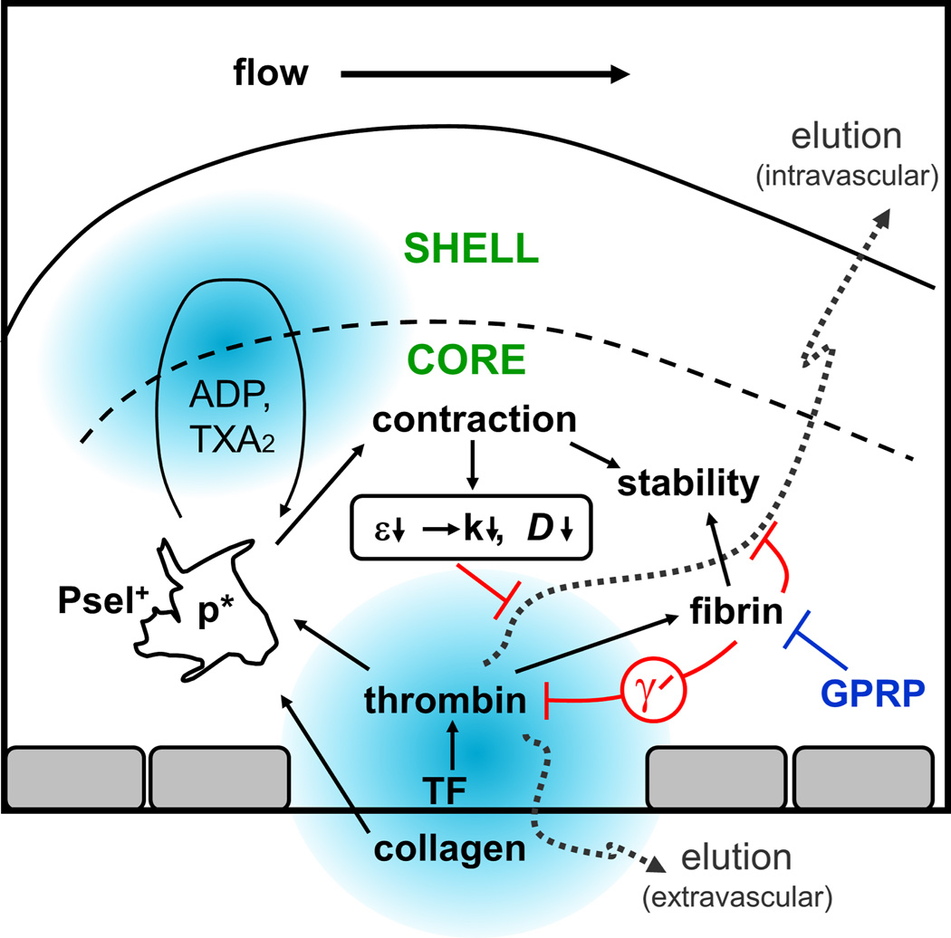
Objective: Biological and physical factors interact to modulate blood response in a wounded vessel, resulting in a hemostatic clot or an occlusive thrombus. Flow and pressure differential (ΔP) across the wound from the lumen to the extravascular compartment may impact hemostasis and the observed core/shell architecture. We examined physical and biological factors responsible for regulating thrombin-mediated clot growth.

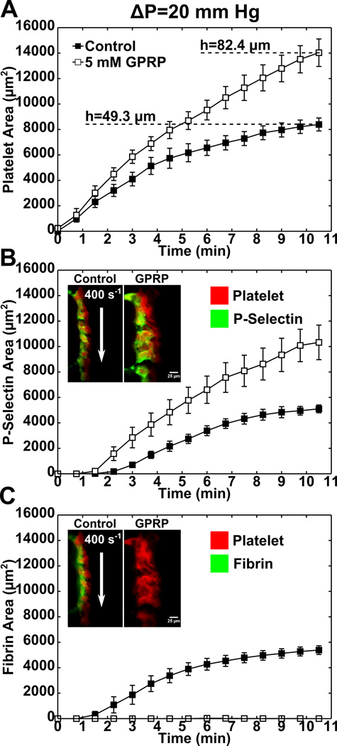
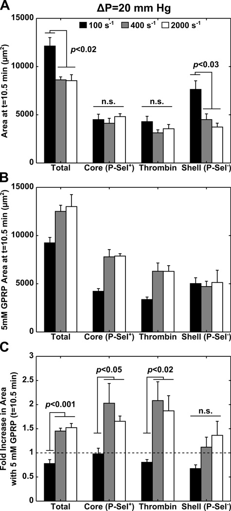
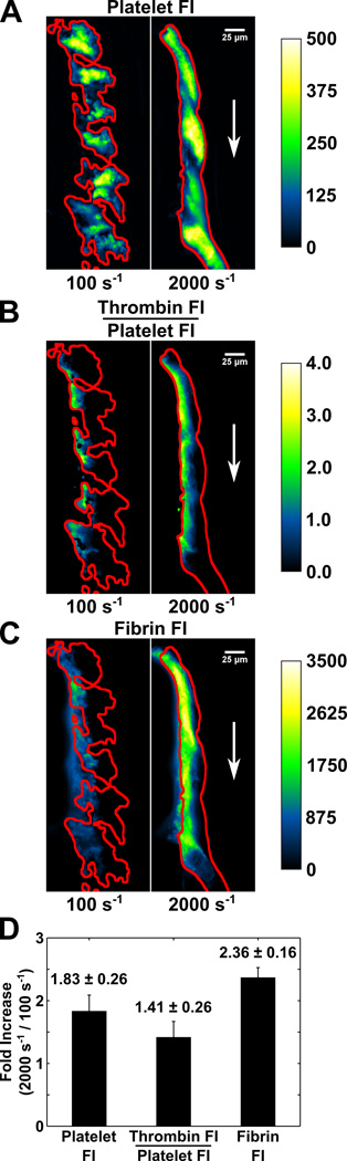
Approach and results: Using factor XIIa-inhibited human whole blood perfused in a microfluidic device over collagen/tissue factor at controlled wall shear rate and ΔP, we found thrombin to be highly localized in the P-selectin(+) core of hemostatic clots. Increasing ΔP from 9 to 29 mm Hg (wall shear rate=400 s(-1)) reduced P-selectin(+) core size and total clot size because of enhanced extravasation of thrombin. Blockade of fibrin polymerization with 5 mmol/L Gly-Pro-Arg-Pro dysregulated hemostasis by enhancing both P-selectin(+) core size and clot size at 400 s(-1) (20 mm Hg). For whole-blood flow (no Gly-Pro-Arg-Pro), the thickness of the P-selectin-negative shell was reduced under arterial conditions (2000 s(-1), 20 mm Hg). Consistent with the antithrombin-1 activity of fibrin implicated with Gly-Pro-Arg-Pro, anti-γ'-fibrinogen antibody enhanced core-localized thrombin, core size, and overall clot size, especially at venous (100 s(-1)) but not arterial wall shear rates (2000 s(-1)). Pathological shear (15 000 s(-1)) and Gly-Pro-Arg-Pro synergized to exacerbate clot growth.

Conclusions: Hemostatic clotting was dependent on core-localized thrombin that (1) triggered platelet P-selectin display and (2) was highly regulated by fibrin and the transclot ΔP. Also, γ'-fibrinogen had a role in venous but not arterial conditions.

Keywords: fibrin; hemodynamics; hemostasis; thrombin.


Figures

Figure 1
Figure 1. Human thrombi develop a core and shell architecture that mimic the architecture of clots developed in vivo
Anticoagulated whole blood was perfused at a constant wall shear rate (Q1) while the transthrombus pressure gradients (ΔP) were independently controlled via downstream buffer infusion Q2 (A). Polymerized collagen ± TF was localized to a post scaffold centered between pressure ports P2 and P1 (B). Spatial and temporal thrombus development was monitored by side-view imaging at controlled wall shear rates and ΔP. Platelet (red) and P-selectin (green) antibodies (anti-CD41 and anti-CD62P) were used to investigate spatial differences in platelet activation under physiologic flow conditions (n=3) (C). A fluorogenic, thrombin cleavable substrate Boc-VPR-AMC was added to whole blood to demonstrate localized thrombin activity within clots developed in the presence of ΔP (n=3) (D). Human clots developed in vitro (n=4) (E) and mouse clots developed in vivo (n=3 mice, 13 clots) (F) were outlined by imaging the clots using platelet (blue) and P-selectin antibodies (red). A heat map was used in both images to illustrate the localization of thrombin sensor cleavage (light blue = low intensity and white = high intensity).
Figure 2
Figure 2. Transthrombus pressure gradients reduce clot size by diminishing thrombin boundary growth
Platelet area (anti-CD41 antibody) was measured for clots (n=3) formed at a constant wall shear rate (400 s−1) in the presence of either a low (ΔP=9.0 mm Hg) or high (ΔP=29.4 mm Hg) transthrombus pressure gradient (A). Clot architecture was investigated by measuring intrathrombus P-selectin (anti-CD62P) (B) and thrombin (C) area for each condition (n=3). The correlation between platelet activation marker P-selectin and thrombin area (thrombin sensing antibody) were quantified for each condition over the duration of the experiment (t=10.8 min) (D).
Figure 3
Figure 3. Fibrin polymerization reduces clot growth at a wall shear rate of 400 s−1 and ΔP=20 mm Hg
Platelet area (n=7, anti-CD41 antibody) was measured in the absence (control) and presence (5 mM GPRP) of fibrin polymerization inhibitor Gly-Pro-Arg-Pro at a constant transthrombus pressure gradient (ΔP=20 mm Hg) and wall shear rate (400 s−1). P-selectin (n=7, B) and fibrin (n=4, C) area were monitored for each condition during 10.5 min of CTI whole blood perfusion. Anti-CD62P and the fibrin specific antibody (T2G1) were used to measure the P-selectin and fibrin area respectively.
Figure 4
Figure 4. Wall shear rate does not change the core or thrombin area but does influence thrombin mediated clot growth in the absence of fibrin polymerization
Total, core (p-sel+), thrombin, and shell (p-sel) areas were measured, at the final time point (t=10.5 min), for clots developed at a constant pressure gradient (ΔP=20 mm Hg) and varying wall shear rate (100 s−1, 400 s−1, 2000 s−1) (n=4 for each shear rate) (A). CTI whole blood in the presence of 5 mM GPRP (fibrin polymerization inhibitor) was run in parallel at all experimental conditions (n=4 for each shear rate) (B). The fold increase in area with the addition of 5 mM GPRP was calculated for total, core, thrombin, and shell area (C). Total platelet and P-selectin+ area were measured with antibodies specific for anti-CD41 and anti-CD62P respectively. Thrombin area was measured with a thrombin sensing antibody and the shell area was calculated as the difference between the total platelet area and the P-selectin+ area.
Figure 5
Figure 5. Inhibition of γ’ fibrin(ogen) with a γ’ specific monoclonal antibody results in larger clots at venous but not arterial shear rates
Whole blood anticoagulated with CTI ± γ’ mAb was perfused over a collagen/TF scaffold at a constant transthrombus pressure gradient (buffer (1.2% final glycerol) ΔP=15.5 mm Hg, γ’ antibody (1.2% final glycerol) ΔP=15.7 mm Hg). Total (anti-CD41), core (p-sel+, anti-CD62P), and thrombin area (thrombin sensing antibody) were measured dynamically at both venous (100 s−1, first column, n=3) and arterial (2000 s−1, second column, n=3) shear rates throughout the duration of the experiment.
Figure 6
Figure 6. Clots formed at arterial shear rates pack tighter, cleave more thrombin sensitive peptide per area, and generate more fibrin per area than venous clots
CTI whole blood was perfused simultaneously at a venous shear rate of 100 s−1 and an arterial shear rate of 2000 s−1. Platelet (A), normalized thrombin sensor (B), and fibrin fluorescence intensity (C) were imaged for 11.5 min. The fold increase in platelet fluorescence intensity per area (n=6) from clots formed at 100 s−1 compared to 2000 s−1 was averaged over the duration of the experiment (D). In addition, the density corrected thrombin sensor cleavage (n=6) and fibrin fluorescence intensity (n=3) were calculated and compared for venous and arterial clots. Platelet, thrombin, and fibrin fluorescence were measured with an anti-CD41 antibody, thrombin sensing antibody, and fibrin specific antibody (T2G1) respectively.
Figure 7
Figure 7. Clot growth at pathological shear rates was abated by fibrin polymerization
PPACK whole blood was perfused across a collagen scaffold at 200 s−1. Clot formation was imaged with a fluorescent platelet specific monoclonal antibody (red, anti-CD41) (A). Following 7 min of perfusion, the shear rate was increased to a pathological shear rate of 15,000 s−1. Images of clot formation were taken at t=10.75 min (B), and t=14.75 min (C). Zones of low shear, caused by rapid platelet aggregation upstream, often formed and contracted at the trailing edge of the developing clot. In a similar experiment either CTI or PPACK whole blood was perfused over a collagen/TF or collagen surface respectively. Perfusion was initially at 2000 s−1 prior to stepping the shear up to 15,000 s−1. Control conditions were compared to GPRP treated samples with inhibited fibrin polymerization. Clot height was recorded throughout the duration of the experiment (D). Average clot heights were calculated for both the control and GPRP samples. The measurements were made for both CTI (n=4 each condition) and PPACK (n=4 each condition) treated whole blood both before and after increasing the shear rate (E). (*, P<0.01)
Figure 8
Figure 8. Thrombin localization within clots is regulated by fibrin, γ’ fibrin(ogen), and transthrombus pressure gradients
A schematic representing the regulation of thrombin and small molecules ADP and TXA2 demonstrates both the physical and biochemical transport regulations observed within clots. (ε, porosity; k, permeability, D, diffusivity, p*, activated platelet)

Similar articles

Cited by

References

    1. Stalker TJ, Traxler EA, Wu J, Wannemacher KM, Cermignano SL, Voronov R, Diamond SL, Brass LF. Hierarchical organization in the hemostatic response and its relationship to the platelet-signaling network. Blood. 2013;121:1875–1885. - PMC - PubMed
    1. Welsh JD, Stalker TJ, Voronov R, Muthard RW, Tomaiuolo M, Diamond SL, Brass LF. A systems approach to hemostasis: 1. The interdependence of thrombus architecture and agonist movements in the gaps between platelets. Blood. 2014;124:1808–1815. - PMC - PubMed
    1. Tomaiuolo M, Stalker TJ, Welsh JD, Diamond SL, Sinno T, Brass LF. A systems approach to hemostasis: 2. Computational analysis of molecular transport in the thrombus microenvironment. Blood. 2014;124:1816–1823. - PMC - PubMed
    1. Stalker TJ, Welsh JD, Tomaiuolo M, Wu J, Colace TV, Diamond SL, Brass LF. A systems approach to hemostasis: 3. Thrombus consolidation regulates intrathrombus solute transport and local thrombin activity. Blood. 2014;124:1824–1831. - PMC - PubMed
    1. Bray PF. Platelet hyperreactivity: predictive and intrinsic properties. Hematol Oncol Clin North Am. 2007;21:633–645. - PMC - PubMed

Publication types

MeSH terms