Review
. 2015 Feb;15(2):104-16.
doi: 10.1038/nri3793.

Cholesterol, inflammation and innate immunity

Affiliations
Review

Cholesterol, inflammation and innate immunity

Alan R Tall et al. Nat Rev Immunol. 2015 Feb.

Abstract

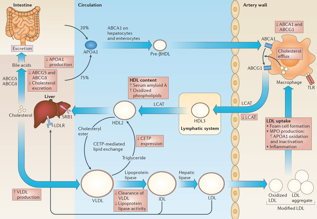
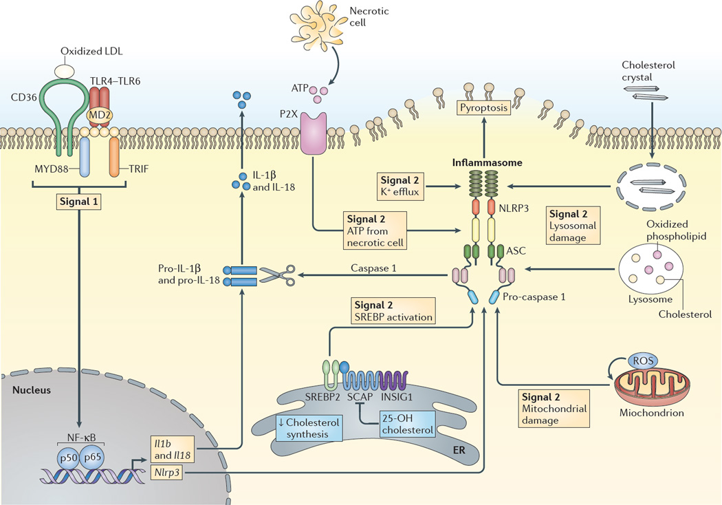
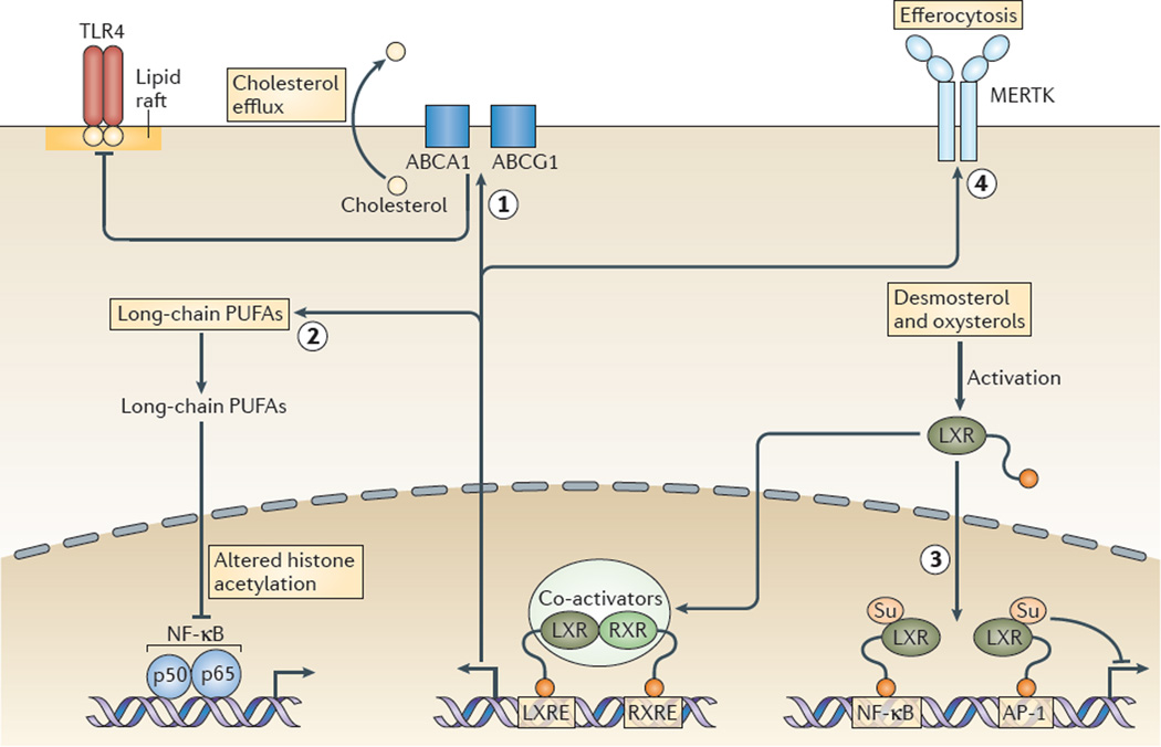
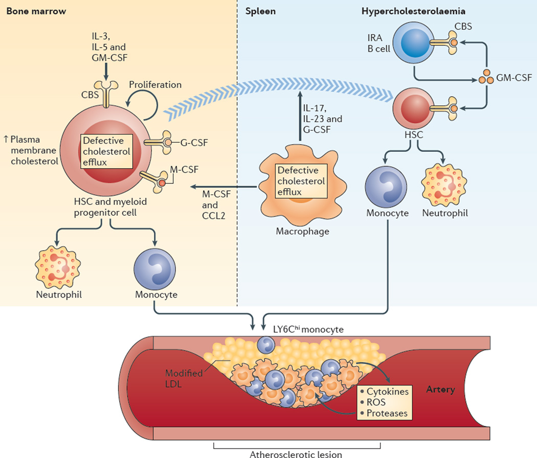
Hypercholesterolaemia leads to cholesterol accumulation in macrophages and other immune cells, which promotes inflammatory responses, including augmentation of Toll-like receptor (TLR) signalling, inflammasome activation, and the production of monocytes and neutrophils in the bone marrow and spleen. On a cellular level, activation of TLR signalling leads to decreased cholesterol efflux, which results in further cholesterol accumulation and the amplification of inflammatory responses. Although cholesterol accumulation through the promotion of inflammatory responses probably has beneficial effects in the response to infections, it worsens diseases that are associated with chronic metabolic inflammation, including atherosclerosis and obesity. Therapeutic interventions such as increased production or infusion of high-density lipoproteins may sever the links between cholesterol accumulation and inflammation, and have beneficial effects in patients with metabolic diseases.


Figures

Figure 1
Figure 1. RCT and its regulation by innate immune responses
The process of reverse cholesterol transport (RCT) is depicted and how inflammation impairs this process is described in the red boxes. Under physiological conditions, apolipoprotein A1 (APOA1), which is the major protein component of high-density lipoprotein (HDL), is secreted by the liver and the intestines, and is assembled into a pre-βHDL particle as a result of its interaction with the ATP-binding cassette transporter ABC subfamily A member 1 (ABCA1) on hepatocytes and enterocytes. ABCA1 on macrophages promotes cholesterol and phospholipid efflux onto these relatively lipid-poor pre-βHDL particles, initiating the process of RCT. ABCG1 promotes further cholesterol efflux onto HDL particles. Free cholesterol in HDL is esterified by the enzyme lecithin–cholesterol acyltransferase (LCAT), which gives rise to cholesteryl esters. Free cholesterol or cholesteryl esters in HDL may be directly cleared in the liver via scavenger receptor B1 (SRB1), which mediates a process of selective free cholesterol or cholesteryl ester uptake in which the lipid moiety of HDL is mostly removed and the protein portion is recycled into the circulation (not shown). Cholesterol deposited in the liver by RCT can either be recycled in the form of secreted triglyceride-rich, very low-density lipoproteins (VLDLs; the main protein component of which is APOB) or can undergo net excretion into the bile via ABCG5 and ABCG8. In humans, plasma cholesteryl ester transfer protein (CETP) mediates the exchange of cholesteryl esters in HDL with triglyceride in VLDL. A lipolytic cascade mediated by lipoprotein lipase and hepatic lipase causes hydrolysis of triglycerides and results in the formation of cholesterol-rich and cholesteryl ester-rich LDL. Although most LDL is cleared in the liver, LDL may supply cholesterol to peripheral tissues and a small proportion is taken up into the arterial wall, where it is modified by oxidation or aggregation, leading to its uptake by macrophages. Modified LDL in the artery wall promotes Toll-like receptor (TLR) signalling in macrophages and it is taken up by these cells, leading to the formation of macrophage foam cells, the production of myeloperoxidase (MPO) and inflammation. IDL, intermediate-density lipoprotein; LDLR, LDL receptor.
Figure 2
Figure 2. Inflammasome activation by sterols
The NLRP3 (NOD-, LRR- and pyrin domain-containing 3) inflammasome is activated by many signals, including infectious agents and stress- or injury-induced host factors, leading to caspase 1 activation, cleavage of pro-interleukin-1β (pro-IL-1β) and pro-IL-18, and the secretion of the active cytokines, as well as in some cases resulting in a form of cell death termed pyroptosis. A priming stimulus (signal 1), acting through nuclear factor-κB (NF-κB), induces the expression of Il1b, Il18 and Nlrp3, and precedes assembly of the inflammasome complex. In atherosclerosis, priming may result from pattern recognition receptor activation; for example, combinatorial Toll-like receptor 4 (TLR4)–TLR6–CD36 signalling induced by oxidized low-density lipoprotein (LDL). The second stimulus (signal 2) may arise from various stressors. In atherosclerosis, one such signal is lysosomal damage or dysfunction, which may result from phagocytosis of extracellular cholesterol crystals via the CD36-mediated uptake of modified LDL (not shown) and free cholesterol release and crystallization in lysosomes. Other signals may result from mitochondrial damage, oxidative stress-induced production of reactive oxygen species (ROS) or ATP release from dying cells. 25-hydroxycholesterol (25-OH cholesterol) suppresses inflammasome activation by reduced sterol regulatory element-binding protein 2 (SREBP2) cleavage mediated by SREBP cleavage-activating protein (SCAP) in the endoplasmic reticulum (ER). INSIG1, insulin-induced gene 1; MYD88, myeloid differentiation primary response protein 88; TRIF, TIR domain-containing adaptor protein inducing IFNβ.
Figure 3
Figure 3. Molecular mechanisms underlying anti-inflammatory effects of LXR activation
Liver X receptors (LXRs) promote macrophage cholesterol efflux via the induction of ABC subfamily A member 1 (ABCA1) and ABCG1 expression, which suppresses Toll-like receptor (TLR)-mediated inflammatory responses, possibly by disrupting membrane lipid rafts (labelled 1 in the figure). LXRs induce the expression of genes mediating elongation and unsaturation of fatty acids, leading to the synthesis of long-chain polyunsaturated fatty acids (PUFAs) including omega 3 fatty acids, as well as specialized pro-resolving lipid mediators. Long-chain PUFAs mediate decreased transcriptional responses of nuclear factor-κB (NF-κB) target genes as a result of altered histone acetylation in their enhancer and/or promoter regions, without changes in nuclear p65 levels (labelled 2 in the figure). Activation of LXRs by desmosterol and oxysterols causes sumoylation (Su) of specific residues in the ligand-binding pocket of LXR, leading to binding of LXR (without retinoid X receptor (RXR)) to NF-κB and AP-1 response elements, blunting the inflammatory responses that are mediated by these transcription factors (labelled 3 in the figure). LXRs increase expression of the tyrosine protein kinase MER (MERTK), which enhances the uptake of apoptotic cells by macrophages (in a process known as efferocytosis; labelled 4 in the figure) and this leads to a suppression of TLR4-mediated inflammatory responses (not shown). Efferocytosis also causes marked LXR-dependent upregulation of ABCA1 and ABCG1, which is probably an important contributor to the anti-inflammatory effect. LXRE, LXR response element; RXRE, RXR response element.
Figure 4
Figure 4. Hypercholesterolaemia and defective cholesterol efflux promote myelopoiesis and atherosclerosis
In the bone marrow, increased plasma membrane cholesterol content in haematopoietic stem cells (HSCs) and myeloid progenitor cells as a result of defective cholesterol efflux promotes increased cell surface levels of the common β-subunit (CBS) of the interleukin-3 (IL-3), IL-5 and granulocyte–macrophage colony-stimulating factor (GM-CSF) receptors and increased proliferation in response to these growth factors. Extramedullary haematopoiesis can also occur after HSCs progressively relocate from the bone marrow to the splenic red pulp. Efferocytosis in the setting of defective cholesterol efflux in macrophages fails to suppress the production of IL-17, IL-23 and granulocyte colony-stimulating factor (G-CSF), and these cytokines can promote HSC relocation to the spleen. In the spleen, the number of GM-CSF-producing innate response activator B cells (IRA B cells) increases in mice with hypercholesterolaemia, which causes increased production of monocytes and neutrophils from HSCs. In addition, defective cholesterol efflux in splenic macrophages can promote the development of monocytes from HSCs and myeloid progenitor cells in the bone marrow via macrophage colony-stimulating factor (M-CSF) and CC-chemokine ligand 2 (CCL2). Monocytes, especially LY6Chi monocytes produced in the bone marrow and the spleen, enter the bloodstream and accumulate in atherosclerotic lesions. LDL, low-density lipoprotein; ROS, reactive oxygen species.

References

    1. Moore KJ, Tabas I. Macrophages in the pathogenesis of atherosclerosis. Cell. 2011;145:341–355. - PMC - PubMed
    1. Stewart CR, et al. CD36 ligands promote sterile inflammation through assembly of a Toll-like receptor 4 and 6 heterodimer. Nature Immunol. 2010;11:155–161. - PMC - PubMed
    1. Yvan-Charvet L, et al. Combined deficiency of ABCA1 and ABCG1 promotes foam cell accumulation and accelerates atherosclerosis in mice. J. Clin. Invest. 2007;117:3900–3908. - PMC - PubMed
    1. Yvan-Charvet L, et al. Increased inflammatory gene expression in ABC transporter-deficient macrophages: free cholesterol accumulation, increased signaling via toll-like receptors, and neutrophil infiltration of atherosclerotic lesions. Circulation. 2008;118:1837–1847. - PMC - PubMed
    1. Fessler MB, Parks JS. Intracellular lipid flux and membrane microdomains as organizing principles in inflammatory cell signaling. J. Immunol. 2011;187:1529–1535. - PMC - PubMed

Publication types